Heparan sulfate in perlecan promotes mouse atherosclerosis: roles in lipid permeability, lipid retention, and smooth muscle cell proliferation
- PMID: 18596265
- PMCID: PMC2765377
- DOI: 10.1161/CIRCRESAHA.108.172833
Heparan sulfate in perlecan promotes mouse atherosclerosis: roles in lipid permeability, lipid retention, and smooth muscle cell proliferation
Erratum in
- Circ Res. 2009 Jan 30;104(2):e24
Abstract
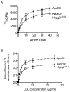
Heparan sulfate (HS) has been proposed to be antiatherogenic through inhibition of lipoprotein retention, inflammation, and smooth muscle cell proliferation. Perlecan is the predominant HS proteoglycan in the artery wall. Here, we investigated the role of perlecan HS chains using apoE null (ApoE0) mice that were cross-bred with mice expressing HS-deficient perlecan (Hspg2(Delta3/Delta3)). Morphometry of cross-sections from aortic roots and en face preparations of whole aortas revealed a significant decrease in lesion formation in ApoE0/Hspg2(Delta3/Delta3) mice at both 15 and 33 weeks. In vitro, binding of labeled mouse triglyceride-rich lipoproteins and human LDL to total extracellular matrix, as well as to purified proteoglycans, prepared from ApoE0/Hspg2(Delta3/Delta3) smooth muscle cells was reduced. In vivo, at 20 minutes influx of human (125)I-LDL or mouse triglyceride-rich lipoproteins into the aortic wall was increased in ApoE0/Hspg2(Delta3/Delta3) mice compared to ApoE0 mice. However, at 72 hours accumulation of (125)I-LDL was similar in ApoE0/Hspg2(Delta3/Delta3) and ApoE0 mice. Immunohistochemistry of lesions from ApoE0/Hspg2(Delta3/Delta3) mice showed decreased staining for apoB and increased smooth muscle alpha-actin content, whereas accumulation of CD68-positive inflammatory cells was unchanged. We conclude that the perlecan HS chains are proatherogenic in mice, possibly through increased lipoprotein retention, altered vascular permeability, or other mechanisms. The ability of HS to inhibit smooth muscle cell growth may also influence development as well as instability of lesions.
Figures


References
-
- Wight TN, Merrilees MJ. Proteoglycans in atherosclerosis and restenosis: key roles for versican. Circ Res. 2004;94:1158–1167. - PubMed
-
- Engelberg H. Endogenous heparin activity deficiency: the ‘missing link’ in atherogenesis? Atherosclerosis. 2001;159:253–260. - PubMed
-
- Pillarisetti S. Lipoprotein modulation of subendothelial heparan sulfate proteoglycans (perlecan) and atherogenicity. Trends Cardiovasc Med. 2000;10:60–65. - PubMed
-
- Hollmann J, Schmidt A, von Bassewitz DB, Buddecke E. Relationship of sulfated glycosaminoglycans and cholesterol content in normal and arteriosclerotic human aorta. Arteriosclerosis. 1989;9:154–158. - PubMed
-
- Murata K, Yokoyama Y. Acidic glycosaminoglycan, lipid and water contents in human coronary arterial branches. Atherosclerosis. 1982;45:53–65. - PubMed
Publication types
MeSH terms
Substances
Grants and funding
LinkOut - more resources
Full Text Sources
Other Literature Sources
Medical
Molecular Biology Databases
Research Materials
Miscellaneous

