

doi: 10.1177/0148607108319795.
Jonathan E Rhoads lecture: Of mice and men... and a few hundred rats
Affiliations
- PMID: 18596320
- PMCID: PMC2596714
- DOI: 10.1177/0148607108319795
Item in Clipboard
Jonathan E Rhoads lecture: Of mice and men... and a few hundred rats
JPEN J Parenter Enteral Nutr.
2008 Jul-Aug.
No abstract available
Figures
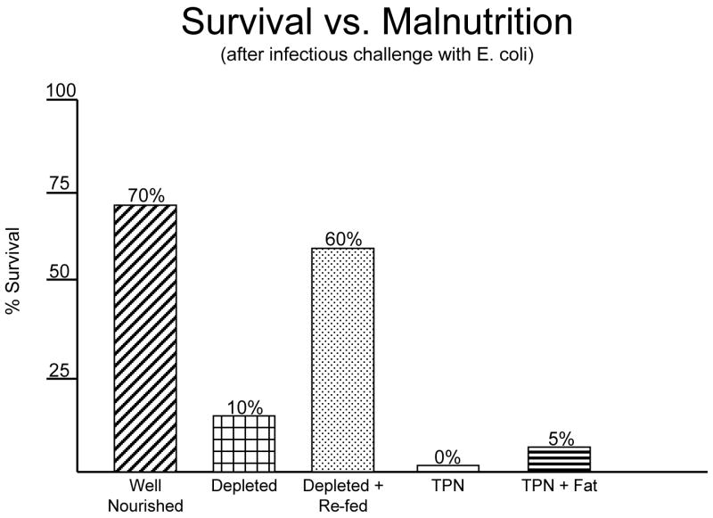
Survival of malnourished animals to an infectious challenge after nutrient manipulation. Protein depletion significantly reduced survival compared to well nourished animals. Refeeding improved the survival of protein depleted rats. Parenteral feeding either with or without fat failed to improve survival.
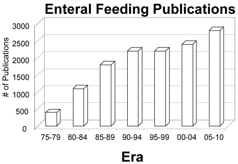
The number of enteral feeding publications in five year blocks from 1975 projected through 2010.
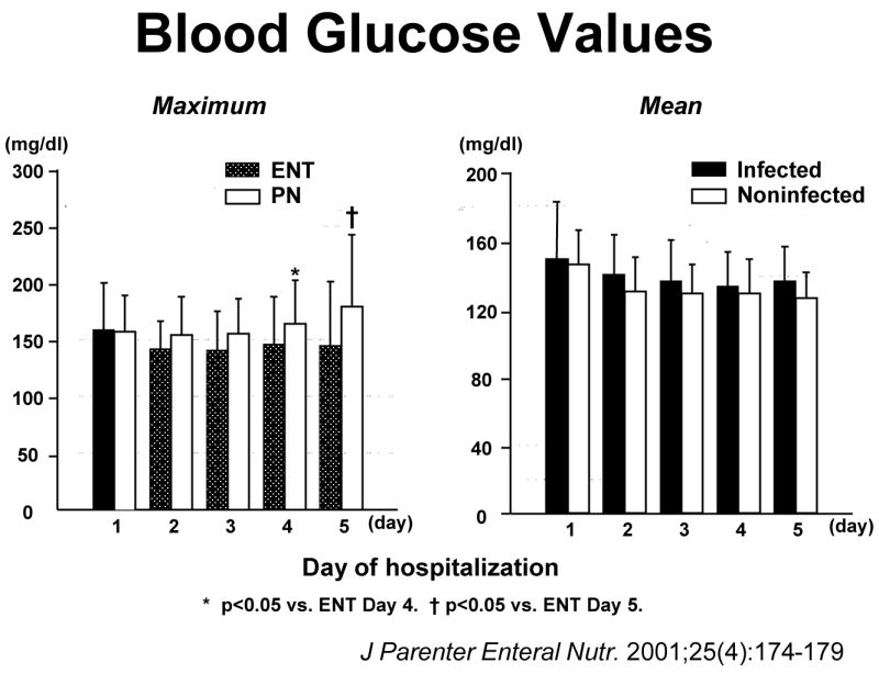
Blood glucose levels in trauma patients randomized to parenteral or enteral nutrition. The figure on the left shows maximal blood glucose levels after enteral or parenteral feeding during the first five days. There were no significant differences in maximal blood glucoses until the fourth or the fifth day when parenterally fed patients developed infectious complications. The right figure shows that there were no significant differences in mean blood glucose values over the first five days in patients who became infected compared to those that remain uninfected. (Reproduced with permission of Kudsk et al. J Parenter Enteral Nutr. 2001;25(4):174–179)
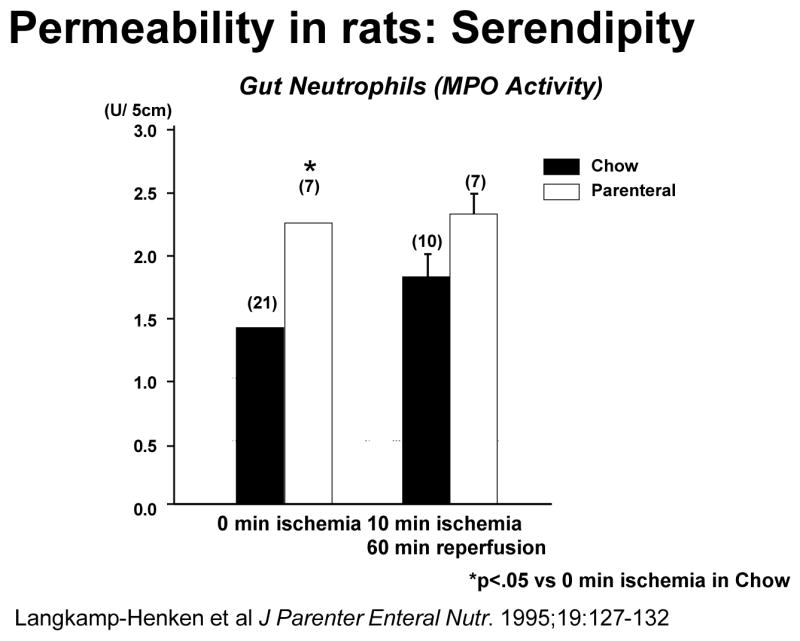
Animals randomized to parenteral feeding significantly increased gut neutrophil accumulation (as measured by myeloperoxidase (MPO) activity) prior to ischemia. (Reproduced with permission of Langkamp-Henken B et al. J Parenter Enteral Nutr. 1995;19:127–132)
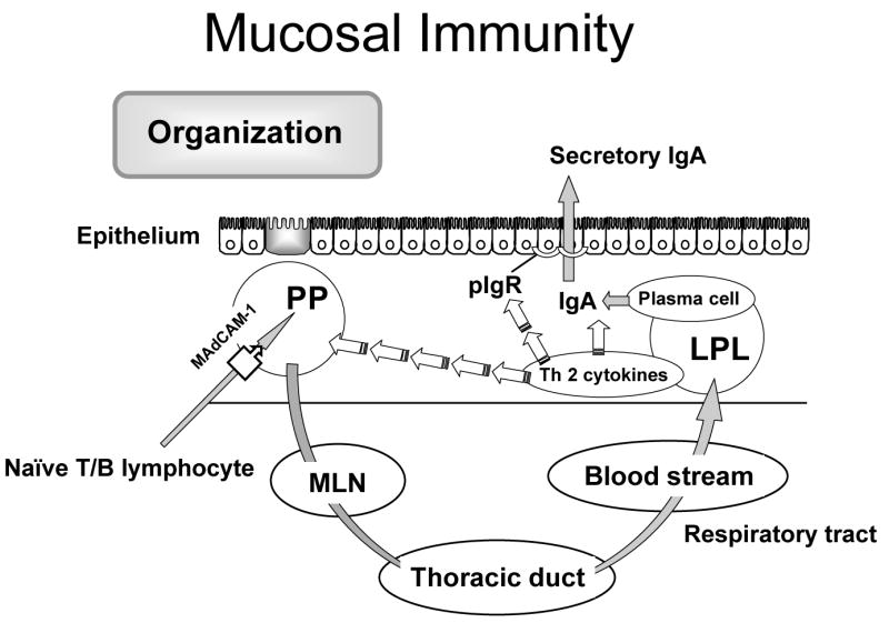
A scheme of the mucosa immune system describing the common mucosa immune hypothesis.
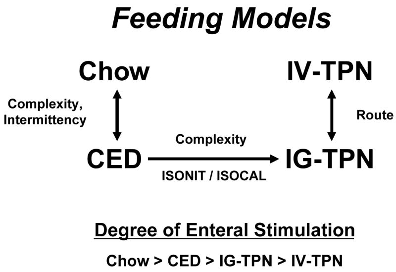
Animals groups used in studies of route and type of nutrition. Intravenous TPN (IV-TPN) and intragastric-TPN (IG-TPN) control for route of nutrition. IG-TPN and complex enteral diets (CED) are isonitrogenous and isocaloric and control for complexity of diets. Both CED and IG-TPN are continuously infused. Chow and CED diets allow comparisons of complexity of diet and intermittency of feeding with Chow representing normal conditions. In general, the beneficial effects of enteral feeding on gut associated lymphoid tissue decreases as the degree of enteral stimulation decreases.
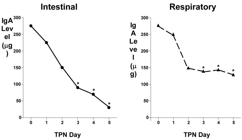
IgA levels in intestinal and the respiratory tract washes drop over time if animals are fed parenteral nutrition (TPN) with no enteral stimulation (Adapted with permission of King et al Arch Surg. 1997;132:1303–1309. Copyright © 1999 American Medical Association. All Rights reserved).
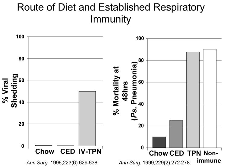
Left figure: In a model of continued viral infection and shedding, enterally fed mice receiving chow or a complex enteral diet (CED) maintain normal immunity while 50% of animals fed parenteral nutrition intravenously (IV-TPN) lost immunity. Right figure: Survival after intra-tracheal Pseudomonas (PS) is increased by previous immunization and reduces mortality from 90% in non-immune animals to 10% in immunized Chow fed mice. Feeding animals a complex enteral diet (CED) after immunization also improves survival but animals fed parenterally (TPN) lose all immunity. (Right figure reproduced with permission of King et al. Ann Surg. 1999;229(2):272–278.).
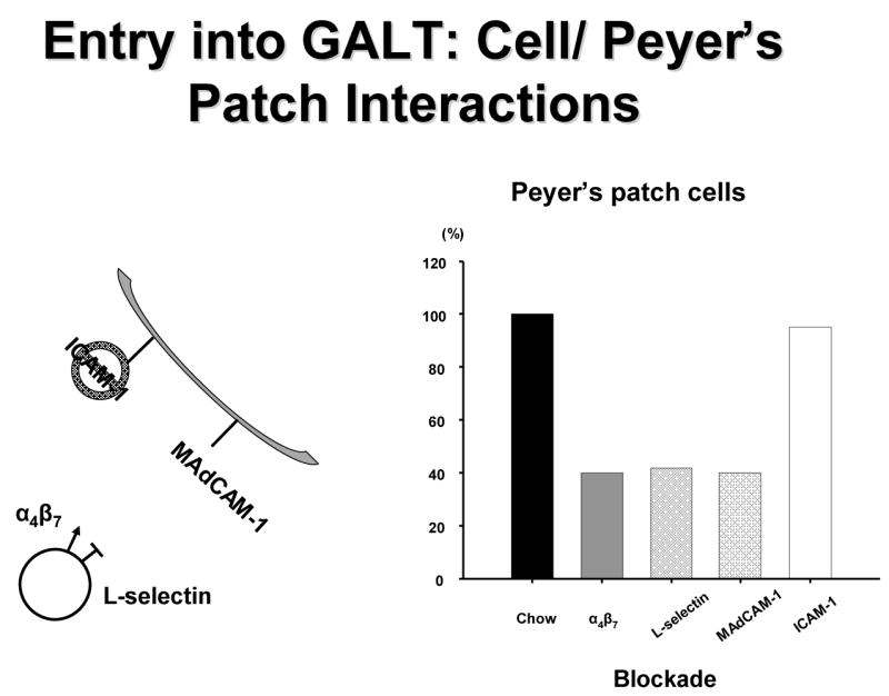
T and B cells enter into the gut associated lymphoid tissue (GALT) through interaction between α4β7 and L-selectin on the cell surfaces with intracellular adhesion molecule-1 (ICAM-1) and mucosal addressin cellular adhesion molecule-1 (MAdCAM-1) on the high endothelial venules on the Peyer’s patches. Blockade of α4β7 , L-selectin or MAdCAM-1 reduced cell entry into Peyer’s patches of chow fed mice whereas ICAM-1 blockade had no effect.
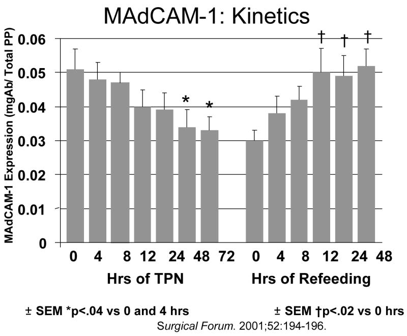
Mucosal addressin cellular adhesion molecule-1 (MAdCAM-1) expression in Peyer’s patches drops after feeding parenteral nutrition (TPN). If animals receive TPN for five days and are then given chow, MAdCAM-1 levels rapidly recover. (Reproduced with permission of Zarzaur et al Surgical Forum. 2001;52:194–196 and the American College of Surgeons).
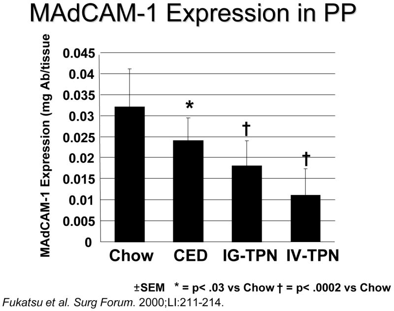
Mucosal addressin cellular adhesion molecule-1 (MAdCAM-1) expression in Peyer’s patches (PP) drops as the amount of enteral stimulation drops from Chow to a complex enteral diet (CED) to intragastric-parenteral nutrition (IG-TPN) to intravenous parenteral nutrition (IV-TPN). (Reproduced with permission of Fukatsu et al. Surgical Forum. 2000;51:211–214 and the American College of Surgeons)
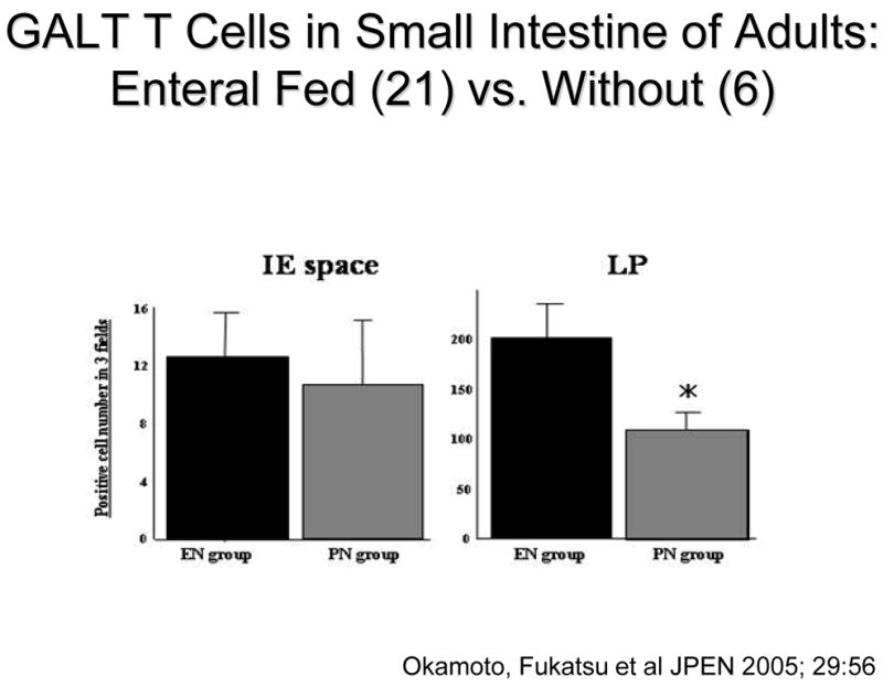
T cells within the gut associated lymphoid tissue (GALT) drop in the lamina propria (LP) of patients fed only parenteral nutrition (PN) preoperatively compared to individuals fed enterally (EN). This reached statistical significance in the Peyer’s patches (reproduced with the permission of Okamato et al, JPEN 2005; 29: 56.)
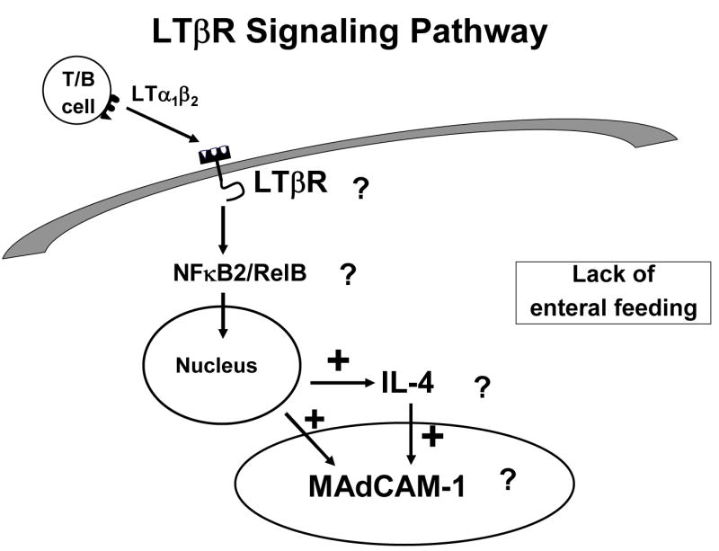
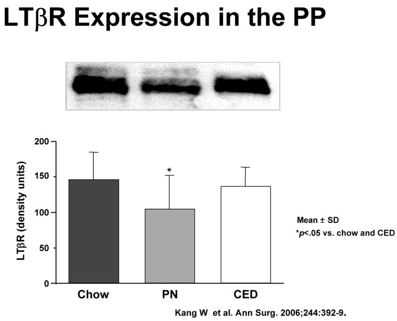
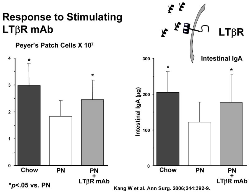
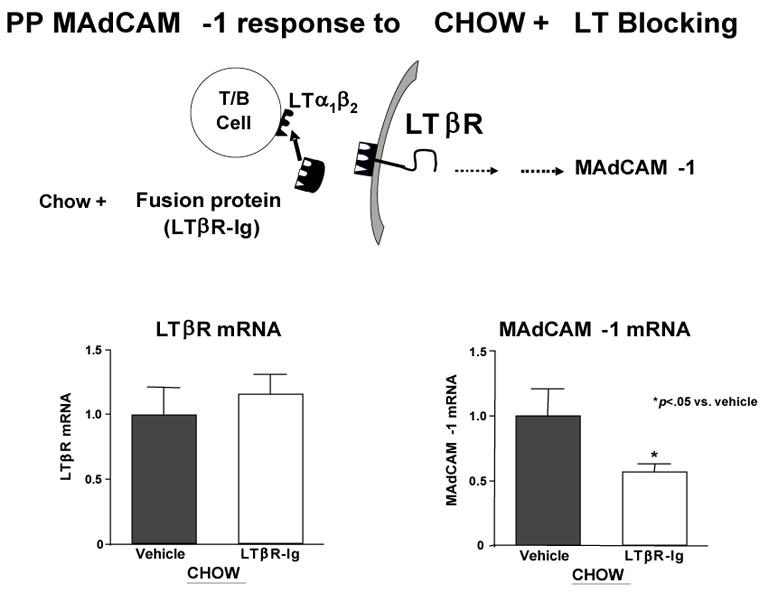
Panel A: The interaction of activated T and B lymphocytes with lymphotoxin beta receptor (LTβR) in the production of mucosal addressin cellular adhesion molecule-1 (MAdCAM-1) and IL-4. Stimulation of LTβR works through NFκB system to stimulate this pathway. Panel B : LTβR expression on Peyer’s patches (PP) is significantly reduced in animals that receive only parenteral nutrition (PN) compared to feeding Chow or a complex enteral diet (CED) (Reproduced with permission of Kang et al Annals of Surgery 2006;244(3):392–399) Panel C: Both Peyer’s patch cell numbers and intestinal IgA significantly increased after LTβR stimulation using a simulating monoclonal antibody (mAb) in PN mice compared to PN fed animals receiving a non-stimulating control antibody. (Reproduced with permission of Kang et al Annals of Surgery 2006;244(3):392–399) Panel D: Chow feeding maintains LTβR mRNA levels while blockade of lymphotoxin α1β1 reduces MAdCAM-1 MRNA production by interfering with LTβR receptor stimulation. (Reproduced with permission of Kang et al JPEN 2007; 31:(5) 358–365)
Panel A: The interaction of activated T and B lymphocytes with lymphotoxin beta receptor (LTβR) in the production of mucosal addressin cellular adhesion molecule-1 (MAdCAM-1) and IL-4. Stimulation of LTβR works through NFκB system to stimulate this pathway. Panel B : LTβR expression on Peyer’s patches (PP) is significantly reduced in animals that receive only parenteral nutrition (PN) compared to feeding Chow or a complex enteral diet (CED) (Reproduced with permission of Kang et al Annals of Surgery 2006;244(3):392–399) Panel C: Both Peyer’s patch cell numbers and intestinal IgA significantly increased after LTβR stimulation using a simulating monoclonal antibody (mAb) in PN mice compared to PN fed animals receiving a non-stimulating control antibody. (Reproduced with permission of Kang et al Annals of Surgery 2006;244(3):392–399) Panel D: Chow feeding maintains LTβR mRNA levels while blockade of lymphotoxin α1β1 reduces MAdCAM-1 MRNA production by interfering with LTβR receptor stimulation. (Reproduced with permission of Kang et al JPEN 2007; 31:(5) 358–365)
Panel A: The interaction of activated T and B lymphocytes with lymphotoxin beta receptor (LTβR) in the production of mucosal addressin cellular adhesion molecule-1 (MAdCAM-1) and IL-4. Stimulation of LTβR works through NFκB system to stimulate this pathway. Panel B : LTβR expression on Peyer’s patches (PP) is significantly reduced in animals that receive only parenteral nutrition (PN) compared to feeding Chow or a complex enteral diet (CED) (Reproduced with permission of Kang et al Annals of Surgery 2006;244(3):392–399) Panel C: Both Peyer’s patch cell numbers and intestinal IgA significantly increased after LTβR stimulation using a simulating monoclonal antibody (mAb) in PN mice compared to PN fed animals receiving a non-stimulating control antibody. (Reproduced with permission of Kang et al Annals of Surgery 2006;244(3):392–399) Panel D: Chow feeding maintains LTβR mRNA levels while blockade of lymphotoxin α1β1 reduces MAdCAM-1 MRNA production by interfering with LTβR receptor stimulation. (Reproduced with permission of Kang et al JPEN 2007; 31:(5) 358–365)
Panel A: The interaction of activated T and B lymphocytes with lymphotoxin beta receptor (LTβR) in the production of mucosal addressin cellular adhesion molecule-1 (MAdCAM-1) and IL-4. Stimulation of LTβR works through NFκB system to stimulate this pathway. Panel B : LTβR expression on Peyer’s patches (PP) is significantly reduced in animals that receive only parenteral nutrition (PN) compared to feeding Chow or a complex enteral diet (CED) (Reproduced with permission of Kang et al Annals of Surgery 2006;244(3):392–399) Panel C: Both Peyer’s patch cell numbers and intestinal IgA significantly increased after LTβR stimulation using a simulating monoclonal antibody (mAb) in PN mice compared to PN fed animals receiving a non-stimulating control antibody. (Reproduced with permission of Kang et al Annals of Surgery 2006;244(3):392–399) Panel D: Chow feeding maintains LTβR mRNA levels while blockade of lymphotoxin α1β1 reduces MAdCAM-1 MRNA production by interfering with LTβR receptor stimulation. (Reproduced with permission of Kang et al JPEN 2007; 31:(5) 358–365)
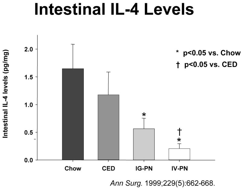
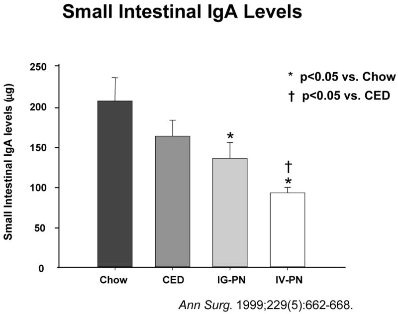
Intestinal interleukin-4 (IL-4) levels drop as the degree of enteral stimulation drops from chow to a complex enteral diet (CED) to intragastric parenteral nutrition (IG-PN) to intravenous PN (IV-PN). Simultaneously, small intestinal immunoglobulin-A (IgA) levels drop in intestinal washings.(Reproduced with the permission of Wu et al, Annals of Surgery 1999: 229 (5): 662–668)
Intestinal interleukin-4 (IL-4) levels drop as the degree of enteral stimulation drops from chow to a complex enteral diet (CED) to intragastric parenteral nutrition (IG-PN) to intravenous PN (IV-PN). Simultaneously, small intestinal immunoglobulin-A (IgA) levels drop in intestinal washings.(Reproduced with the permission of Wu et al, Annals of Surgery 1999: 229 (5): 662–668)
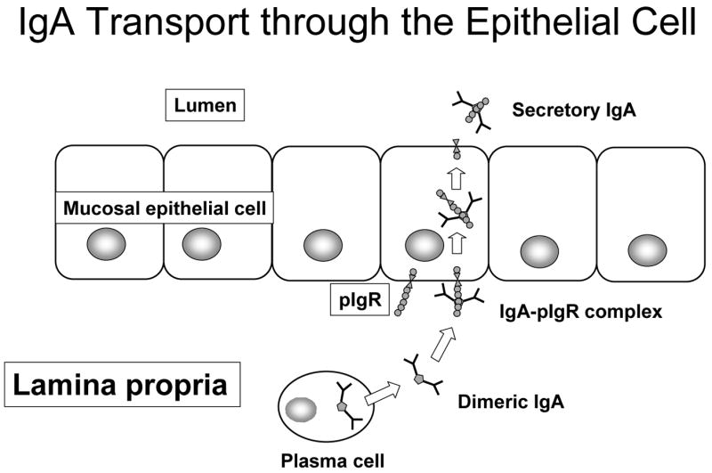
Immunoglobulin-A transport through the epithelial cell is dependant on polymeric immunoglobulin receptor (pIgR) expressed on the basal surface of the mucosal epithelial cells. After pIgR forms a complex with dimeric IgA, the complex is transported across cell and released into the lumen as secretory IgA (sIgA). A small component of the pIgR molecule remains attached to the IgA to distinguish dimeric IgA from secretory IgA.
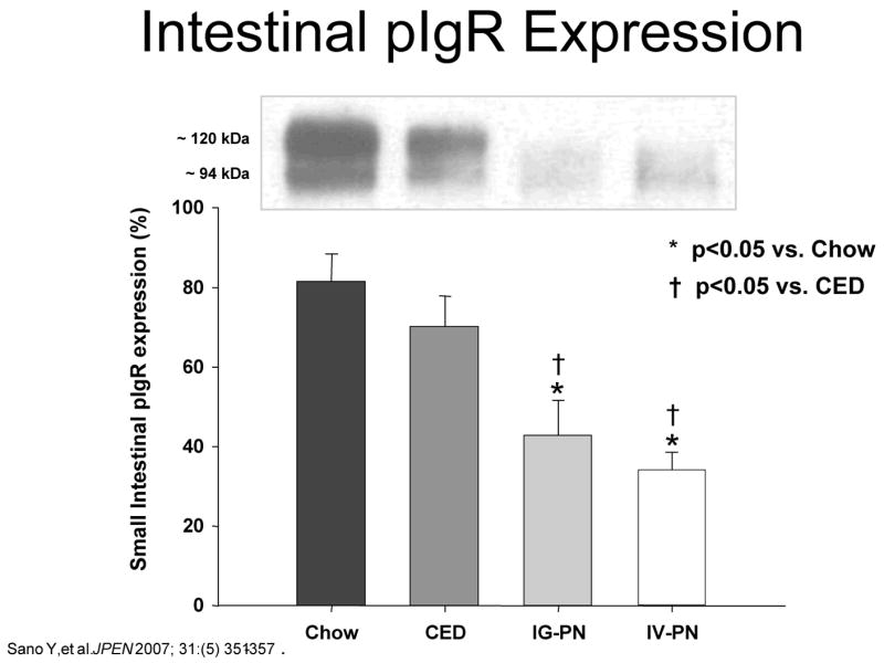
Intestinal polymeric immunoglobulin receptor (pIgR) expression drops as the degree of enteral stimulation decreases from Chow to a complex enteral diet (CED) to intragastric parenteral nutrition (IG-PN) to intravenous PN (IV-PN). (Reproduced with the permission of Sano et al JPEN 2007; 31:(5) 351–357)
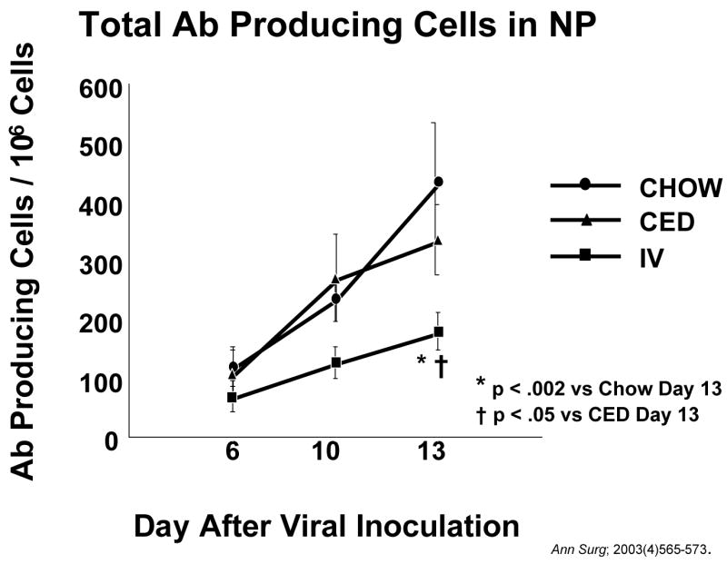
After inoculation with a new viral challenge, antibody (AB) producing cells increase in the nasal passages (NP) of animals fed chow or a complex enteral diet (CED). Accumulation of antibody producing cells is significantly depressed in animals fed parenteral nutrition (IV-TPN). (Reproduced with permission of Johnson et al Ann Surg; 2003(4)565–573)
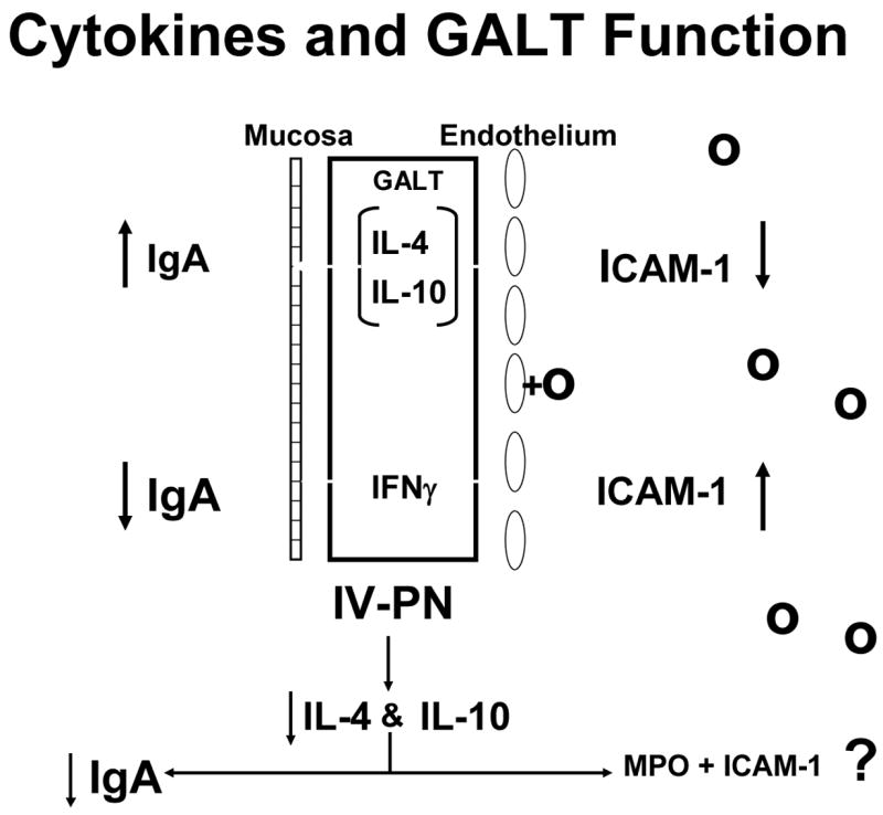
Figure 18a. The interaction between cytokines and gut associated lymphoid tissue (GALT) function is shown. Interleukin-4 (IL-4) and interleukin-10 (IL-10) stimulate IgA production with release of IgA into the lumen while inhibiting intercellular adhesion molecule-1 (ICAM-1) expression on endothelial cells of the vasculature. Interferon gamma (IFNγ) counteracts IL-4 to inhibit IgA production and augment ICAM-1expression. Figure 18b: Intravenous parenteral nutrition (IV-PN) reduces IL-4 and IL-10 leading to an increase in ICAM-1 expression and lower IgA. (Figures 18 a and b reproduced with permission of Kudsk et al. Am J Surg. 2003;185(1)16–21 and The American Journal of Surgery)
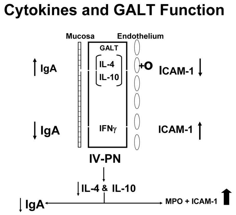
Figure 18a. The interaction between cytokines and gut associated lymphoid tissue (GALT) function is shown. Interleukin-4 (IL-4) and interleukin-10 (IL-10) stimulate IgA production with release of IgA into the lumen while inhibiting intercellular adhesion molecule-1 (ICAM-1) expression on endothelial cells of the vasculature. Interferon gamma (IFNγ) counteracts IL-4 to inhibit IgA production and augment ICAM-1expression. Figure 18b: Intravenous parenteral nutrition (IV-PN) reduces IL-4 and IL-10 leading to an increase in ICAM-1 expression and lower IgA. (Figures 18 a and b reproduced with permission of Kudsk et al. Am J Surg. 2003;185(1)16–21 and The American Journal of Surgery)

Fifteen minutes of gut ischemia/reperfusion (IR) increases the mortality of animals fed intravenous parenteral nutrition (IV-TPN) compared to animals fed chow, a complex enteral diet (CED) or intragastric PN (IG-TPN).
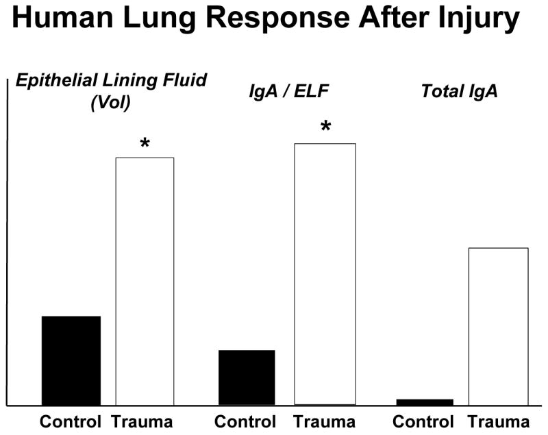
Compared to normal patients undergoing elective surgery, severely injured trauma patients significantly increase the volume of epithelial lining fluid (ELF) recovered after bronchoalveolar lavage and increase the concentration of immunoglobulin-A in the ELF. Overall, there is an increase in total IgA recovered. (adapted with permission of Kudsk et al J Trauma 2008;64(2)316–325)
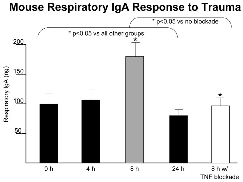
After the minor stress of neck and abdominal incisions in mice, respiratory IgA levels significantly increase eight hours after injury compared to control. This response appears to be driven by tumor necrosis factor (TNF) since TNF blockade eliminates the response. (adapted with permission of Kudsk et al J Trauma 2008;64(2)316–325).
Similar articles
-
1996 Jonathan E. Rhoads Lecture. Mechanism, mechanism, mechanism.JPEN J Parenter Enteral Nutr. 1996 Sep-Oct;20(5):319-24. doi: 10.1177/0148607196020005319. JPEN J Parenter Enteral Nutr. 1996. PMID: 8887899 No abstract available.
-
Jonathan E. Rhoads Lecture. Medicine, nutrition, and patient care: a panoramic view.JPEN J Parenter Enteral Nutr. 1994 Sep-Oct;18(5):387-95. doi: 10.1177/0148607194018005387. JPEN J Parenter Enteral Nutr. 1994. PMID: 7815667 Review. No abstract available.
-
First annual Jonathan E. Rhoads lecture: Dedicated to Jonathan E. Rhoads for 45 years the purveyor of opportunities for students young and old--in grateful appreciation.JPEN J Parenter Enteral Nutr. 1978 May;2(2):75-6. doi: 10.1177/014860717800200201. JPEN J Parenter Enteral Nutr. 1978. PMID: 121755 No abstract available.
-
Clinical aspects of essential fatty acid metabolism: Jonathan Rhoads Lecture.JPEN J Parenter Enteral Nutr. 2003 May-Jun;27(3):168-75. doi: 10.1177/0148607103027003168. JPEN J Parenter Enteral Nutr. 2003. PMID: 12757109 Review.
-
Jonathan E. Rhoads lecture. Intravenous hyperalimentation and cancer. A historical perspective.JPEN J Parenter Enteral Nutr. 1986 Jul-Aug;10(4):337-42. doi: 10.1177/0148607186010004337. JPEN J Parenter Enteral Nutr. 1986. PMID: 3091858 Review. No abstract available.
Cited by
-
The change in the amount of immunoglobulins as a response to stress experienced by soldiers on a peacekeeping mission.Int Arch Occup Environ Health. 2014 Aug;87(6):615-22. doi: 10.1007/s00420-013-0899-0. Epub 2013 Aug 13. Int Arch Occup Environ Health. 2014. PMID: 23943194
-
Nutrition and gut immunity.Surg Clin North Am. 2011 Aug;91(4):755-70, vii. doi: 10.1016/j.suc.2011.04.007. Epub 2011 Jun 8. Surg Clin North Am. 2011. PMID: 21787966 Free PMC article. Review.
-
The enteric nervous system neuropeptide, bombesin, reverses innate immune impairments during parenteral nutrition.Ann Surg. 2014 Sep;260(3):432-43; discussion 443-4. doi: 10.1097/SLA.0000000000000871. Ann Surg. 2014. PMID: 25115419 Free PMC article.
References
-
- Rombeau JL, Muldoon D, Jonathan E, Rhoads MD. Quaker sense and sensibility in the world of surgery. Philadelphia: Hanley & Belfus; 1997.
-
- Kudsk KA. Dear Miss Milk Toast (1998 Presidential Address - ASPEN) J Parenter Enteral Nutr. 1998;22(4):191–198. - PubMed
-
- Peterson SR, Kudsk KA, Carpenter G, Sheldon GF. Malnutrition and Immunocompetence: Increased mortality following an infectious challenge during hyperalimentation. J Trauma. 1981;21:528–533. - PubMed
-
- Kudsk KA, Carpenter BS, Peterson S, Sheldon GF. Effect of Enteral and Parenteral Feeding in Malnourished Rats with E. coli-Hemoglobin Adjuvant Peritonitis. J Surg Res. 1981;31:105–110. - PubMed
-
- Kudsk KA, Stone JM, Carpenter BA, Sheldon GF. Enteral and parenteral feeding influences mortality after hemoglobin-E. coli peritonitis in normal rats. J Trauma. 1983;23:605–609. - PubMed
Publication types
MeSH terms
Grants and funding
LinkOut - more resources
Full Text Sources
Medical
