

Oxidative injury in the cerebral cortex and subplate neurons in periventricular leukomalacia
- PMID: 18596545
- PMCID: PMC2831221
- DOI: 10.1097/NEN.0b013e31817e5c5e
Oxidative injury in the cerebral cortex and subplate neurons in periventricular leukomalacia
Abstract
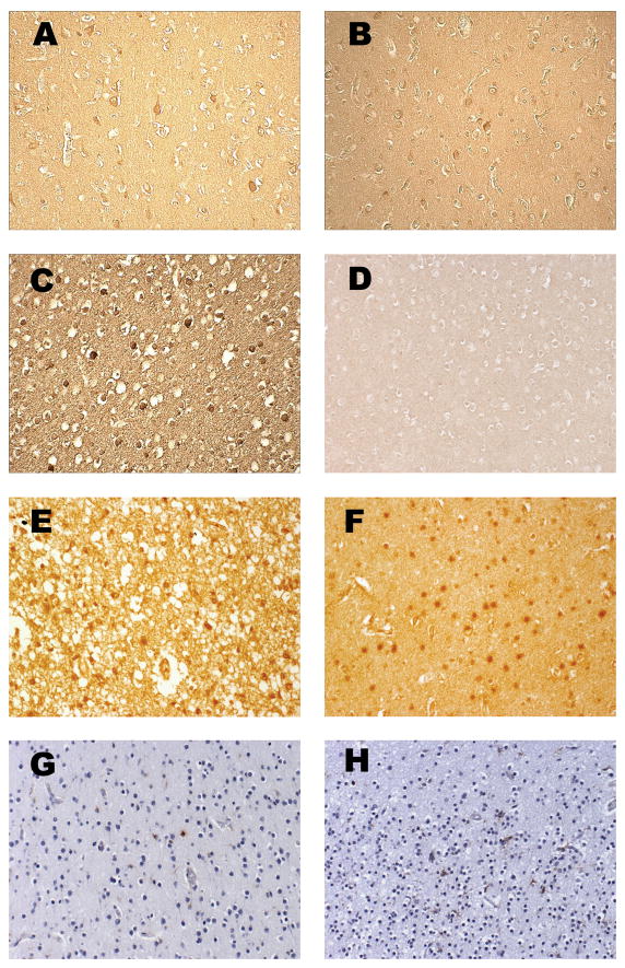
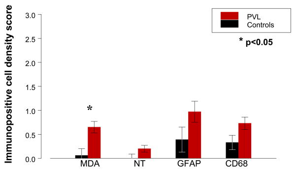
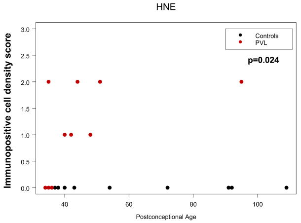
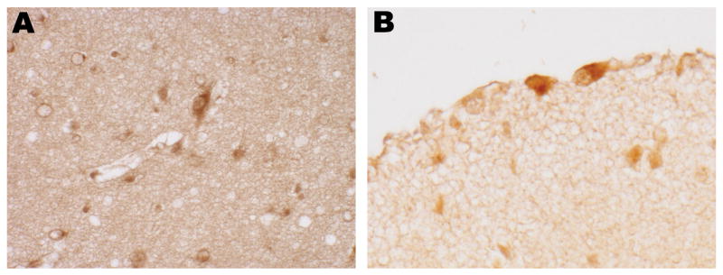
We previously identified immunocytochemical evidence of nitrative and oxidative injury in premyelinating oligodendrocytes in periventricular leukomalacia (PVL). Here, we tested the hypothesis that free radical injury occurs in the overlying cerebral cortex and subplate neurons in PVL. We immunostained for nitrotyrosine, malondialdehyde, and hydroxynonenal adducts and scored neuron staining density in PVL (n = 11) and non-PVL (n = 15) cases (postconceptional ages from 34 to 109 weeks). Analysis of covariance controlled for age. Mean malondialdehyde scores in PVL cases were increased over controls (p = 0.005). Hydroxynonenal scores increased with age only in PVL cases (diagnosis vs age interaction; p = 0.024). Nitrotyrosine scores were not significantly increased. In 11 PVL and 23 control cases between 20 and 183 postconceptional weeks, cells morphologically consistent with subplate and Cajal-Retzius neurons showed qualitatively increased free radical modification in PVL over control cases with statistically significant odds ratios for hydroxynonenal and nitrotyrosine in both subplate neurons and Cajal-Retzius cells. Glial fibrillary acidic protein and CD68 scores for reactive astrocytes and microglia, respectively, were not significantly increased, suggesting a minimal inflammatory response. Thus, oxidative/nitrative damage to cortical and "pioneer" neurons, although mild overall, may contribute to cortical volume loss and cognitive/behavioral impairment in survivors of prematurity.
Figures
References
-
- Volpe JJ. Cerebral white matter injury of the premature infant-more common than you think. Pediatrics. 2003;112:176–80. - PubMed
-
- Kinney HC, Panigrahy A, Newburger JW, Jonas RA, Sleeper LA. Hypoxic-ischemic brain injury in infants with congenital heart disease dying after cardiac surgery. Acta Neuropathol (Berl) 2005;110:563–78. - PubMed
-
- Inder TE, Huppi PS, Warfield S, Kikinis R, Zientara P, Barnes PD, et al. Periventricular white matter injury in the premature infant is associated with a reduction in cerebral cortical gray matter volume at term. Ann Neurol. 1999;46:755–60. - PubMed
-
- Inder TE, Warfield SK, Wang H, Huppi PS, Volpe JJ. Abnormal cerebral structure is present at term in premature infants. Pediatrics. 2005;115:286–94. - PubMed
