Review
. 2008 Jul;67(7):635-48.
doi: 10.1097/NEN.0b013e31817d751c.

Update on recent molecular and genetic advances in frontotemporal lobar degeneration

Affiliations
Review

Update on recent molecular and genetic advances in frontotemporal lobar degeneration

Eileen H Bigio. J Neuropathol Exp Neurol. 2008 Jul.

Abstract

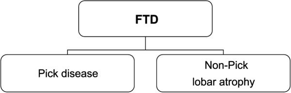
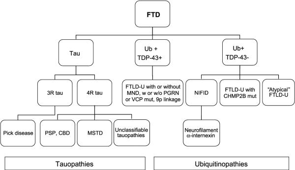
Great strides have been made in the last 2 years in the field of frontotemporal lobar degeneration (FTLD), particularly with respect to the genetics and molecular biology of FTLD with ubiquitinated inclusions. It is now clear that most cases of familial FTLD with ubiquitinated inclusions have mutations in the progranulin gene, located on chromosome 17. It is also clear that most ubiquitinated inclusions in FTLD with ubiquitinated inclusions are composed primarily of TAR DNA-binding protein-43. Thus, FTLDs can be separated into 2 major groups (i.e. tauopathies and ubiquitinopathies), and most of the ubiquitinopathies can now be defined as TAR DNA-binding protein-43 proteinopathies. Many of the familial FTLDs are linked to chromosome 17, including both the familial tauopathies and the familial TAR DNA-binding protein-43 proteinopathies with progranulin mutations. This review highlights the neuropathologic features and the most important discoveries of the last 2 years and places these findings into the historical context of FTLD.


Figures

FIGURE 1
FIGURE 1
Frontotemporal dementia (FTD) pathologic subtypes, 1987.
FIGURE 2
FIGURE 2
Pick disease pathology. (A, B) Circumscribed frontal and temporal atrophy. (C) Pick bodies in dentate gyrus seen on hematoxylin and eosin (top; 40×) and with paired helical filament 1 immunohistochemistry (IHC) (bottom; 60×). (D) Pick bodies seen with paired helical filament 1 IHC (top; 60×) in frontal cortical layer II, which also shows microvacuolation and gliosis on hematoxylin and eosin (bottom; 10×).
FIGURE 3
FIGURE 3
Dementia lacking distinctive histology. Pathology (circumscribed frontal and temporal atrophy, as in Pick disease, is also present). (A) Caudate atrophy. (B) Pallor of the substantia nigra. (C) Superficial microvacuolation and gliosis, cortical layer II, frontal and temporal lobes (hematoxylin and eosin: 20×). (D) Neuronal loss and gliosis, caudate nucleus (hematoxylin and eosin: 40×). (E) Ubiquitin immunohistochemistry (IHC) of frontal lobe shows no inclusions (40×). (F) Ubiquitin IHC of dentate gyrus shows no inclusions (60×). (G) Neuronal loss and gliosis in substantia nigra (hematoxylin and eosin: 20×).
FIGURE 4
FIGURE 4
Familial tauopathy with dementia linked to chromosome 17 pathology. (A) Dentate gyrus with Pick-like bodies, L266V tau mutation (paired helical filament 1 immunohistochemistry [IHC]; 40×). (B) Progressive supranuclear palsy (PSP)-like pathology in frontal cortex, with neuronal PSP-type tangle (arrow) and tufted astrocyte (arrowhead; AT8 IHC; 60×). (C) Corticobasal degeneration-like cortical pathology with 2 large astrocytic plaques and abundant thread pathology (Gallyas stain; 20×). (D) Cortical gray-white junction with unique tau pathology consisting predominantly of globular white matter (oligodendroglial)-insoluble tau deposits (AT8 IHC; 20×).
FIGURE 5
FIGURE 5
Frontotemporal lobar degeneration with ubiquitinated inclusions pathology. (A) Superficial frontal cortex with neuronal cytoplasmic inclusions (arrow), neuronal intranuclear inclusions (NIIs; solid arrowhead), and dystrophic neurites (open arrowhead; 40×). (B) Dentate gyrus with neuronal cytoplasmic inclusions (60×). (C) Putamen neuron with NII (100×). (D) Dentate gyrus neuron with NII (60×). All are ubiquitin immunohistochemistry (Dako polyclonal, Carpinteria, CA).
FIGURE 6
FIGURE 6
Ubiquitin and TAR-DNA binding protein-43 (TDP-43) immunohistochemistry (IHC) in frontotemporal lobar degeneration with ubiquitinated inclusions (FTLD-U). Ubiquitin (A; ubiquitin IHC, Dako polyclonal; 60×) and TDP-43 (B; TDP-43 IHC, Proteintech) in frontal cortex of FTLD-U have similar labeling patterns; (A) Ubiquitin IHC (Dako polyclonal). (B) TDP-43 (Proteintech).
FIGURE 7
FIGURE 7
Typical and “atypical” frontotemporal lobar degeneration with ubiquitinated inclusions (FTLD-U). (A, B) Typical FTLD-U labeled with ubiquitin (A; 40×) and TAR-DNA binding protein-43 (TDP-43; B; 40×); both label cytoplasmic inclusions (CIs). (C, D) “Atypical” FTLD-U labeled with ubiquitin (C; 60×) and TDP-43 (D; 60×). Inclusions are ubiquitin positive but TDP-43 negative (note arrows pointing to unlabeled CIs).
FIGURE 8
FIGURE 8
Hippocampal sclerosis, ubiquitin, and TAR-DNA binding protein-43 immunohistochemistry (IHC). (A) Ubiquitin IHC of dentate gyrus in hippocampal sclerosis shows 2 equivocal inclusions noted retrospectively (arrows; 60×). (B) TAR-DNA binding protein-43 of same case clearly shows 2 positive cytoplasmic inclusions (CIs; arrows; 40×). Note that all nuclei are labeled except those in neurons with CIs.
FIGURE 9
FIGURE 9
Ubiquitin and TAR-DNA binding protein-43 immunohistochemistry (IHC) in familial amyotrophic lateral sclerosis (FALS) with and without superoxide dismutase 1 (SOD1) mutations. Familial amyotrophic lateral sclerosis cases without SOD1 mutation (A, B) and with SOD1 mutation (C, D). Ubiquitin (A, C) clearly labels Lewy-like bodies (A) and skein-like inclusions (C) in FALS cases with (C) and without (A) SOD1 mutations. TAR-DNA binding protein-43 (B, D) labels FALS without SOD1 mutation (B) but is negative in FALS with SOD1 mutation (D) (all magnifications: 60×).
FIGURE 10
FIGURE 10
Frontotemporal lobar degeneration (FTLD) pathologic subtypes, 2008. The pathology of frontotemporal dementia (FTD) has become increasingly complex, and pathologic diagnoses now incorporate molecular information compared with Figure 1. CBD, corticobasal degeneration; CHMP2B, charged multivesicular body protein 2B; MND, motor neuron disease; MSTD, multiple system taupathy with presenile dementia; NIFID, neuronal intermediate filament inclusion disease; PGRN, progranulin; PSP, progressive supranuclear palsy; TDP-43, TAR-DNA binding protein-43; VCP, volosin-containing protein.

Similar articles

Cited by

References

    1. Pick A. Über die Beziehungen der senilen Hirnatrophie zur Aphasie. Prager Med Wochenschr. 1892;17:165–67.
    1. Alzheimer A. Über eigenartige Krankheitsfȧlle des späteren Alters. Z Gesamte Neurol Psychiat. 1911;4:356–85.
    1. Gans A. Betrachtungen über art und Ausbreitung des krankhaften Prozesses in einem Fall van Pickscher Atrophie des Stirnhins. Zeitschr f d ges Neurol u Psychiatr (Berl) 1922;80:10–28.
    1. Brun A, Gustafson L, Risberg J, et al. Frontal lobe dementia of non-Alzheimer type. Arch Gerontol Geriatr. 1987;6:193–208. - PubMed
    1. Knopman DS, Mastri AR, Frey WH, 2nd, et al. Dementia lacking distinctive histologic features: A common non-Alzheimer degenerative dementia. Neurology. 1990;40:251–56. - PubMed

Publication types

Substances