Review
. 2008 Jul;15(7):690-8.
doi: 10.1038/nsmb.1456.

Viral membrane fusion

Affiliations
Review

Viral membrane fusion

Stephen C Harrison. Nat Struct Mol Biol. 2008 Jul.

Abstract

Infection by viruses having lipid-bilayer envelopes proceeds through fusion of the viral membrane with a membrane of the target cell. Viral 'fusion proteins' facilitate this process. They vary greatly in structure, but all seem to have a common mechanism of action, in which a ligand-triggered, large-scale conformational change in the fusion protein is coupled to apposition and merger of the two bilayers. We describe three examples--the influenza virus hemagglutinin, the flavivirus E protein and the vesicular stomatitis virus G protein--in some detail, to illustrate the ways in which different structures have evolved to implement this common mechanism. Fusion inhibitors can be effective antiviral agents.


Figures

Figure 1
Figure 1. Sequence of events in membrane fusion promoted by a viral fusion protein.
Ambiguities remain in some aspects of this scheme (see main text). (a) The protein in the pre-fusion conformation, with its fusion peptide or loop (light green) sequestered. The representation is purely schematic, and various features of specific proteins are not incorporated—for example, the displacement of the N-terminal fragment of proteins that are cleaved from a precursor or the dimer-to-trimer rearrangement on the surface of flaviviruses. (b) Extended intermediate. The protein opens up, extending the fusion peptide or loop to interact with the target bilayer. The part of the protein that bears the fusion peptide forms a trimer cluster. (c) Collapse of the extended intermediate: a C-terminal segment of the protein folds back along the outside of the trimer core. The segments from the three subunits fold back independently, so that at any point in the process they can extend to different distances along the trimer axis, and the entire trimer can bow outward, away from the deforming membrane. (d) Hemifusion. When collapse of the intermediate has proceeded far enough to bring the two bilayers into contact, the apposed, proximal leaflets merge into a hemifusion stalk. (e) Fusion pore formation. As the hemifused bilayers open into a fusion pore, the final zipping up of the C-terminal ectodomain segments snaps the refolded trimer into its fully symmetric, post-fusion conformation, preventing the pore from resealing.
Figure 2
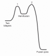
Figure 2. Schematic diagram illustrating the (free) energy changes during fusion of two bilayers.
The relative heights of the various barriers are arbitrary. Fusion proteins accelerate the process by coupling traversal of these barriers to energetically favorable conformational changes.
Figure 3
Figure 3. Influenza virus hemagglutinin: proposed sequence of fusogenic conformational changes.
(a) The pre-fusion conformation. Each subunit is shown in a different color. The binding site for the receptor, sialic acid, is at the top of each subunit, but contact with a receptor molecule is not shown. Red asterisk, the sequestered fusion peptide of the red subunit, at the N terminus of HA2. (b) HA1 dissociates from its tightly docked position in response to proton binding. Each HA1 remains flexibly tethered to the corresponding HA2 by a disulfide bond (near the bottom of the ectodomain, in the orientation shown here). (c) The extended intermediate. The loop between the shorter and longer helices in HA2 (for example, the two red helices and the loop connecting them, in b) becomes a helix, thereby translocating the fusion peptide toward the target membrane. The fusion peptides (asterisk) are shown interacting as amphipathic helices with the target bilayer. The loop-to-helix transition creates a long, three-chain coiled coil at the core of the trimer. (d) Collapse of the extended intermediate to generate the post-fusion conformation. The lower parts of the protein (as seen in the orientation in c) fold back along the outside of the three-chain coiled coil. The collapse is complete only when the two membranes have fused completely. The post-fusion conformation is shown in a 'horizontal' orientation, to correspond to the sequence in Figure 1. (e) Detail illustrating some features of the membrane-proximal region of influenza virus HA2 after fusion is complete. The N termini of the coiled-coil helices are capped by contacts with amino acid residues in the link between the fusion peptide and the coiled coil, as well as with residues near the C terminus of the ectodomain, proximal to the transmembrane helices. This cap locks into place all the membrane-proximal components of the structure. The fusion peptides at the N termini of three HA2 chains are shown as cylinders (possible amphipathic helices) lying partly immersed in the outer leaflet of the membrane bilayer, as suggested by NMR and EPR studies. The transmembrane segments, likely to be α-helices, are also shown as cylinders. The relationships in this drawing among the fusion peptides and the transmembrane helices, chosen to illustrate the scale of the structures and the approximate distances between them, are purely schematic, as there is no single structure yet determined experimentally that contains all the elements included here. Only the crystallographically determined components are in ribbon representation.
Figure 4
Figure 4. Flavivirus E: proposed sequence of fusogenic conformational changes.
(a) The packing of 180 E subunits (90 dimers) in an icosahedral array on the surface of a flavivirus particle. The red, yellow and blue parts of each subunit correspond respectively to domains I, II and III of the ectodomain. (b) 'Side view' of the pre-fusion, dimeric conformation of the E protein, based on the crystal structure of dengue E (residues 1–395), supplemented by a representation of the 'stem' segment (two helices linked by a short loop, lying in the plane of the membrane head groups) and the transmembrane anchor (a helical hairpin), derived from a cryo-EM reconstruction of the virion. The domains in one of the two subunits are colored as in a; the other subunit is in gray. The fusion loop is at the tip of domain II, on the far right of the colored subunit, buried at the contact with domain III of the dimer partner. (c) Monomeric transition between the pre-fusion dimer and the trimeric extended intermediate. The three subunits that will associate into the extended intermediate in d are not yet in contact. The drawing embodies the suggestion that domains I and II have swung outward, while domain III and the stem remain oriented against the membrane roughly as in the pre-fusion state. The fusion loop is now at the top of the diagram and is shown already interacting with the target bilayer. (d) Extended intermediate. Domains I and II have associated into the trimeric core of the post-fusion conformation, but domain III has not yet flipped over (upper arrows) to dock against them. To indicate that the stem segment must then zip back along the trimer core (lower arrows), the stem is represented by loops 'poised' to reconfigure. (e) Post-fusion conformation. Domain III has reoriented, and the stem (dashed line, as there is no direct structural information on its conformation or exact position in the post-fusion trimer) connects it to the transmembrane anchor, now brought together with the fusion loop in the single, fused bilayer. The post-fusion conformation is shown in a 'horizontal' orientation, to correspond to the sequence in Figure 1.
Figure 5
Figure 5. VSV-G: proposed fusogenic conformational changes.
(a) Pre-fusion trimer. The three subunits are in red, blue and green. The fusion loops (asterisk) are held away from the target membrane. The crystal structure does not include about 40 residues, represented here for each subunit by a slightly wavy line, that connect to the transmembrane anchor. (b) Pre-fusion conformation of one subunit (in the orientation of the red subunit in a). Core domain, red; α-helix at the trimer contact, light blue; two-part fusion apparatus, dark blue; C-terminal segment, dark green. Dashed line, the part of the C-terminal segment that is missing from the crystal structure; letter N, the N terminus. (c) Suggested extended intermediate conformation of one subunit, colored as in b. The fusion domains have reoriented (curved arrow in b), with the fusion loops (asterisk) now in contact with the target membrane; the reorientation seems to be driven in part by a loop-to-helix transition that elongates the helix at the trimer contact. The C-terminal segment still connects to the viral membrane (dashed arrow), but it must fold back along the outside of the trimer (curved arrow) to complete the transition to the post-fusion conformation. (d) Post-fusion conformation of one subunit, in the orientation and colors of the subunit in b and c. The C-terminal segment has folded back, and it now projects toward the fusion loops. (e) Post-fusion conformation of the trimer, with colors as in a. It is shown in a 'horizontal' orientation, to correspond to the sequence in Figure 1.
Figure 6
Figure 6. The transition between the trimeric extended intermediate and the post-fusion conformation of the HIV gp41 ectodomain.
In the extended intermediate (left), the HR1 segment of each of the three subunits is shown as an α-helix, and the HR2 segment as an extended chain. The fusion peptides are imagined to be inserted into or against the target-cell membrane (top) and the transmembrane anchors pass into the viral membrane (bottom). In the post-fusion conformation (right), the HR2 segment has zipped up into a helix along the outside of the HR1 three-chain coiled-coil, creating a 'trimer of hairpins', and the two membranes have fused. Courtesy Gaël McGill (see http://www.molecularmovies.org).

Similar articles

Cited by

References

    1. Chernomordik LV, Kozlov MM. Protein-lipid interplay in fusion and fission of biological membranes. Annu. Rev. Biochem. 2003;72:175–207. doi: 10.1146/annurev.biochem.72.121801.161504. - DOI - PubMed
    1. Chernomordik LV, Zimmerberg J, Kozlov MM. Membranes of the world unite! J. Cell Biol. 2006;175:201–207. doi: 10.1083/jcb.200607083. - DOI - PMC - PubMed
    1. Helenius A, Kartenbeck J, Simons K, Fries E. On the entry of Semliki Forest virus into BHK-21 cells. J. Cell Biol. 1980;84:404–420. doi: 10.1083/jcb.84.2.404. - DOI - PMC - PubMed
    1. White J, Helenius A. pH-dependent fusion between Semliki Forest virus membrane and liposomes. Proc. Natl. Acad. Sci. USA. 1980;77:3273–3277. doi: 10.1073/pnas.77.6.3273. - DOI - PMC - PubMed
    1. Kuzmin PI, Zimmerberg J, Chizmadzhev YA, Cohen FS. A quantitative model for membrane fusion based on low-energy intermediates. Proc. Natl. Acad. Sci. USA. 2001;98:7235–7240. doi: 10.1073/pnas.121191898. - DOI - PMC - PubMed

Publication types

MeSH terms

Substances