The fusion pores of Ca2+ -triggered exocytosis
- PMID: 18596819
- PMCID: PMC2914174
- DOI: 10.1038/nsmb.1449
The fusion pores of Ca2+ -triggered exocytosis
Abstract
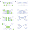
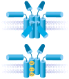
The aqueous compartment inside a vesicle makes its first connection with the extracellular fluid through an intermediate structure termed the exocytotic fusion pore. Progress in exocytosis can be measured in terms of the formation and growth of the fusion pore. The fusion pore has become a major focus of research in exocytosis; sensitive biophysical measurements have provided various glimpses of what it looks like and how it behaves. Some of the principal questions about the molecular mechanism of exocytosis can be cast explicitly in terms of properties and transitions of fusion pores. This Review will present current knowledge about fusion pores in Ca(2+)-triggered exocytosis, highlight recent advances and relate questions about fusion pores to broader issues concerning how cells regulate exocytosis and how nerve terminals release neurotransmitter.
Figures


References
-
- Lindau M, Almers W. Structure and function of fusion pores in exocytosis and ectoplasmic membrane fusion. Curr Opin Cell Biol. 1995;7:509–517. - PubMed
-
- Klyachko VA, Jackson MB. Capacitance steps and fusion pores of small and large-dense-core vesicles in nerve terminals. Nature. 2002;418:89–92. This study revealed capacitance steps of small clear vesicles and showed that they undergo kiss-and-run by forming much smaller fusion pores. - PubMed
-
- Breckenridge LJ, Almers W. Currents through the fusion pore that forms during exocytosis of a secretory granule. Nature. 1987;328:814–817. The first measurement of fusion pore conductance gave a value in the range of ion-channel conductances. - PubMed
Publication types
MeSH terms
Substances
Grants and funding
LinkOut - more resources
Full Text Sources
Other Literature Sources
Miscellaneous

