. 2008 Nov;15(21):1446-53.
doi: 10.1038/gt.2008.101. Epub 2008 Jul 3.

Cancer gene therapy using mesenchymal stem cells expressing interferon-beta in a mouse prostate cancer lung metastasis model

Affiliations

Cancer gene therapy using mesenchymal stem cells expressing interferon-beta in a mouse prostate cancer lung metastasis model

C Ren et al. Gene Ther. 2008 Nov.

Abstract

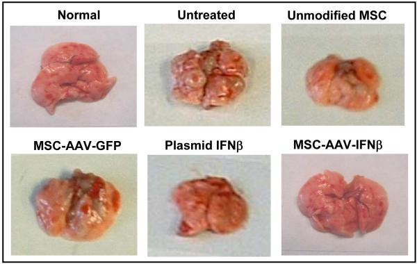
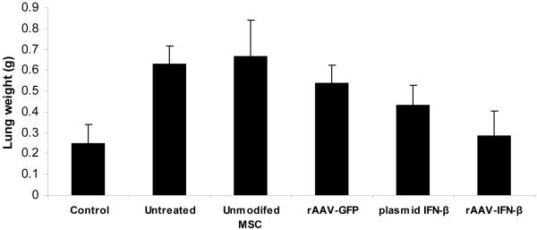
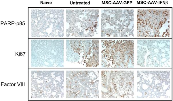
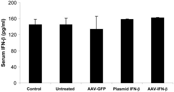
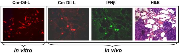
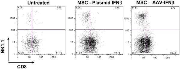
Cell-based therapy for cancer is a promising new field. Among cell types that can be used for this purpose, mesenchymal stem cells (MSCs) appear to hold great advantage for reasons including easier propagation in culture, possible genetic modification to express therapeutic proteins and preferential homing to sites of cancer growth upon in vivo transfer. The present study evaluated the potential of genetically modified MSC, constitutively expressing interferon (IFN)-beta, in an immunocompetent mouse model of prostate cancer lung metastasis. A recombinant adeno-associated virus (rAAV) encoding mouse IFN-beta was constructed and initially tested in vitro for high-level expression and bioactivity of the transgenic protein. MSCs were transduced by the rAAV-IFN-beta or green fluorescent protein ex vivo and used as cellular vehicles to target lung metastasis of TRAMP-C2 prostate cancer cells in a therapy model. Cohorts of mice were killed on days 30 and 75 to determine the effect of therapy by measurement of tumor volume, histology, immunohistochemistry, enzyme-linked immunosorbent assay and flow cytometry. Results indicated a significant reduction in tumor volume in lungs following IFN-beta-expressing MSC therapy. Immunohistochemistry of the lung demonstrated increased tumor cell apoptosis and decreased tumor cell proliferation and blood vessel counts. A significant increase in the natural kill cell activity was observed following IFN-beta therapy correlating the antitumor effect. Systemic level of IFN-beta was not significantly elevated from this targeted cell therapy. These data demonstrate the potential of MSC-based IFN-beta therapy for prostate cancer lung metastasis.


Figures

Figure 1
Figure 1. Recombinant-AAV 6 expressing murine IFN-β
An rAAV encoding human IFN-β under the control of CMV-chicken beta actin promoter was constructed using pSub201 vector (A). Expression of IFN-β was confirmed by transducing the vector in 293 cells and analyzing the culture supernatant by Western blot using an IFN-β antibody (B). Transduction efficiency of mouse MSC by rAAV- IFN-β was determined by in situ staining of the fixed cells using a rat, anti-mouse IFN-β antibody (C).
Figure 2
Figure 2. Effects of rAAV produced IFN-β on cell proliferation
The bioactivity of rAAV produced IFN-β was determined in vitro in TRAMP-C2 cells by incubating the cells in conditioned media obtained from MSC cultures that were either mock-transduced or transduced with rAAV encoding IFN-β or transfected with a plasmid vector encoding IFN-β. (*p<0.004 compared to mock).
Figure 3
Figure 3. In vivo studies for anti-tumor activity
Cohorts of C57BL/6 mice were injected with 5×10⁵ TRAMP-C2 cells by tail vain. Ten days later, mice were given no treatment or MSC (2×10⁵) that was transduced with rAAV-IFN-β or GFP. Cohorts of mice from each group (n=10) were sacrificed at day 30 to determine the effect of therapy remaining mice were sacrificed on day 75. Images represent lungs from indicated treatment group on day 30 (A) and mean lung volume on day 75 (B).
Figure 4
Figure 4. Immunochemistry of lungs with TRAMP-C2 cells for apoptosis, cell proliferation and neovasculature and systemic IFN-β level following therapy
For immunohistochemical analysis of tumor cell proliferation, apoptosis and microvessel density, lungs were paraffin-embedded and sectioned. Staining was performed with anti-PARP-p85 fragment polyclonal antibody for apoptosis, anti-Ki67 antibody for proliferation, and Factor-VIII antibody for endothelial cells. Slides were slightly counterstained with haematoxylin (A). An ELISA was performed with serum samples from mice in different groups, obtained on day-20 following MSC administration (B).
Figure 5
Figure 5. Immunofluorescence analysis of MSC homing to tumors in the lung, and expression of IFN-β
MSC were labeled with the cell tracker dye CM-DiI (Red) prior to in vivo administration. C57BL/6 mice bearing TRAMP-C2 tumors in the lungs were injected with 2×10⁵MSC transduced with rAAV-IFN-β and labeled with CM-DiI. Twenty days later, mice were sacrificed and lungs harvested, and paraffin sections made. The sections were stained with IFN-β antibody (green). Tumor-homed MSC were identified from CM-DiI staining (Red). H&E staining indicates areas of the tumor in the lungs.
Figure 6
Figure 6. Determination of immune effectors in the lung following IFN-β therapy
MSC, transduced with rAAV-IFN-β or transfected with a plasmid encoding IFN-β were injected intravenously into C57BL/6 mice with lung metastases of TRAM-C2 cells. Twenty days later, mononuclear cells from the lung were isolated and stained with anti-CD8 and anti-NK1.1 antibodies and analyzed by flow cytometry.

References

    1. Jemal A, Siegel R, Ward E, Murray T, Xu J, Thun MJ. Cancer Statistics. CA Cancer J Clin. 2007;57:43–66. - PubMed
    1. Miller DC, Spencer BA, Ritchey J, et al. Treatment choice and quality of care for men with localized prostate cancer. Med Care. 2007;45:401–409. - PubMed
    1. Webster WS, Small EJ, Rini BI, Kwon ED. Prostate cancer immunology: biology, therapeutics, and challenges. J Clin Oncol. 2005;23:8262–8269. - PubMed
    1. Pestka S, Langer JA, Zoon KC, Samuel CE. Interferons and their actions. Annu Rev Biochem. 1987;56:727–777. - PubMed
    1. Dong Z, Greene G, Pettaway C, et al. Suppression of angiogenesis, tumorigenicity, and metastasis by human prostate cancer cells engineered to produce interferon-β. Cancer Research. 1999;59:872–879. - PubMed

Publication types

MeSH terms

Substances