

Human brain myelination and amyloid beta deposition in Alzheimer's disease
- PMID: 18596894
- PMCID: PMC2442864
- DOI: 10.1016/j.jalz.2007.01.019
Human brain myelination and amyloid beta deposition in Alzheimer's disease
Abstract
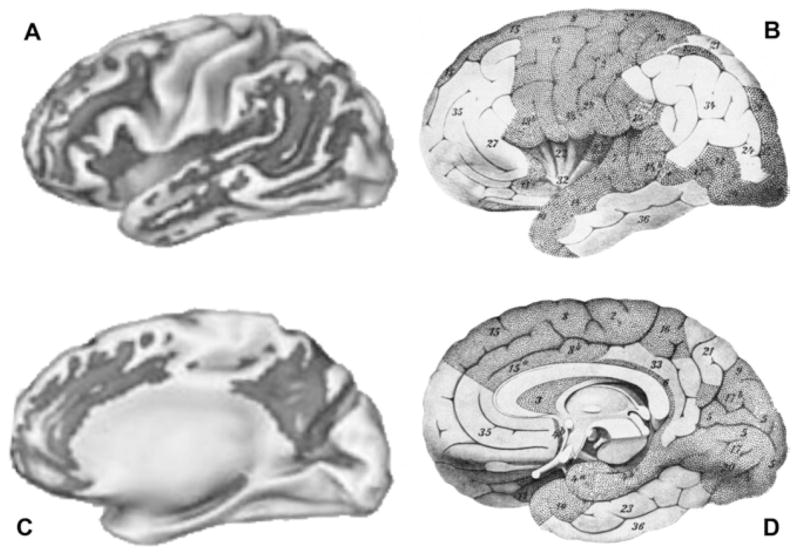
We hypothesized that myelin breakdown in vulnerable late-myelinating regions releases oligodendrocyte- and myelin-associated iron that promotes amyloid beta (A beta) oligomerization, its associated toxicity, and the deposition of oligomerized A beta and iron in neuritic plaques observed in Alzheimer's disease (AD). The model was tested by using published maps of cortical myelination from 1901 and recent in vivo imaging maps of A beta deposits in humans. The data show that in AD, radiolabeled ligands detect A beta deposition in a distribution that matches the map of late-myelinating regions. Furthermore, the strikingly lower ability of this imaging ligand to bind A beta in animal models is consistent with the much lower levels of myelin and associated iron levels in rodents when compared with humans. The hypotheses derived from the "myelin model" are testable with current imaging methods and have important implications for therapeutic interventions that should be expanded to include novel targets such as oligodendrocytes, myelin, and brain iron.
Keywords: Aging; Alzheimer’s disease; Amyloid; Degeneration; Dementia; Iron; Medications; Myelin; Oligodendrocyte; PIB; Prevention; Treatment; White matter.
Figures
References
-
- Semendeferi K, Lu A, Schenker N, Damasio H. Humans and great apes share a large frontal cortex. Nat Neurosci. 2002;5:272–6. - PubMed
-
- Schoenemann PT, Sheehan MJ, Glotzer LD. Prefrontal white matter volume is disproportionately larger in humans than in other primates. Nat Neurosci. 2005;8:242–52. - PubMed
-
- Bartzokis G. Age-related myelin breakdown: a developmental model of cognitive decline and Alzheimer’s disease. Neurobiol Aging. 2004;25:5–18. - PubMed
-
- Bartzokis G. Quadratic trajectories of brain myelin content: unifying construct for neuropsychiatric disorders. Neurobiol Aging. 2004;25:49–62.
Grants and funding
LinkOut - more resources
Full Text Sources
Other Literature Sources
