Comparative Study
. 2008 Jul 2;3(7):e2539.
doi: 10.1371/journal.pone.0002539.

Autoimmune pancreatocholangitis, non-autoimmune pancreatitis and primary sclerosing cholangitis: a comparative morphological and immunological analysis

Affiliations
Comparative Study

Autoimmune pancreatocholangitis, non-autoimmune pancreatitis and primary sclerosing cholangitis: a comparative morphological and immunological analysis

Irene Esposito et al. PLoS One. .

Abstract

Background: Autoimmune pancreatocholangitis (AIPC) is an emerging, not completely characterized disease. Aim of this study was the comprehensive evaluation of a series of AIPC patients, who were diagnosed and treated in a European institution between January 2003 and July 2006.

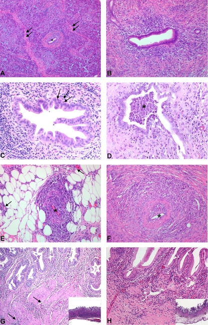
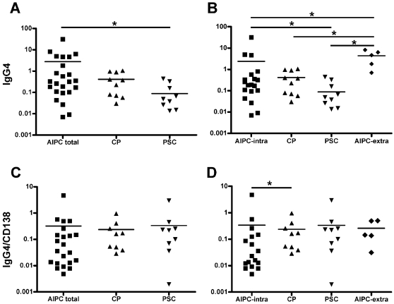
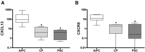
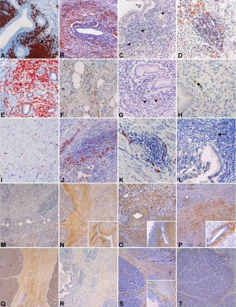
Methodology/principal findings: Thirty-three patients with histologically confirmed AIPC were analyzed and compared to 20 patients with non-autoimmune chronic pancreatitis (CP) and 14 patients with primary sclerosing cholangitis (PSC). Clinical features and conventional histopathology were taken into account. Immunohistochemistry and real-time quantitative PCR were used for the characterization of the inflammatory infiltrate and the stromal reaction. AIPC was localized in the pancreatic head in 94% of the patients. Intra- and/or extrapancreatic biliary tract involvement was present in 64% of the cases. The number of infiltrating T-lymphocytes, macrophages and total plasma cells was significantly higher in AIPC than in CP (3-, 4- and 8-fold increase, respectively). The absolute number of IgG4-positive plasma cells was higher in AIPC than in CP and PSC (7-fold and 35-fold increase, respectively), but significance was only reached in comparison with PSC. CXCR5- and CXCL13-positive cells were almost exclusively detected in AIPC.

Conclusions/significance: AIPC is mainly a disease of the pancreatic head with possible extension into the periphery of the gland and/or into the biliary tract/gallbladder. The morphology of AIPC, as well as the immune- and stromal reaction is characteristic and comparable between cases with and without biliary tract involvement. Immunological markers (IgG4, CXCR5, CXCL13) can be of diagnostic relevance in specific settings.


Conflict of interest statement

Competing Interests: The authors have declared that no competing interests exist.

Figures

Figure 1
Figure 1. Histopathological features of AIPC.
(A) Overview of pancreatic parenchyma with typical AIPC changes. In the center, a duct (*) with periductal mononuclear infiltration and fibrosis. Interlobular fibrosis is shown (arrows). (B) Interlobular duct with moderate periductal mononuclear infiltration, fibrosis and beginning stenosis of the lumen. (C) Higher magnification of an interlobular duct with early granulocytic epithelial lesion (GEL). Note the few neutrophils that infiltrate through the basal membrane into the epithelium (arrows). (D) Advanced GEL with abscess formation (*) in the lumen of an interlobular duct. (E) Lymphoplasmocytic inflammation of the wall of a small peripancreatic vein (*). Note the extension of the inflammatory infiltrate into the peripancreatic fat (arrows). (F) Lymphoplasmocytic inflammation of the wall of a small-sized intrapancreatic artery (*). (G) Involvement of the gallbladder in a case of AIPC. In the mucosa, a dense lymphoplasmacytic infiltrate below an almost intact epithelium. Note the thickening of the wall, due to the fibrosis and the extension of the inflammation into the deeper layers (arrows, inset). (H) Involvement of the gallbladder in a case of primary sclerosing cholangitis. Here, also a predominant lymphoplasmacytic inflammation with focal erosions of the epithelium and only superficial fibrosis. The deeper portions of the gallbladder wall are intact (inset).
Figure 2
Figure 2. Quantitative evaluation of the IgG4-positive cells and of the IgG4/plasma cell ratio.
(A) Total number of IgG4-positive cells and (C) IgG4/CD138 ratio in the disease groups of AIPC total, CP and PSC. (B) Total number of IgG4-positive cells and (D) IgG4/CD138 ratio when the AIPC cases are subclassified in those with and without extrapancreatic biliary tract involvement (AIPC extrapancreatic and AIPC intrapancreatic, respectively). Y-axis indicates the number of positive cells/mm2 in a logarithmic scale for better representation. Horizontal bars represent mean. * indicates statistical significance (p≤0.05).
Figure 3
Figure 3. Real-time quantitative PCR analysis of CXCR5 and CXCL13.
Real-time quantitative RT-PCR analysis of pancreatic tissues showing significantly higher levels of CXCL13 (A) and CXCR5 (B) mRNA in AIPC than in CP and PSC. RNA input was normalized to the average expression of the two housekeeping genes HPRT and cyclophilin B as described in Methods . Y-axis indicates the relative amount of mRNA in a logarithmic scale for better representation. Box and whiskers blot with 10–90 percentile are shown. * indicates statistical significance (p≤0.05).
Figure 4
Figure 4. Immunohistochemical characterization of AIPC, CP and PSC.
(A–H) Immunohistochemical characterization of the inflammatory infiltrate of AIPC. (A) CD20-positivity of B-lymphocytes in periductal localization and with tendency to form lymph follicles (arrow). (B) CD5-positive T-lymphocytes with diffuse arrangement around an interlobular duct. (C) Scattered CXCR5-positive cells (arrowheads) around the main pancreatic duct. (D) CXCL13-positive cells arranged in small clusters in the pancreatic tissue. (E) Numerous CD138-positive plasma cells in a diffuse arrangement in the pancreatic parenchyma. (F–G) IgG4-positive cells diffusely distributed in the peripancreatic adipose tissue (F) and in a periductal localization (G). (H) Isolated IgG4-positive cells (arrow) below the epithelium of the gallbladder in a case of AIPC with extrapancreatic biliary involvement. (I–K) Immunohistochemical characterization of the inflammatory infiltrate in CP. (I) A few CD138-positive plasma cells in a diffuse arrangement in pancreatic scar tissue. (J–K) IgG4-positive cells in large (J) and small (K) clusters in the pancreatic parenchyma. (L) Isolated IgG4-positive cells (arrow) in the gallbladder of a patient affected by PSC. (M–P) Immunohistochemical characterization of the stromal reaction in AIPC. (M) Low-power view of pancreatic tissue shows a weak (+) to moderate (++) interstitial staining for collagen I. (N) Moderate (++) to strong (+++) interlobular and periductal (inset) collagen V staining. (O) Strong (+++) diffuse and periductal (inset) α-smooth muscle actin (α-SMA) staining. (P) Diffuse interlobular and strong (+++) periductal (inset) positivity for Tenascin C. (Q–T) Immunohistochemical characterization of the stromal reaction in CP. (Q) Moderate (++) interlobular collagen I staining. (R) Very weak (−/+) interlobular collagen V staining. (S) Moderate (++) positivity for α-SMA in the interlobular connective tissue, without periductal arrangement (inset, here notice the extensive squamous cell metaplasia of the duct epithelium). (T) Moderate (++) staining for Tenascin C at the interface between the pancreatic lobules and the interlobular connective tissue.

Similar articles

Cited by

References

    1. Sarles H, Sarles JC, Muratore R, Guien C. Chronic inflammatory sclerosis of the pancreas–an autonomous pancreatic disease? Am J Dig Dis. 1961;6:688–698. - PubMed
    1. Yoshida K, Toki F, Takeuchi T, Watanabe S, Shiratori K, et al. Chronic pancreatitis caused by an autoimmune abnormality. Proposal of the concept of autoimmune pancreatitis. Dig Dis Sci. 1995;40:1561–1568. - PubMed
    1. Finkelberg DL, Sahani D, Deshpande V, Brugge WR. Autoimmune pancreatitis. N Engl J Med. 2006;355:2670–2676. - PubMed
    1. van Buuren HR, Vleggaar FP, Willemien Erkelens G, Zondervan PE, Lesterhuis W, et al. Autoimmune pancreatocholangitis: a series of ten patients. Scand J Gastroenterol. 2006;(Suppl):70–78. - PubMed
    1. Zen Y, Fujii T, Harada K, Kawano M, Yamada K, et al. Th2 and regulatory immune reactions are increased in immunoglobin G4-related sclerosing pancreatitis and cholangitis. Hepatology. 2007;45:1538–1546. - PubMed

Publication types