Case Reports
. 2008 Aug;118(8):2822-31.
doi: 10.1172/JCI34538.

Decreased FGF8 signaling causes deficiency of gonadotropin-releasing hormone in humans and mice

Affiliations
Case Reports

Decreased FGF8 signaling causes deficiency of gonadotropin-releasing hormone in humans and mice

John Falardeau et al. J Clin Invest. 2008 Aug.

Abstract

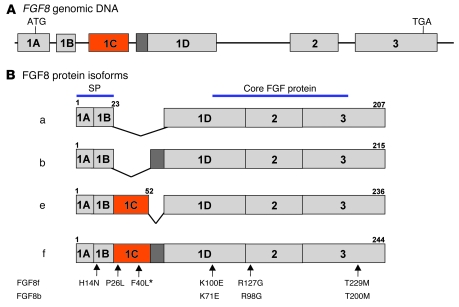
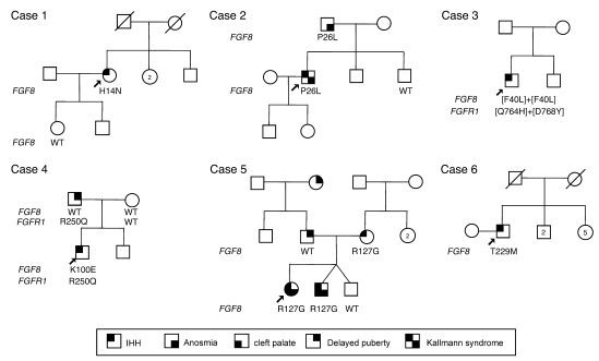
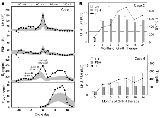
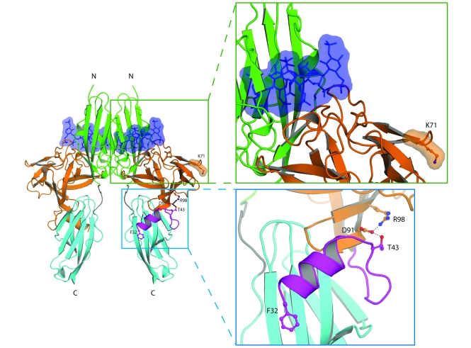
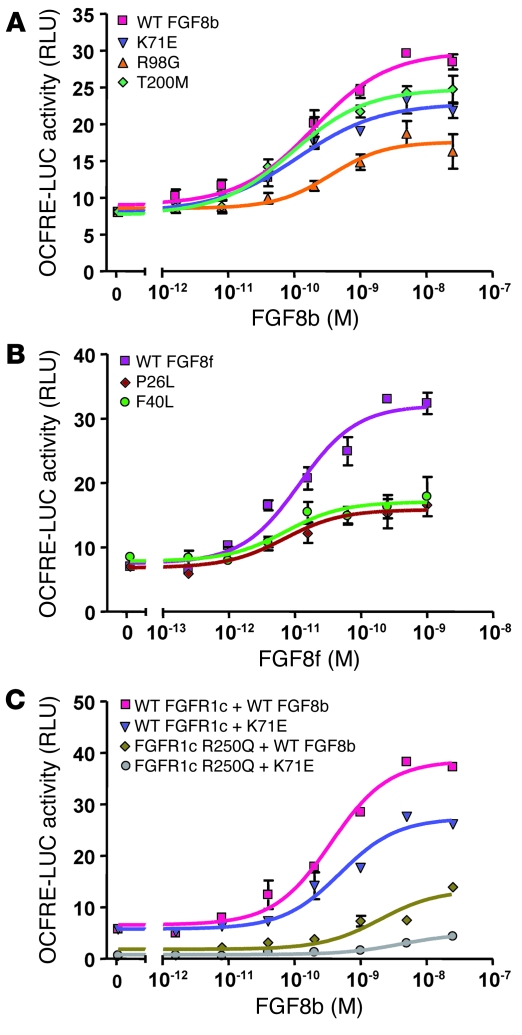
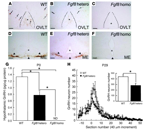
Idiopathic hypogonadotropic hypogonadism (IHH) with anosmia (Kallmann syndrome; KS) or with a normal sense of smell (normosmic IHH; nIHH) are heterogeneous genetic disorders associated with deficiency of gonadotropin-releasing hormone (GnRH). While loss-of-function mutations in FGF receptor 1 (FGFR1) cause human GnRH deficiency, to date no specific ligand for FGFR1 has been identified in GnRH neuron ontogeny. Using a candidate gene approach, we identified 6 missense mutations in FGF8 in IHH probands with variable olfactory phenotypes. These patients exhibited varied degrees of GnRH deficiency, including the rare adult-onset form of hypogonadotropic hypogonadism. Four mutations affected all 4 FGF8 splice isoforms (FGF8a, FGF8b, FGF8e, and FGF8f), while 2 mutations affected FGF8e and FGF8f isoforms only. The mutant FGF8b and FGF8f ligands exhibited decreased biological activity in vitro. Furthermore, mice homozygous for a hypomorphic Fgf8 allele lacked GnRH neurons in the hypothalamus, while heterozygous mice showed substantial decreases in the number of GnRH neurons and hypothalamic GnRH peptide concentration. In conclusion, we identified FGF8 as a gene implicated in GnRH deficiency in both humans and mice and demonstrated an exquisite sensitivity of GnRH neuron development to reductions in FGF8 signaling.


Figures

Figure 1
Figure 1. Genomic structure and differential splicing of the human FGF8 gene.
(A) Structure of the FGF8 gene. Boxes denote exons; lines denote introns. (B) Schematic of the 4 FGF8 isoforms identified in humans, which differ with regard to the inclusion of exon 1C and part of exon 1D. Most of the conserved FGF core is encoded by exons 2 and 3. Numbers above exons denote aa numberings for each isoform. The mutations identified to date are indicated by arrows numbered according to the FGF8f and FGF8b protein isoforms. Asterisk denotes the homozygous change.
Figure 2
Figure 2. Pedigrees of probands carrying an FGF8 mutation.
Arrows indicate the probands. Circles denote females; squares denote males; struck-through symbols denote deceased subjects; numbers within symbols denote number of individuals. Phenotypes are as described in Results.
Figure 3
Figure 3. Response to GnRH therapy in 3 probands harboring an FGF8 mutation.
(A) Pulsatile GnRH was administered at a dose of 100 ng/kg i.v. at a physiologic frequency to the proband described in case 1, demonstrating a normal increase in LH and FSH, an increase in E2 consistent with development of 2 follicles, and an increase in progesterone (Prog) consistent with ovulation. Data are centered to the day of ovulation (0 d); for E2, follicle diameters are indicated; shaded areas represent mean ± 1 SD hormone levels in 109 control women; boxed regions at top denote GnRH pulse frequency. (B) LH, FSH, and T responses to GnRH therapy in 2 male patients (cases 2 and 6). Horizontal lines denote lowest value of the normal range for adult serum T levels. To convert serum T values from ng/dl to nmol/l, divide by 28.84.
Figure 4
Figure 4. Structural modeling of K71E and R98G FGF8 mutations predicts that they are loss-of-function mutations.
The locations of the mutated FGF8 residues are mapped onto the ribbon diagram of 2:2:2 FGF8b/FGFR2c/heparin complex. Orange and purple denote the core and N terminus, respectively, of FGF8b. Green, cyan, and gray denote D2, D3, and D2-D3 linker, respectively, of the extracellular FGFR2c ligand-binding region. The 2 heparin oligosaccharides are rendered as blue sticks and surface representations. Also shown are detailed views of the regions where the mutated FGF8 residues are located. Selected residues are shown as ball-and-stick representations, as is F32 of FGF8b N terminus to underscore the importance of the FGF8b N terminus in FGF8b-FGFR binding. Blue balls, nitrogen atoms; red balls, oxygen atoms. Hydrogen bonds are shown as dashed lines. The N and C termini of polypeptide chains are indicated.
Figure 5
Figure 5. FGF8b mutants are loss-of-function mutations, consistent with structural prediction.
L6 myoblasts were transiently transfected with WT and mutant FGFR1 cDNA along with the OCFRE luciferase reporter, and then stimulated with increasing doses of WT or mutant FGF8. Luciferase activity was plotted as mean ± SEM of 3 triplicates, and a dose-response curve was fitted as described in Methods. The results of a representative experiment is shown. (A) Reporter activity of FGF8b WT and mutants. (B) Reporter activity of FGF8f WT and mutants. (C) Reporter activity of FGF8b WT and K71E mutant assayed on cell transfected with either WT or R250Q mutant FGFR1c.
Figure 6
Figure 6. Study of GnRH neurons in Fgf8 hypomorphic mice.
(AF) GnRH ICC illustrating the complete absence of GnRH neuron fibers and cell bodies in P0 homozygous Fgf8 hypomorphic mice (C and F) in the preoptic area and median eminence (ME) compared with WT controls (A and D) and heterozygous mice (B and E). Arrows denote GnRH neuron cell bodies; arrowheads denote GnRH fibers. Asterisks and dotted lines delineate the medial aspects of the brain. OVLT, organum vasculosum of the lamina terminalis. Scale bars: 100 μm. (G and H) The level of immunoreactive GnRH markedly decreased in Fgf8 heterozygotes at all postnatal ages measured. (H) Distribution of GnRH neurons in the preoptic-hypothalamic area of P29 WT controls and Fgf8 heterozygotes. Numbers in the x axis indicate increments of 40-μm sections. Negative and positive numbers indicate sections rostral and caudal to the organum vasculosum of the lamina terminalis (denoted 0), respectively. The distribution of GnRH neurons was largely normal in the transgenic mice, except that there were fewer GnRH neurons. Fgf8 heterozygotes had markedly fewer GnRH neurons in the preoptic-hypothalamic area compared with age-matched controls. ND, not detected. Data are mean ± SEM. *P < 0.05.

Similar articles

Cited by

References

    1. Herbison, A.E. 2006. Physiology of the GnRH neuronal network. InKnobil and Neill’s physiology of reproduction. 3rd edition. J.D. Neill, editor. Academic Press. San Diego, California, USA. 1415–1482.
    1. Knobil E., Neill J.D., Johansson E.D. Influence of hypophysectomy, sham hypophysectomy and other surgical procedures on luteal function in the rhesus monkey. Endocrinology. 1968;82:410–415. - PubMed
    1. Bless E.P., et al. Live view of gonadotropin-releasing hormone containing neuron migration. Endocrinology. 2005;146:463–468. doi: 10.1210/en.2004-0838. - DOI - PubMed
    1. Gonzalez-Martinez D., Hu Y., Bouloux P.M. Ontogeny of GnRH and olfactory neuronal systems in man: novel insights from the investigation of inherited forms of Kallmann’s syndrome. Front. Neuroendocrinol. 2004;25:108–130. doi: 10.1016/j.yfrne.2004.06.001. - DOI - PubMed
    1. Seminara S.B., Hayes F.J., Crowley W.F., Jr. Gonadotropin-releasing hormone deficiency in the human (idiopathic hypogonadotropic hypogonadism and Kallmann’s syndrome): pathophysiological and genetic considerations. Endocr. Rev. . 1998;19:521–539. doi: 10.1210/er.19.5.521. - DOI - PubMed

Publication types

MeSH terms