. 2008 Aug;118(8):2711-21.
doi: 10.1172/JCI34780.

KLF6-SV1 overexpression accelerates human and mouse prostate cancer progression and metastasis

Affiliations

KLF6-SV1 overexpression accelerates human and mouse prostate cancer progression and metastasis

Goutham Narla et al. J Clin Invest. 2008 Aug.

Abstract

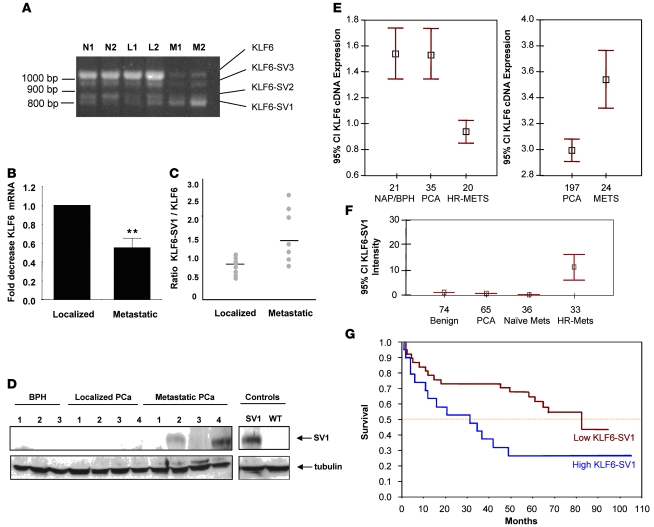
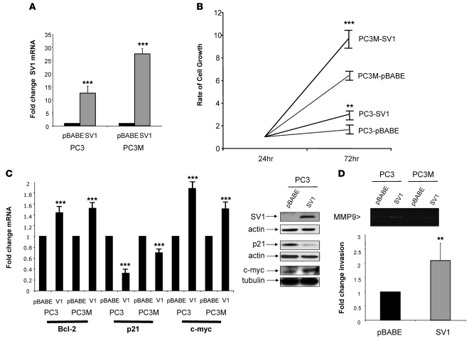
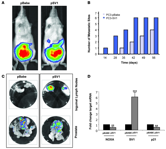
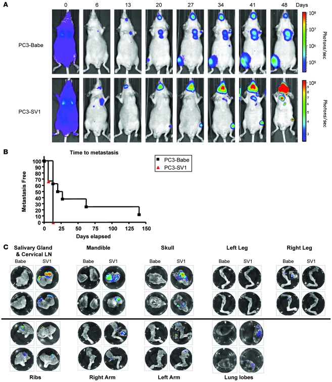
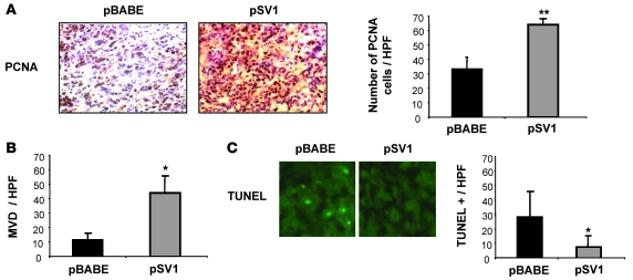
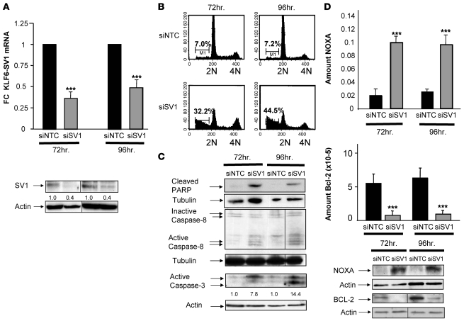
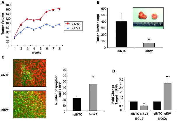
Metastatic prostate cancer (PCa) is one of the leading causes of death from cancer in men. The molecular mechanisms underlying the transition from localized tumor to hormone-refractory metastatic PCa remain largely unknown, and their identification is key for predicting prognosis and targeted therapy. Here we demonstrated that increased expression of a splice variant of the Kruppel-like factor 6 (KLF6) tumor suppressor gene, known as KLF6-SV1, in tumors from men after prostatectomy predicted markedly poorer survival and disease recurrence profiles. Analysis of tumor samples revealed that KLF6-SV1 levels were specifically upregulated in hormone-refractory metastatic PCa. In 2 complementary mouse models of metastatic PCa, KLF6-SV1-overexpressing PCa cells were shown by in vivo and ex vivo bioluminescent imaging to metastasize more rapidly and to disseminate to lymph nodes, bone, and brain more often. Interestingly, while KLF6-SV1 overexpression increased metastasis, it did not affect localized tumor growth. KLF6-SV1 inhibition using RNAi induced spontaneous apoptosis in cultured PCa cell lines and suppressed tumor growth in mice. Together, these findings demonstrate that KLF6-SV1 expression levels in PCa tumors at the time of diagnosis can predict the metastatic behavior of the tumor; thus, KLF-SV1 may represent a novel therapeutic target.


Figures

Figure 1
Figure 1. Expression of KLF6 and its splice variants in PCa.
(A) RT-PCR of representative prostate-derived cDNAs with KLF6-specific primers. N, normal prostate; L, localized PCa; M, metastatic PCa. (B) qRT-PCR analysis of localized and metastatic PCa cDNAs for wild-type KLF6 expression. Metastatic tumors expressed significantly less wild-type KLF6 mRNA compared with localized tumors. **P < 0.001. (C) qRT-PCR of localized and metastatic PCa samples using wild-type KLF6– and KLF6-SV1–specific PCR primers (refs. , , and our unpublished observations). (D) Increased KLF6-SV1 expression in metastatic PCa. Western blot analysis using a KLF6-SV1–specific monoclonal antibody (ref. and our unpublished observations). Transfected KLF6-SV1 and transfected wild-type KLF6 controls were run on the same gel but were noncontiguous. Tubulin was used as the loading control for all lanes. (E) Left: DNA microarray analysis of PCa demonstrated downregulation of KLF6 mRNA in hormone-refractory metastatic PCa (HR-MET) compared with both noncancerous prostate tissue and localized PCa. NAP, normal adjacent prostate tissue; PCA, localized PCa. Right: Tissue microarray analysis of KLF6 expression using KLF6 monoclonal antibody 2A2. Data points and error bars represent mean KLF6 protein expression and 95% confidence intervals, respectively. (F) Immunohistochemistry of high-density tissue microarray analyses with the KLF6-SV1–specific monoclonal antibody demonstrated marked upregulation of KLF6-SV1 expression in hormone-refractory metastatic PCa compared with naive metastatic PCa, localized PCa, and benign prostate tissue (P < 0.001). Data points and error bars represent mean KLF6-SV1 protein expression and 95% confidence intervals, respectively. (G) Median survival, as measured using biochemical recurrence and assessed by qRT-PCR, in men whose localized prostate tumors expressed high levels of KLF6-SV1 (blue) was 30 mo compared with 80 mo in men with low KLF6-SV1–expressing tumors (P < 0.01).
Figure 2
Figure 2. Overexpression of KLF6-SV1 in PCa cell lines.
(A) qRT-PCR analysis of pBABE and KLF6-SV1 vector–retrovirally infected PC3 and P3M cell lines demonstrated 12- and 27-fold overexpression, respectively, of KLF6-SV1 in pBABE-SV1 vector–infected cell lines compared with control. (B) KLF6-SV1–overexpressing cell lines proliferated significantly more than control cell lines, assessed by tritiated thymidine incorporation at 24 and 72 h of the PC3 and PC3M cell lines. Mean change in cell growth rate from 3 independent experiments is shown. (C) Increased expression of the oncogene c-myc and antiapoptotic Bcl-2, with concomitant reduction of the cyclin-dependent inhibitor p21, in KLF6-SV1–expressing cell lines. RNA and protein were harvested from 3 independent experiments, and qRT-PCR and Western blotting were performed. Overexpression of KLF6-SV1 increased Bcl-2 expression 50%, increased c-myc expression 80%, and reduced p21 expression 50% in PCa cell lines at the mRNA and protein levels. Actin was used as loading control for KLF6-SV1 and p21; tubulin was used for c-myc. (D) Increased invasion in KLF6-SV1 cell lines was associated with increased expression of MMP9. Gelatin zymography of the supernatant isolated from PC3 and PC3M cell lines is shown. Overexpression of KLF6-SV1 increased MMP9 expression in both PC3 and PC3M lines. KLF6-SV1–expressing stable PC3 cell lines (PC3-SV1) were 2-fold more invasive through a Matrigel basement membrane. The number of invasive cells was counted from 4 fields from 3 independent experiments. **P < 0.01, ***P < 0.001 versus control.
Figure 3
Figure 3. Overexpression of KLF6-SV1 in an orthotopic mouse model of PCa progression results in increased metastasis.
(A) Male SCID beige mice were anesthetized, and the dorsolateral aspect of the prostate was injected with 1 × 10⁶ PC3 cells in 25 μl PBS. Tumors were imaged every week to determine local tumor growth and evidence of tumor cell dissemination. A representative image of 2 mice is illustrated. Local tumor growth, as determined by fluorescent intensity, was equal between control and KLF6-SV1 mice. (B and C) KLF6-SV1–overexpressing cells metastasized more frequently than did control cells. The number of metastatic lesions was determined using a combination of whole-body imaging and ex vivo histological analysis of all mice upon sacrifice (n = 8 [pBABE]; 9 [pSV1]); representative images of ex vivo tissue analysis are shown. (D) pSV1 vector–derived tumors expressed significantly less NOXA and p21 than did control tumors. qRT-PCR of pSV1 vector–derived tumors using real-time primers specific to NOXA, KLF6-SV1, and p21 demonstrated marked overexpression of KLF6-SV1 in pSV1 vector–derived tumors with concomitant reduction in p21 and NOXA expression. **P < 0.01, ***P < 0.001 versus control.
Figure 4
Figure 4. Intracardiac model of PCa dissemination and metastasis.
(A) Female nu/nu mice were injected with 1 × 10⁶ PC3 cells in 25 μl PBS. In vivo BLI was performed weekly 10–15 minutes after animals received the substrate d-luciferin at 150 mg/kg in PBS by i.p. injection. The bioluminescent signals from metastatic lesions in the KLF6-SV1–expressing PC3 cell group was 3 orders of magnitude higher than the control pBABE vector–expressing PC3 cell–injected mice, demonstrating that the former developed larger metastatic tumors. (B) pSV1 vector–expressing cells demonstrated detectable metastasis significantly earlier than did control pBABE vector–expressing cells lines (P < 0.01). The median time to metastatic development was 3.5 times more rapid in the mice with KLF6-SV1–expressing PC3 cells (7 d) compared with controls (24 d; P < 0.009). (C) Ex vivo imaging and histology was performed on 20 different tissues excised from a subset of mice after final in vivo imaging. Representative images are shown.
Figure 5
Figure 5. Changes in KLF6-SV1 metastatic tumor behavior correlates with markers of cellular proliferation, angiogenesis, and apoptosis.
Metastatic tumors were analyzed for their expression of PCNA, CD31, and TUNEL. (A) Immunohistochemistry for PCNA in pBABE vector– and KLF6-SV1–derived tumors. KLF6-SV1 caused increased PCNA staining in vivo. Eight independent high-power fields for each pBABE (n = 4) and KLF6-SV1 (n = 6) tumor were counted, assessing both the total number of cells and PCNA-positive cells. The graph represents the average percentage of PCNA-positive cells for each group. (B) CD31 staining of pBABE and KLF6-SV1 cell line–derived tumors. Overexpression of KLF6-SV1 protein caused a 4-fold increase in microvessel density (MVD), as measured by the number of CD31-positive endothelial cells per ×400 high-power field (HPF). (C) TUNEL staining of pBABE and KLF6-SV1 tumors. Representative images are shown. Overexpression of KLF6-SV1 decreased apoptosis by 70%. For each tumor, 6 high-power fields were counted and the total number of TUNEL-positive cells was determined. *P < 0.01, **P < 0.001 versus control. Original magnification, ×400.
Figure 6
Figure 6. Targeted KLF6-SV1 inhibition dramatically increases spontaneous apoptosis.
(A) qRT-PCR and Western blot analysis of the PC3M cell line transfected with si-NTC and si-SV1 demonstrated significant downregulation of KLF6-SV1 mRNA and protein at 72 and 96 h. (B) Fluorescence-activated cell sorting analysis of si-NTC– and si-SV1–transfected PC3 cells at 72 and 96 h. On average, si-SV1 treatment increased cell death 5-fold. Data represent the average of 3 independent experiments. Numbers within plots indicate percent hypodiploid (i.e., apoptotic) cells. (C) Western blot analysis demonstrated marked upregulation of caspase-3 and -8 and increased poly(ADP-ribose) polymerase (PARP) cleavage in si-SV1–transfected cell lines. (D) qRT-PCR and Western blot analysis of si-NTC– and si-SV1–transfected cells for NOXA and Bcl-2 demonstrated marked upregulation of the proapoptotic NOXA with concomitant downregulation of Bcl-2. Numbers above actin blots in A and C represent densitometric analysis of the protein bands, expressed as fold change relative to control and normalized to actin. In A, B, and D, lanes were run on the same gel but were noncontiguous (black lines). Each Western blot shown is representative of 3 independent experiments. ***P < 0.0001 versus control.
Figure 7
Figure 7. Direct intratumoral injection of si-SV1 induces apoptosis and reduces tumor growth in vivo.
(A) Intratumoral injection of si-SV1 reduced tumor growth in a PCa xenograft model. PC3M cells were injected subcutaneously into nude mice. After 3 weeks of growth, mice were randomized into 2 treatment groups, and si-NTC or si-SV1 was injected directly into the tumors twice a week for a total of 3 weeks. Mice were sacrificed, tumor volume was determined, and RNA and tissue was harvested. (B) Photographs of representative si-NTC– and si-SV1–treated tumors, which had identical volumes prior to treatment. Three weeks after treatment, si-SV1–injected tumors were significantly smaller than si-NTC–treated tumors. n = 9 (si-SV1); 8 (si-NTC). (C) TUNEL staining of si-SV1– and si-NTC–treated tumors revealed a significant increase in the number of apoptotic cells per high-power field in si-SV1–derived tumors. Six tumors per high-power field were counted. (D) qRT-PCR of si-NTC (n = 7) and si-SV1 (n = 8) derived tumors using PCR primers specific to human Bcl-2 and NOXA demonstrate a 50% reduction in Bcl-2 expression with a concomitant 3 fold increase in NOXA expression in si-SV1 treated tumors. *P < 0.01, **P < 0.001, ***P < 0.0001 versus control. Original magnification, ×400.

Similar articles

Cited by

References

    1. Jemal A., et al. Cancer statistics, 2007. CA Cancer J. Clin. 2007;57:43–66. - PubMed
    1. Narla G., et al. KLF6, a candidate tumor suppressor gene mutated in prostate cancer. Science. 2001;294:2563–2566. doi: 10.1126/science.1066326. - DOI - PubMed
    1. Chen C., et al. Deletion, mutation, and loss of expression of KLF6 in human prostate cancer. Am. J. Pathol. 2003;162:1349–1354. - PMC - PubMed
    1. Reeves H.L., et al. Kruppel-like factor 6 (KLF6) is a tumor-suppressor gene frequently inactivated in colorectal cancer. Gastroenterology. 2004;126:1090–1103. doi: 10.1053/j.gastro.2004.01.005. - DOI - PubMed
    1. Kremer-Tal S., et al. Frequent inactivation of the tumor suppressor kruppel-like factor 6 (KLF6) in hepatocellular carcinoma. Hepatology. 2004;40:1047–1052. doi: 10.1002/hep.20460. - DOI - PubMed

Publication types

MeSH terms

Substances