. 2008 Jul 2;3(7):e2603.
doi: 10.1371/journal.pone.0002603.

Expulsion of symbiotic algae during feeding by the green hydra--a mechanism for regulating symbiont density?

Affiliations

Expulsion of symbiotic algae during feeding by the green hydra--a mechanism for regulating symbiont density?

Yelena Fishman et al. PLoS One. .

Abstract

Background: Algal-cnidarian symbiosis is one of the main factors contributing to the success of cnidarians, and is crucial for the maintenance of coral reefs. While loss of the symbionts (such as in coral bleaching) may cause the death of the cnidarian host, over-proliferation of the algae may also harm the host. Thus, there is a need for the host to regulate the population density of its symbionts. In the green hydra, Chlorohydra viridissima, the density of symbiotic algae may be controlled through host modulation of the algal cell cycle. Alternatively, Chlorohydra may actively expel their endosymbionts, although this phenomenon has only been observed under experimentally contrived stress conditions.

Principal findings: We show, using light and electron microscopy, that Chlorohydra actively expel endosymbiotic algal cells during predatory feeding on Artemia. This expulsion occurs as part of the apocrine mode of secretion from the endodermal digestive cells, but may also occur via an independent exocytotic mechanism.

Significance: Our results demonstrate, for the first time, active expulsion of endosymbiotic algae from cnidarians under natural conditions. We suggest this phenomenon may represent a mechanism whereby cnidarians can expel excess symbiotic algae when an alternative form of nutrition is available in the form of prey.


Conflict of interest statement

Competing Interests: The authors have declared that no competing interests exist.

Figures

Figure 1
Figure 1. Many symbiotic algae are observed in the apical part of Chlorohydra viridissima endodermal digestive cells following feeding with Artemia.
The symbiotic algae can be seen as small (∼10 µm) green spheres, and those found in the apical part of the cells are marked by arrowheads. A) Unfed Chlorohydra; B) Chlorohydra 15 minutes after feeding; C) Chlorohydra 5 hours after feeding. en = endoderm, ec = ectoderm, gvc = gastrovascular cavity, art = artemia. The dashed line between the endoderm and the mesoderm denotes the mesoglea. Bar = 50 µm. D) A quantitative analysis of the number of symbiotic algae in the apical third of the endoderm, at different times after feeding. Values and error bars represent averages and SE from 4–6 animals. There number of symbionts in the apical part of the cell was weakly affected by time (ANOVA, F = 5.067, p = 0.025) and time by treatment (F = 4.267, p = 0.04) but strongly affected by the treatment (F = 26.667, p<0.001). The difference between the fed and unfed animals is highly significant 15 minutes after feeding (Student's two-tailed t-test, p = 0.0048, represented by * in the figure), and is not significant at other time points. E) Reduction in the number of algal symbionts per endodermal cell in fed compared to unfed animals, 15 minutes after feeding. Values and error bars represent averages and SE from 30 endodermal cells per time point. The number of symbionts was not affected by time (ANOVA, F = 0.438, p = 0.781), and the effect of treatment was marginally significant (F = 3.76, p = 0.053). There was a significant difference between fed and unfed animals after 15 minutes (Student's two tailed t-test, t = 2.686, p = 0.009, represented by * in the figure) but this difference was not significant at other time points.
Figure 2
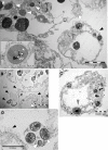
Figure 2. Expulsion of symbiotic algae by apocrine secretion and exocytosis during feeding by Chlorohydra.
en = endoderm, gvc = gastrovascular cavity. A) A general view of the apical part of Chlorohydra endoderm, 15 minutes after feeding. A large membrane-bound “aposome” is seen within the GVC, adjacent to the apical membrane of an endodermal digestive cell. (black arrowhead). A symbiotic alga is in the process of exocytosis from the aposome (enlarged in the inset), and several others are seen in the apical part of the endoderm (white arrowheads). Bar = 10 µm (1 µm in the inset). B) Expulsion of alga during apocrine secretion, 15 minutes after feeding. Note heterogenous aposomes within the GVC (black arrowheads), one of which contains an alga (algae are marked by white arrowheads). Bar = 10 µm. C) Enlargement of the aposome marked by an square in B. The aposome contains one intact alga (in the process of exocytosis, white arrowhead), as well as possibly another being digested (grey arrowhead). * = mitochondria. Bar = 5 µm. D) An aposome containing four symbiotic algae within the GVC (white arrowheads). Note the microvilli seen on the apical membrane of an endodermal cell (black arrowhead). Bar = 10 µm.

References

    1. Rosenberg E, Koren O, Reshef L, Efrony R, Zilber-Rosenberg I. The role of microorganisms in coral health, disease and evolution. Nat Rev Microbiol. 2007;5(5):355–362. - PubMed
    1. Douglas AE. Coral bleaching - how and why? Mar Pol bull. 2003;46:385–392. - PubMed
    1. Merle PL, Sabourault C, Richier S, Allemand D, Furla P. Catalase characterization and implication in bleaching of a symbiotic sea anemone. Free Radic Biol Med. 2007;42(2):236–246. - PubMed
    1. Richier S, Sabourault C, Courtiade J, Zucchini N, Allemand D, et al. Oxidative stress and apoptotic events during thermal stress in the symbiotic sea anemone, Anemonia viridis. FEBS J. 2006;273(18):4186–4198. - PubMed
    1. McAuley PJ. The cell cycle of symbiotic Chlorella. III. Numbers of algae in green hydra digestive cells are regulated at digestive cell division. J Cell Sci. 1986;85:63–71. - PubMed

Publication types