. 2008 Jul;118(7):2482-95.
doi: 10.1172/JCI34363.

Implantation of olfactory ensheathing cells promotes neuroplasticity in murine models of stroke

Affiliations

Implantation of olfactory ensheathing cells promotes neuroplasticity in murine models of stroke

Woei-Cherng Shyu et al. J Clin Invest. 2008 Jul.

Abstract

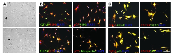
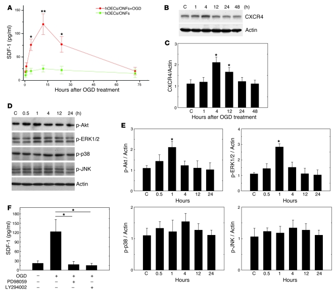
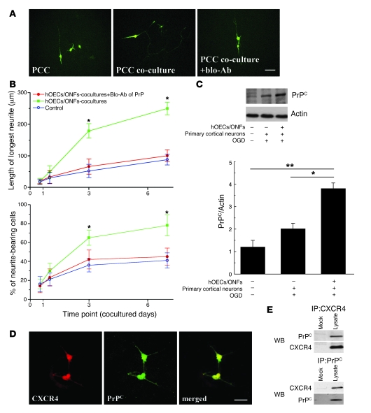
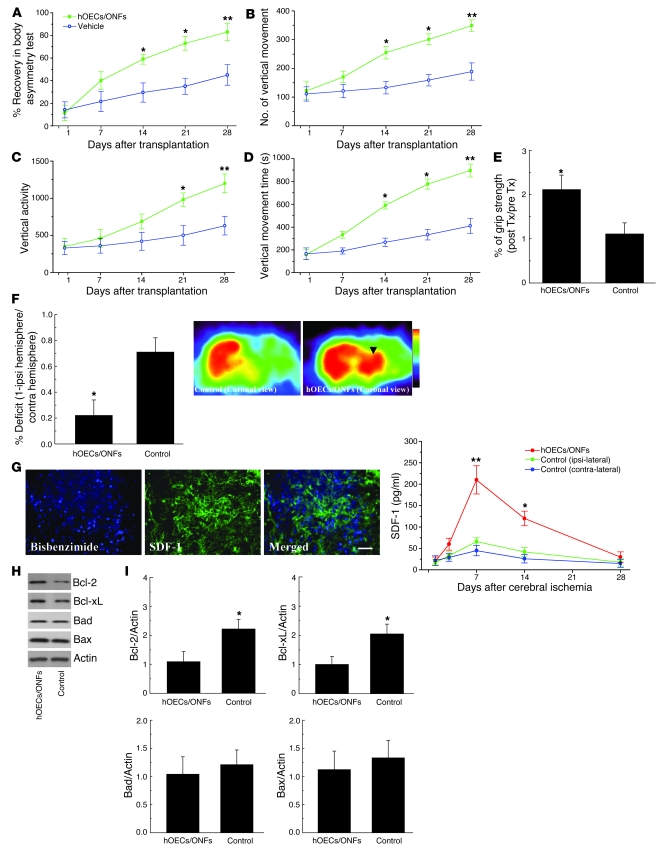
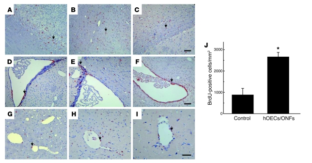
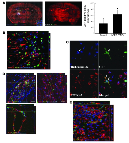
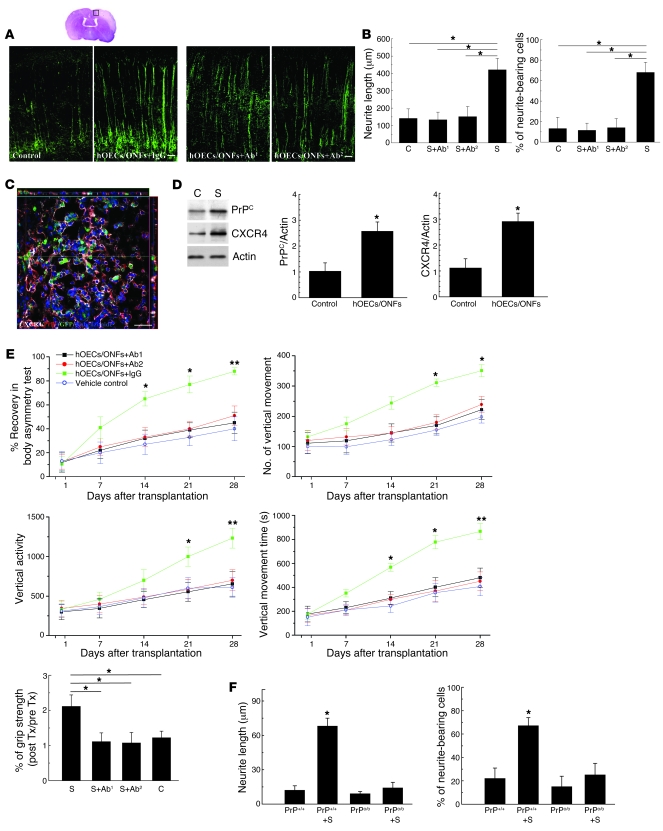
Murine olfactory ensheathing cells (OECs) promote central nervous system axonal regeneration in models of spinal cord injury. We investigated whether OECs could induce a neuroplastic effect to improve the neurological dysfunction caused by hypoxic/ischemic stress. In this study, human OECs/olfactory nerve fibroblasts (hOECs/ONFs) specifically secreted trophic factors including stromal cell-derived factor-1alpha (SDF-1alpha). Rats with intracerebral hOEC/ONF implantation showed more improvement on behavioral measures of neurological deficit following stroke than control rats. [18F]fluoro-2-deoxyglucose PET (FDG-PET) showed increased glucose metabolic activity in the hOEC/ONF-treated group compared with controls. In mice, transplanted hOECs/ONFs and endogenous homing stem cells including intrinsic neural progenitor cells and bone marrow stem cells colocalized with specific neural and vascular markers, indicating stem cell fusion. Both hOECs/ONFs and endogenous homing stem cells enhanced neuroplasticity in the rat and mouse ischemic brain. Upregulation of SDF-1alpha and CXCR4 in hOECs/ONFs promoted neurite outgrowth of cocultured primary cortical neurons under oxygen glucose deprivation conditions and in stroke animals through upregulation of cellular prion protein (PrP C) expression. Therefore, the upregulation of SDF-1alpha and the enhancement of CXCR4 and PrP C interaction induced by hOEC/ONF implantation mediated neuroplastic signals in response to hypoxia and ischemia.


Figures

Figure 1
Figure 1. Phenotypic characterization of hOECs/ONFs.
(A) The appearance of hOECs/ONFs by phase-contrast microscopy was either spindle shaped (arrow) or astrocyte-like (arrowhead). (B) Immunofluorescence colocalization analysis of hOECs/ONFs showed coexpression of p75 and GFAP, p75 and FN, p75 and S100, and GFAP and S100. (C) The proteins identified in our study as being produced by hOECs/ONFs including SDF-1α and its receptor, CXCR4, were coexpressed with p75 and GFAP in a double immunofluorescence study in hOECs/ONFs. Scale bars: 50 μm.
Figure 2
Figure 2. hOECs/ONFs upregulated the synthesis of soluble factors via the specific signaling pathway.
(AC) Under conditions of OGD, significantly increased expression of SDF-1α and CXCR4 was found by ELISA and Western blot analyses, respectively. (D and E) Expression of signal transduction proteins (p-Akt and p-ERK1/2) increased significantly by 1 hour after treatment. (F) The upregulation of SDF-1α could be blocked by specific inhibitors of p-Akt and p-ERK1/2 (LY294002 and PD98059). Data are expressed as mean ± SEM. *P < 0.05 and **P < 0.01 versus control.
Figure 3
Figure 3. In coculture, hOECs/ONFs promoted neurite regeneration and survival of PCC under OGD.
(A) Representative images of neurite regeneration in PCC with or without hOEC/ONF cocultivation. Blo-Ab, blocking Ab. (B) Neurite length and the number of surviving neurons increased in the hOECs/ONFs cocultured with PCC compared with the control. The enhancement of neurite length and number of surviving neurons was inhibited by the neutralization peptide of PrPC. (C) Upregulation of PrPC expression was found in PCC cocultured with hOECs/ONFs. (D) In IHC, subcellular colocalization of CXCR4 and PrPC was shown by costaining with CXCR4 and PrPC antibodies in PCCs cocultured with hOEC/ONFs. (E) Using coimmunoprecipitation analysis, the cell lysates were immunoprecipitated (IP) with anti-CXCR4 or anti-PrPC, and the signal of both proteins was detected by Western blotting (WB). Data are expressed as mean ± SEM. *P < 0.05 and **P < 0.01 versus control. Scale bars: 50 μm.
Figure 4
Figure 4. Intracerebral hOEC/ONF transplantation improves neurological behavior after cerebral ischemia.
(A) A body asymmetry trial was used to assess body swing before and after MCA ligation. Between 14 and 28 days after cerebral ischemia, rats treated intracerebrally with hOECs/ONFs exhibited significantly less body asymmetry than controls. (BD) Locomotor activity of all experimental rats was examined. Vertical activity, vertical movement time, and the number of vertical movements significantly increased between 14 and 28 days after cerebral ischemia in rats receiving hOECs/ONFs in comparison with vehicle-treated control rats. (E) Grip strength measurement of the grasping power of forelimbs before (pre Tx) and 28 days after each of the 2 treatments (post Tx). There was a higher postischemia/preischemia strength ratio in the hOEC/ONF transplants than in vehicle control rats. (F) Representative result (coronal view) of FDG-PET of the right cortex (black arrowhead) of hOEC/ONF-treated and control groups. Semiquantitative measurement showed that relative glucose metabolic activity in the right cortex was much greater in the hOEC/ONF-treated than the control group (ipsi, ipsilateral; contra, contralateral). (G) In immunohistochemical and ELISA analyses, brain samples of the hOEC/ONF-treated rats showed significantly increased expression of SDF-1α compared with the contralateral cortex and other areas of control rat brains. (H and I) In Western blot assay at 3 days after treatment, antiapoptotic proteins (Bcl-2 and Bcl-xL) were present at higher levels in the hOEC/ONF-treated than the control rat. Data are expressed as mean ± SEM. *P < 0.05 and **P < 0.01 versus control. Scale bar: 50 μm.
Figure 5
Figure 5. Immunohistochemical staining for BrdU in rats treated with hOECs/ONFs after cerebral ischemia.
A few BrdU-immunoreactive cells (arrows) were detected around the infarct boundary (AC), in the subventricular area (DF), and over the perivascular region (GI) in the ipsilateral cortex. (J) Quantitative analysis revealed that the number of BrdU-immunoreactive cells in the ipsilateral hemisphere of rats treated with hOECs/ONFs was significantly increased at 28 days after treatment in comparison with rats treated with vehicle control. Data are expressed as mean ± SEM. *P < 0.05 versus control. Scale bars: 50 μm.
Figure 6
Figure 6. Biological mechanism of neuroplastic effects on the ischemic brain after intracerebral transplantation of hOECs/ONFs.
(A) In a representative brain section of a GFP-chimeric mouse treated with or without hOECs/ONFs (white arrow indicates the injection site), GFP+ cells are seen dispersed over the periphery of the transplanted hOECs/ONFs and were significantly increased in quantity in the hOEC/ONF-treated mice in comparison with controls. In FISH analysis (white arrow, 2 red spots), hOECs/ONFs were shown to be of human origin (inset square in left panel). (B) IHC of hOEC/ONF treatment in the BrdU-labeled mice. Many BrdU+nestin+ cells were distributed around the transplanted hOECs/ONFs. (C) Interestingly, 1 cell with 2 nuclei (cell fusion) was found in the implanted hOECs/ONFs (white arrows, blue nucleus) and GFP+ cells (white arrowheads, red nucleus). The nucleic dye TOTO-3 (red) was used to define the outline of all nuclei in the section. (D) In a colocalization study (3D image) some bis-benzimide–labeled cells and some GFP+ cells colocalized with MAP-2+, vWF+, and GFAP+ cells in the penumbra of hOEC/ONF-treated ischemic rat brains. (E) SDF-1α–immunoreactive cells colocalized with a few bis-benzimide–labeled hOECs/ONFs and GFP+ cells. Data are expressed as mean ± SEM. *P < 0.05 versus control. Scale bars: 50 μm.
Figure 7
Figure 7. Intracerebral transplantation of hOECs/ONFs enhanced neurite regeneration through upregulation of PrPC and CXCR4 expression.
(A) In immunohistochemical analysis, hOEC/ONF + IgG (control human IgG) implantation significantly improved neurite outgrowth in the penumbral area of rats in comparison with hOECs/ONFs plus Ab1 (PrPC-blocking antibody), hOECs/ONFs plus Ab2 (CXCR4-neutralizing antibody), and vehicle (Control) treatment. (B) hOEC/ONF + IgG (S) implantation produced neurites significantly longer than those found in rats treated with S + Ab1, S + Ab2, and vehicle (control [C]). Moreover, hOEC/ONF + IgG–treated rats had more neurite-bearing neurons than the control rats. (C) In colocalization analyses, PrPC and CXCR4 coexpressed around the region of bis-benzimide–labeled hOECs/ONFs and GFP+ cells on the cerebral ischemic microenviroment. (D) In a Western blot analysis, PrPC and CXCR4 expression was upregulated in the hOEC/ONF-treated rats compared with control rats. (E) The neurological behavior measurement modalities showed no significant difference between the 3 therapeutic groups (C, S + Ab1, S + Ab2). (F) In evaluating neurite regeneration, hOEC/ONF implantation in the PrPo/o mice did not increase the neurite length and number of neurite-bearing cells in contrast to those of PrP+/+ mice after cerebral ischemia. Data are expressed as mean ± SEM. *P < 0.05 and **P < 0.01 versus control. Scale bars: 50 μm.

Similar articles

Cited by

References

    1. Li Y., Decherchi P., Raisman G. Transplantation of olfactory ensheathing cells into spinal cord lesions restores breathing and climbing. J. Neurosci. 2003;23:727–731. - PMC - PubMed
    1. Field P., Li Y., Raisman G. Ensheathment of the olfactory nerves in the adult rat. J. Neurocytol. 2003;32:317–324. doi: 10.1023/B:NEUR.0000010089.37032.48. - DOI - PubMed
    1. Kato T., Honmou O., Uede T., Hashi K., Kocsis J.D. Transplantation of human olfactory ensheathing cells elicits remyelination of demyelinated rat spinal cord. Glia. 2000;30:209–218. doi: 10.1002/(SICI)1098-1136(200005)30:3<209::AID-GLIA1>3.0.CO;2-8. - DOI - PMC - PubMed
    1. Agrawal A.K., et al. Olfactory ensheathing cell transplantation restores functional deficits in rat model of Parkinson’s disease: a cotransplantation approach with fetal ventral mesencephalic cells. Neurobiol. Dis. 2004;16:516–526. doi: 10.1016/j.nbd.2004.04.014. - DOI - PubMed
    1. Devon R., Doucette R. Olfactory ensheathing cells myelinate dorsal root ganglion neurites. Brain Res. 1992;589:175–179. doi: 10.1016/0006-8993(92)91182-E. - DOI - PubMed

Publication types

MeSH terms