. 2008 Dec;17(6):1053-63.
doi: 10.1089/scd.2008.0117.

Defining stem and progenitor cells within adipose tissue

Affiliations

Defining stem and progenitor cells within adipose tissue

Guiting Lin et al. Stem Cells Dev. 2008 Dec.

Abstract

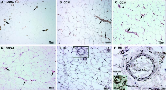
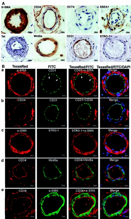
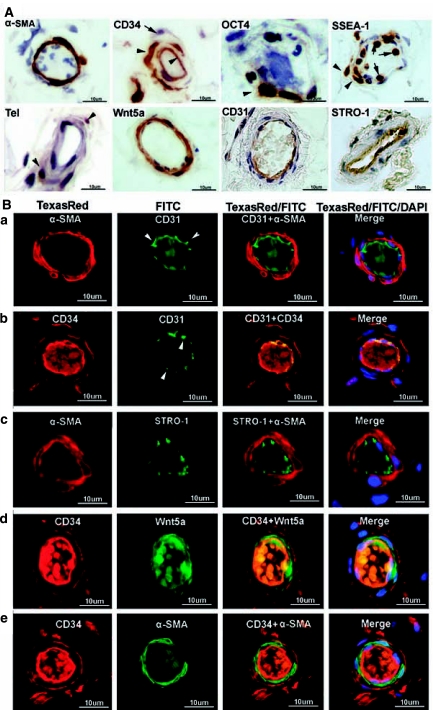
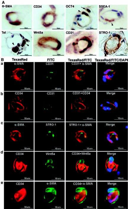
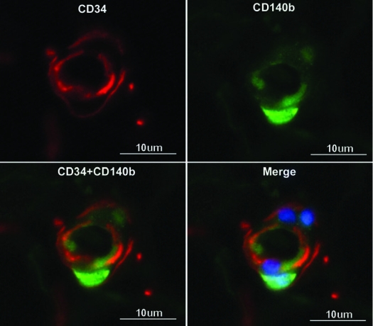
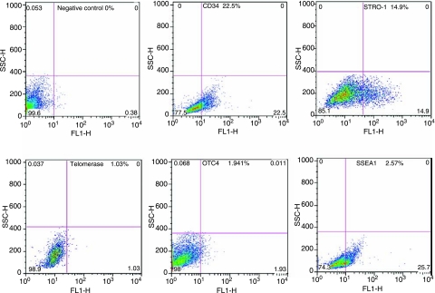
Adipose tissue-derived stem cells (ADSC) are routinely isolated from the stromal vascular fraction (SVF) of homogenized adipose tissue. Freshly isolated ADSC display surface markers that differ from those of cultured ADSC, but both cell preparations are capable of multipotential differentiation. Recent studies have inferred that these progenitors may reside in a perivascular location where they appeared to coexpress CD34 and smooth muscle actin (alpha-SMA) but not CD31. However, these studies provided only limited histological evidence to support such assertions. In the present study, we employed immunohistochemistry and immunofluorescence to define more precisely the location of ADSC within human adipose tissue. Our results show that alpha-SMA and CD31 localized within smooth muscle and endothelial cells, respectively, in all blood vessels examined. CD34 localized to both the intima (endothelium) and adventitia neither of which expressed alpha-SMA. The niche marker Wnt5a was confined exclusively to the vascular wall within mural smooth muscle cells. Surprisingly, the widely accepted mesenchymal stem cell marker STRO-1 was expressed exclusively in the endothelium of capillaries and arterioles but not in the endothelium of arteries. The embryonic stem cell marker SSEA1 localized to a pericytic location in capillaries and in certain smooth muscle cells of arterioles. Cells expressing the embryonic stem cell markers telomerase and OCT4 were rare and observed only in capillaries. Based on these findings and evidence gathered from the existing literature, we propose that ADSC are vascular precursor (stem) cells at various stages of differentiation. In their native tissue, ADSC at early stages of differentiation can differentiate into tissue-specific cells such as adipocytes. Isolated, ADSC can be induced to differentiate into additional cell types such as osteoblasts and chondrocytes.


Figures

FIG. 1.
FIG. 1.
Immunohistochemical localization of vascular and stem cell markers in the adipose tissue. Human adipose tissues were examined by immnuohistochemistry for the expression of α-SMA (A), CD31 (B), CD34 (C), and SSEA1 (D). Positively stained structures are marked with arrows. Control specimen stained with HE (without antibody) is shown in (E), and two areas of which (a and b) are enlarged and displayed in (F). The small artery, the arteriole, and the capillary are approximately 70, 25, and 10 μm in diameter, respectively.
FIG. 2.
FIG. 2.
Immunohistochemical and immunofluorescence localization of vascular and stem cell markers in small arteries of adipose tissue. Human adipose tissues were immunostained for the indicated markers and a small artery (∼70 μm in diameter) from each stained specimen was chosen for display. (A) Note the absence of staining for OCT4, telomerase, and STRO-1. Also note the double-ring staining pattern of CD34 (intima and adventitia), as opposed to the single-ring staining of CD31 (intima only). (B) Each row (a, b, c, d, and e) displays a selected artery, while the columns are arranged as follows: From left to right, columns 1 and 2 show images of arteries stained for the indicated markers. Column 3 shows merged images of columns 1 and 2. Column 4 shows merged images of columns 1 and 2 plus an image of DAPI (nuclear) staining. Arrowheads indicate the punctuated staining pattern of CD31.
FIG. 3.
FIG. 3.
Immunohistochemical and immunofluorescence localization of vascular and stem cell markers in arterioles of adipose tissue. Human adipose tissues were immunostained for the indicated markers and an arteriole (∼25 μm in diameter) from each stained specimen was chosen for display. (A) The two arrowheads in CD34 point to positively stained cells in the endothelium and adventitia, respectively, while the lone arrow points to an unstained cell in the adventitia. The arrowheads in SSEA1 point to two positively stained cells in the muscle layer, while the arrows point to two white blood cells. The arrowheads in telomerase point to two positively stained cells. Also note the specific STRO-1 staining in the endothelium possibly of a venule. (B) Each row (a, b, c, d, and e) displays a selected arteriole, while the columns are arranged as follows: From left to right, columns 1 and 2 show images of arteries stained for the indicated markers. Column 3 shows merged images of columns 1 and 2. Column 4 shows merged images of columns 1 and 2 plus an image of DAPI (nuclear) staining. Arrowheads indicate the punctuated staining pattern of CD31. The luminal materials are blood clots.
FIG. 4.
FIG. 4.
Immunohistochemical and immunofluorescence localization of vascular and stem cell markers in capillaries of adipose tissue. Human adipose tissues were immunostained for the indicated markers and a capillary in each stained specimen was chosen for display. In (A) the arrowhead and arrow in OCT4 point to two positively stained cells in the capillary's inner and outer regions, respectively. These cells are probably precursors of endothelial cells and pericytes, respectively. Cells positively stained for SSEA1 and telomerase are also indicated by arrowheads. In (B) each row (a, b, c, d, and e) displays a selected capillary, while the columns are arranged as follows: From left to right, columns 1 and 2 show images of capillaries stained for the indicated markers. Column 3 shows merged images of columns 1 and 2. Column 4 shows merged images of columns 1 and 2 plus an image of DAPI (nuclear) staining. The luminal materials are blood clots.
FIG. 5.
FIG. 5.
Immunofluorescence localization of pericytes in capillaries of adipose tissue. Human adipose tissues were immunostained for pericyte marker CD140b and CD34. Cell nucleus was stained with DAPI.
FIG. 6.
FIG. 6.
Cytometric analysis of primary ADSC. SVF cells were isolated from human adipose tissue, allowed to attach to a plastic culture dish, and then harvested for flow cytometric analysis for the indicated markers. The results are presented next to the markers as a percent of positively stained cells.

Similar articles

Cited by

References

    1. Gimble JM. Katz AJ. Bunnell BA. Adipose-derived stem cells for regenerative medicine. Circ Res. 2007;100:1249–1260. - PMC - PubMed
    1. Helder MN. Knippenberg M. Klein-Nulend J. Wuisman PI. Stem cells from adipose tissue allow challenging new concepts for regenerative medicine. Tissue Eng. 2007;13:1799–1808. - PubMed
    1. Schaffler A. Buchler C. Concise review: adipose tissue-derived stromal cells–basic and clinical implications for novel cell-based therapies. Stem Cells. 2007;25:818–827. - PubMed
    1. Housman TS. Lawrence N. Mellen BG. George MN. Filippo JS. Cerveny KA. DeMarco M. Feldman SR. Fleischer AB. The safety of liposuction: results of a national survey. Dermatol Surg. 2002;28:971–978. - PubMed
    1. Strem BM. Hicok KC. Zhu M. Wulur I. Alfonso Z. Schreiber RE. Fraser JK. Hedrick MH. Multipotential differentiation of adipose tissue-derived stem cells. Keio J Med. 2005;54:132–141. - PubMed

Publication types

Substances