

Transdermal lovastatin enhances fracture repair in rats
- PMID: 18597639
- PMCID: PMC2685484
- DOI: 10.1359/jbmr.080603
Transdermal lovastatin enhances fracture repair in rats
Abstract
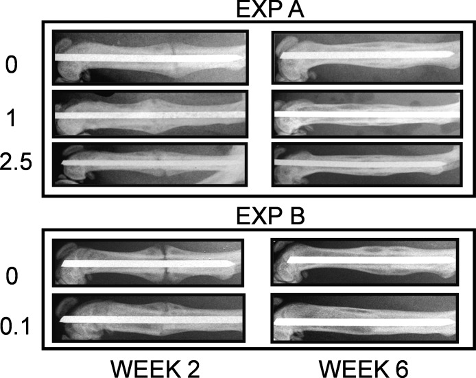
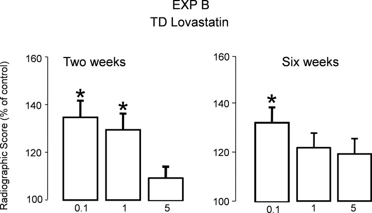
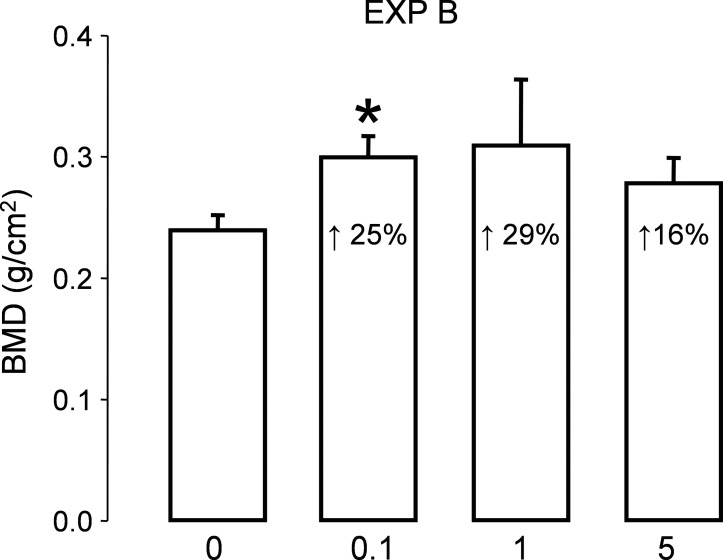
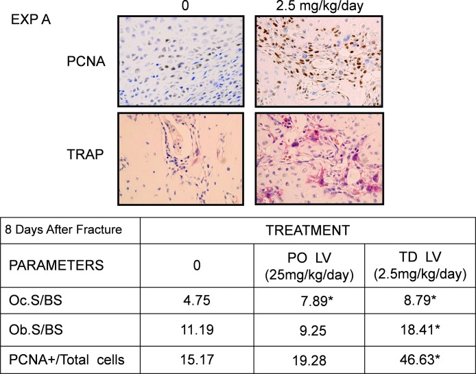
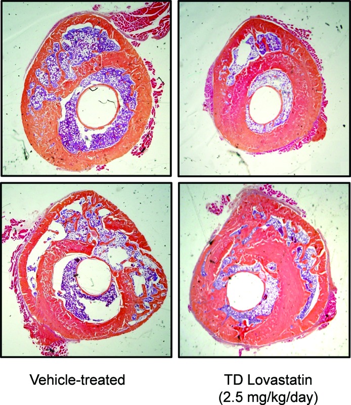
Statins have been shown to stimulate BMP2 transcription and bone formation. This raises the possibility that they could be useful for enhancing rates of fracture repair. Observational studies in patients treated with oral statins for lipid-lowering have been controversial. The likely reason for their inconsistent effects is that the statin concentration reaching the periphery was too low after oral administration to produce a reproducible biologic effect. Thus, we examined the effects of lovastatin (LV) given transdermally in a well-described preclinical model of fracture repair. Effects on the healing fracture callus were assessed by biomechanical strength, radiographs, and quantitative morphology. LV was administered transdermally (TD) for 5 days after fracture in several doses (0.1-5 mg/kg/d) and compared with vehicle-treated control rats and rats treated with LV by oral gavage (PO) at 5-25 mg/kg/d for 5 days from the day of fracture. Radiological evaluation of bones treated with TD LV showed enhanced fracture repair at 2 and 6 wk. BMD in the callus area at 6 wk was also increased in the TD group compared with vehicle-treated controls (p < 0.05). The force required to break TD-treated bones (0.1 mg/kg/d for 5 days) was 42% greater than vehicle-treated controls (p < 0.02), and there was a 90% increase in stiffness (p < 0.01). PO LV at much higher doses (10 and 25 mg/kg/d) showed increased stiffness but no change in other biomechanical properties. By histological examination, a significant increase was also observed in the size of the callus, surrounding proliferating cell nuclear antigen-positive cells, and osteoblast and osteoclast number in TD-treated rats compared with controls at day 8 after fracture (n = 6). In summary, we found that TD LV in low doses accelerates fracture healing, whereas 10-fold the lipid-lowering dose was required to produce any effect when it was administered orally. These studies provide valuable information on the potential of statins and TD delivery as a new and effective therapeutic modality in fracture repair.
Figures
References
-
- Bax BE, Wozney JM, Ashhurst DE. Bone morphogenetic protein-2 increases the rate of callus formation after fracture of the rabbit tibia. Calcif Tissue Int. 1999;65:83–89. - PubMed
-
- Schmidmaier G, Wildemann B, Cromme F, Kandziora F, Haas NP, Raschke M. Bone morphogenetic protein-2 coating of titanium implants increases biomechanical strength and accelerates bone remodeling in fracture treatment: A biomechanical and histological study in rats. Bone. 2002;30:816–822. - PubMed
-
- Einhorn TA, Majeska RJ, Mohaideen A, Kagel EM, Bouxsein ML, Turek TJ, Wozney JM. A single percutaneous injection of recombinant human bone morphogenetic protein-2 accelerates fracture repair. J Bone Joint Surg Am. 2003;85:1425–1435. - PubMed
-
- Hollinger JO, Leong K. Poly(alpha-hydroxy acids): Carriers for bone morphogenetic proteins. Biomaterials. 1996;17:187–194. - PubMed
-
- Mundy G, Garrett R, Harris S, Chan J, Chen D, Rossini G, Boyce B, Zhao M, Gutierrez G. Stimulation of bone formation in vitro and in rodents by statins. Science. 1999;286:1946–1949. - PubMed
