Review
. 2009 Feb 6;158(3):1098-111.
doi: 10.1016/j.neuroscience.2008.05.033. Epub 2008 Jul 1.

Effect of experimental stroke on peripheral immunity: CNS ischemia induces profound immunosuppression

Affiliations
Review

Effect of experimental stroke on peripheral immunity: CNS ischemia induces profound immunosuppression

H Offner et al. Neuroscience. .

Abstract

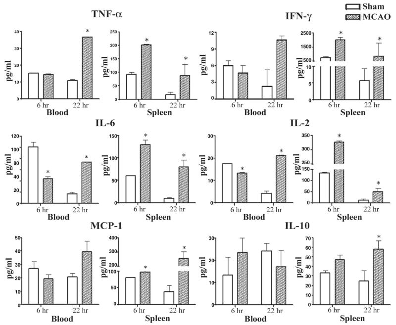
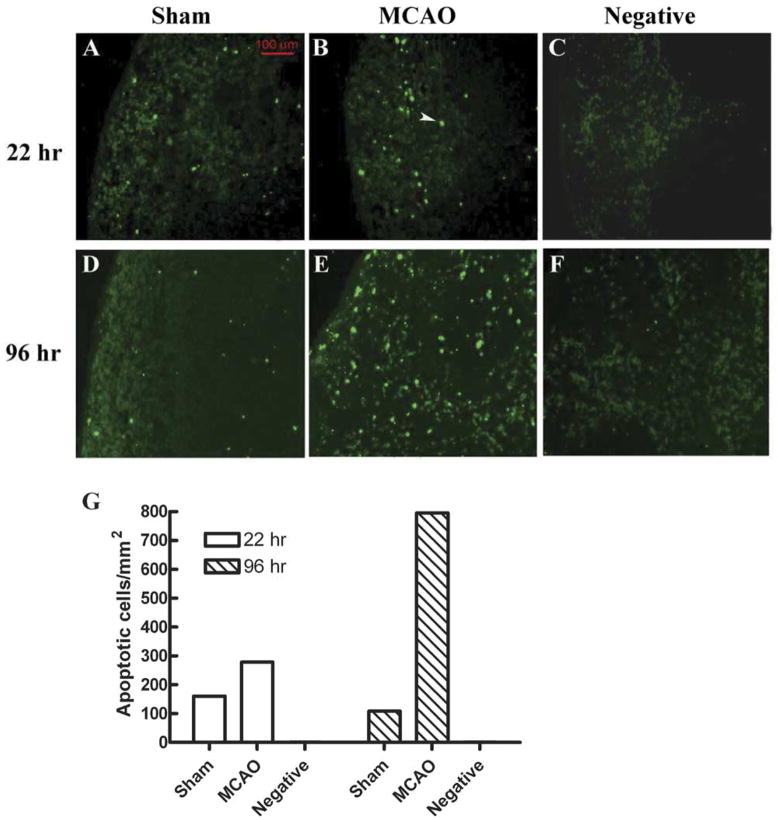
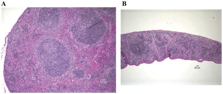
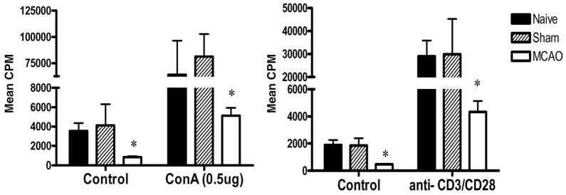
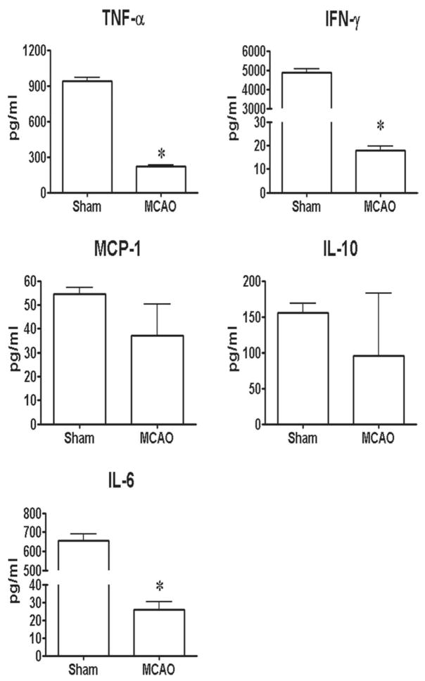
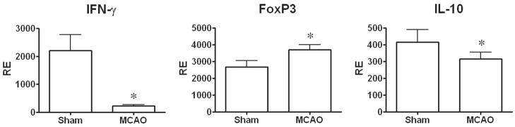
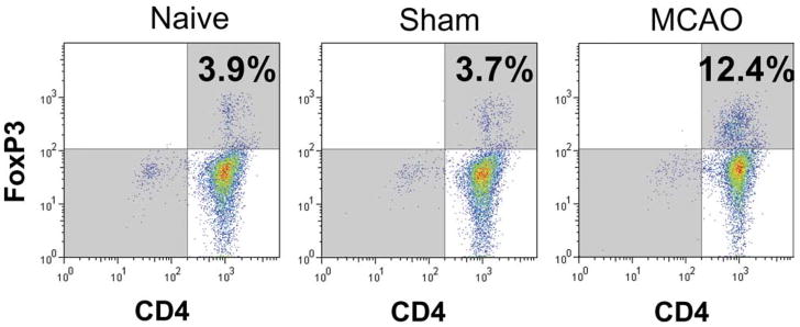
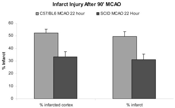
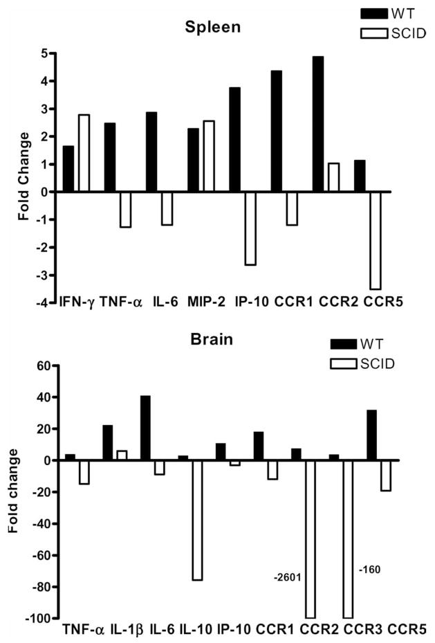
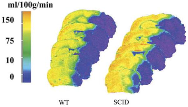
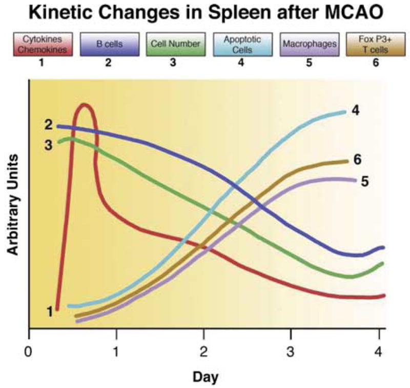
The profound damage to the CNS caused by ischemic lesions has been well documented. Yet, relatively little is known about the contribution to and effects on the immune system during stroke. We have focused on both early and late events in the peripheral immune system during stroke in mice and have observed an early activation of splenocytes that conceivably could result in immune-mediated damage in the developing CNS lesion, followed by global immunosuppression that affects the spleen, thymus, lymph nodes and circulation. While this second immunosuppressive phase may not directly enhance infarction size, it without doubt leads to an inability to respond to antigenic challenges, thereby enhancing the risk for crippling systemic infection and septicemia in stroke survivors. These novel findings advocate the need to develop or effectively utilize agents that can block early neural splenic activation and modulate immune cells specific for brain antigens as a means to prevent mobilization of T and B cells carrying a cytokine death warrant to the brain. Equally important for the recovering stroke patient are approaches that can derail the second phase of immune dysfunction and restore the ability to mount a defense against systemic infectious insults.


Figures

Fig. 1
Fig. 1
(Previously published, Journal of Cerebral Blood Flow and Metabolism 26:654, 2006.) Effects of stroke on cytokines secreted from stimulated splenocytes and blood cells. Spleens and blood were collected 6 h and 22 h after vascular occlusion and immune cells were stimulated for 48 h with plate-bound anti-CD3/CD28 antibodies. Supernatants were evaluated for levels of secreted factors, including TNF-α, IFN-γ, IL-6, MCP-1, IL-2, and IL-10. * Indicates a significant difference in expression in stroke mice versus sham-treated mice.
Fig. 2
Fig. 2
(Previously published, Journal of Immunology 176:6523, 2006.) In situ TUNEL assay showed more apoptotic cells in MCAO than in Sham mouse at 22 h and 96 h. (A, B, D, E) Fluorescence microscopic detection of apoptotic cells in mouse splenic tissues from Sham (A, D) or MCAO (B, E) mice, or in a section that was not treated with TdT enzyme ((C, F) negative control). (G) Quantification of the TUNEL-positive cell density in different splenic sections. Splenic tissue sections were prepared and assayed with In Situ Cell Death Detection Kit, Fluorescein (Roche). One section from an MCAO mouse was treated with the same kit but not incubated with TdT enzyme in the labeling step, to serve as negative control. The section was viewed under a fluorescence microscope equipped with a digital camera. Apoptotic cells intensely stained (green) by the TUNEL treatment were counted and their densities were calculated by dividing the total number of apoptotic cells by the total area of the splenic section. A typical apoptotic cell is indicated by the white arrow.
Fig. 3
Fig. 3
(Previously published, Journal of Immunology 176:6523, 2006.) Comparison of spleens and thymi obtained from Sham-MCAO, i.e. identical surgical treatment without actual vascular occlusion (A, C) and MCAO (B, D) treated mice 96 h after occlusion.
Fig. 4
Fig. 4
(Previously published, Journal of Immunology 176:6523, 2006.) Histopathology of spleens 96 h after A) Sham-MCAO treatment, and B) MCAO treatment of C57BL/6 mice. Representative section of MCAO spleen exhibits marked depletion of lymphoid tissue with lack of germinal centers. There is marked diffuse loss of normal extramedullary hematopoietic elements in the red pulp. Original magnification 100×. Representative section of Sham MCAO spleen exhibits abundant lymphoid tissue and abundant extramedullary hematopoiesis. Central follicle contains a germinal center. Original magnification 100×.
Fig. 5
Fig. 5
(Previously published, Journal of Immunology 176:6523, 2006.) Strongly reduced T cell proliferation 96 h after MCAO. Splenocytes were obtained from naïve, sham MCAO, and MCAO mice 96 h after stroke and evaluated for proliferation responses 3 days after stimulation with 0.5 μg ConA, anti-CD3 mAb or medium. * Significant reduction in response compared with naïve or sham MCAO mice.
Fig. 6
Fig. 6
(Previously published, Journal of Immunology 176:6523, 2006.) Effects of stroke on cytokines secreted from stimulated splenocytes. Spleens were collected 96 h after vascular occlusion and immune cells were stimulated for 48 h with plate-bound anti-CD3/CD28 antibodies. Supernatants were evaluated for levels of secreted factors, including TNF-α, IFN-γ, IL-6, MCP-1 and IL-10. * Indicates a significant difference in expression in MCAO versus sham MCAO mice.
Fig. 7
Fig. 7
(Previously published, Journal of Immunology 176:6523, 2006.) Effect of stroke on expression of IFN-γ, FoxP3 and IL-10 in spleen tissue. Spleens were collected from MCAO and sham MCAO mice 96 h after occlusion, and mRNA evaluated by RT-PCR for gene expression. * Indicates a significant difference in expression in MCAO versus sham MCAO mice.
Fig. 8
Fig. 8
(Previously published, Journal of Immunology 176:6523, 2006.) Increase in CD4+FoxP3± Treg cells in spleen 96 h after MCAO. Splenocytes were obtained from naïve, sham MCAO, and MCAO mice 96 h after stroke and evaluated for staining with antibodies to CD4 and FoxP3, a marker for Treg cells. Note increase in CD4+FoxP3+ T cells in MCAO mice (upper right quadrant).
Fig. 9
Fig. 9
(Previously published, Journal of Cerebral Blood Flow and Metabolism 27:1798, 2007.) Infarct size in 22 h MCAO and sham MCAO SCID mice.
Fig. 10
Fig. 10
(Previously published, Journal of Cerebral Blood Flow and Metabolism 27L1798, 2007.) Effects of stroke on expression of cytokines and chemokines/receptors in CNS tissue and spleen in MCAO vs. sham MCAO WT and SCID mice. Brains and spleen were collected from MCAO and Sham MCAO mice 22 h after occlusion, and mRNA prepared from spleen and the occluded hemispheres of brain tissue for RT-PCR analysis. Data are presented as fold changes of relative expression for indicated cytokines and chemokines/receptors in spleens (upper panel) and brain (lower panel) of MCAO vs. Sham MCAO WT (filled bars) and SCID (open bars) mice. Each figure represents data from at least two separate experiments. Each experiment included two sham MCAO and three MCAO mice.
Fig. 11
Fig. 11
C14 iodoantipyrine radiography shows that CBF at end-occlusion is equivalent in slices from brains of SCID vs. WT mice. In addition, there were no differences in the volume of tissue experiencing blood flow in the 1–10, 11–20, 21–30 ml/min/100 g ranges in SCID vs. WT mice. These data suggest that differences in intra-ischemic CBF (MCAO) do not explain the remarkably low infarction volume in SCID mice without T and B lymphocytes, as compared with their normal WT counterparts.
Fig. 12
Fig. 12
Summary of key preliminary findings and present working hypothesis of the molecular events leading to damage of lymphoid tissue and loss of peripheral immune competence.

References

    1. Ajmo CT, Vernon DOL, Collier L, Hall AA, Garbuzova-Davis S, Willing A, Pennypacker KR. The spleen contributes to stroke-induced neurodegeneration. J Neurosci Res. 2008 doi: 10.1002/jnr.21661. published online: April 1, 2008. - DOI - PMC - PubMed
    1. Allan SM, Rothwell NJ. Cytokine and acute neurodegeneration. Nat Rev. 2001;2:734–744. - PubMed
    1. Allan SM, Rothwell NJ. Inflammation in central nervous system injury. Phil Trans R Soc Lond. 2003;358:1669–1677. - PMC - PubMed
    1. Barone FC, Feuerstein GZ. Inflammatory mediators and stroke: New opportunities for novel therapies. J Cereb Blood Flow Metab. 1999;19:819–834. - PubMed
    1. Becker KJ, McCarron RM, Ruetzler C, Laban O, Sternberg E, Flanders KC, Hallenbeck JM. Immunologic tolerance to myelin basic protein decreases stroke size after transient focal cerebral ischemia. Proc Natl Acad Sci U S A. 1997;94:10873–10878. - PMC - PubMed

Publication types