

Age-related changes in dopamine transporters and accumulation of 3-nitrotyrosine in rhesus monkey midbrain dopamine neurons: relevance in selective neuronal vulnerability to degeneration
- PMID: 18598263
- PMCID: PMC3391583
- DOI: 10.1111/j.1460-9568.2008.06307.x
Age-related changes in dopamine transporters and accumulation of 3-nitrotyrosine in rhesus monkey midbrain dopamine neurons: relevance in selective neuronal vulnerability to degeneration
Abstract
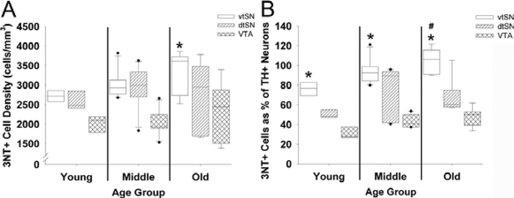
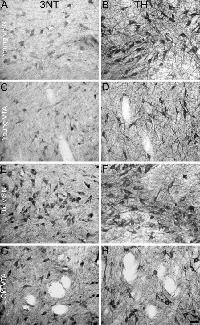
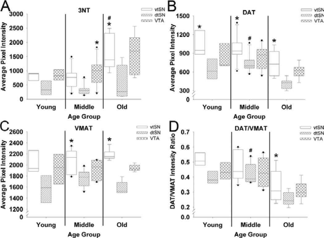
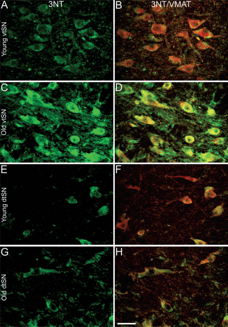
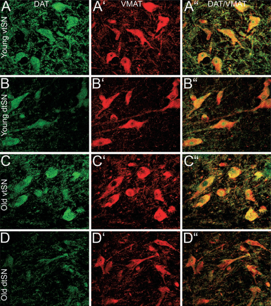
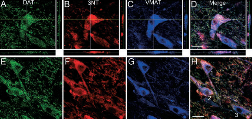
Aging is the strongest risk factor for developing Parkinson's disease (PD). There is a preferential loss of dopamine (DA) neurons in the ventral tier of the substantia nigra (vtSN) compared to the dorsal tier and ventral tegmental area (VTA) in PD. Examining age-related and region-specific differences in DA neurons represents a means of identifying factors potentially involved in vulnerability or resistance to degeneration. Nitrative stress is among the factors potentially underlying DA neuron degeneration. We studied the relationship between 3-nitrotyrosine (3NT; a marker of nitrative damage) and DA transporters [DA transporter (DAT) and vesicular monoamine transporter-2 (VMAT)] during aging in DA subregions of rhesus monkeys. The percentage of DA neurons containing 3NT increased significantly only in the vtSN with advancing age, and the vtSN had a greater percentage of 3NT-positive neurons when compared to the VTA. The relationship between 3NT and DA transporters was determined by measuring fluorescence intensity of 3NT, DAT and VMAT staining. 3NT intensity increased with advancing age in the vtSN. Increased DAT, VMAT and DAT/VMAT ratios were associated with increased 3NT in individual DA neurons. These results suggest nitrative damage accumulates in midbrain DA neurons with advancing age, an effect exacerbated in the vulnerable vtSN. The capacity of a DA neuron to accumulate more cytosolic DA, as inferred from DA transporter expression, is related to accumulation of nitrative damage. These findings are consistent with a role for aging-related accrual of nitrative damage in the selective vulnerability of vtSN neurons to degeneration in PD.
Figures
Similar articles
-
Age-related accumulation of Marinesco bodies and lipofuscin in rhesus monkey midbrain dopamine neurons: relevance to selective neuronal vulnerability.J Comp Neurol. 2007 Jun 10;502(5):683-700. doi: 10.1002/cne.21333. J Comp Neurol. 2007. PMID: 17436290
-
Age-related changes in glial cells of dopamine midbrain subregions in rhesus monkeys.Neurobiol Aging. 2010 Jun;31(6):937-52. doi: 10.1016/j.neurobiolaging.2008.07.006. Epub 2008 Aug 19. Neurobiol Aging. 2010. PMID: 18715678 Free PMC article.
-
Expression of dopamine and vesicular monoamine transporters and differential vulnerability of mesostriatal dopaminergic neurons.J Comp Neurol. 2004 Nov 8;479(2):198-215. doi: 10.1002/cne.20323. J Comp Neurol. 2004. PMID: 15452855
-
Aging and Parkinson's disease: Different sides of the same coin?Mov Disord. 2017 Jul;32(7):983-990. doi: 10.1002/mds.27037. Epub 2017 May 18. Mov Disord. 2017. PMID: 28520211 Free PMC article. Review.
-
Reward and aversion in a heterogeneous midbrain dopamine system.Neuropharmacology. 2014 Jan;76 Pt B(0 0):351-9. doi: 10.1016/j.neuropharm.2013.03.019. Epub 2013 Apr 8. Neuropharmacology. 2014. PMID: 23578393 Free PMC article. Review.
Cited by
-
Lack of functional relevance of isolated cell damage in transplants of Parkinson's disease patients.J Neurol. 2009 Aug;256 Suppl 3:310-6. doi: 10.1007/s00415-009-5242-z. J Neurol. 2009. PMID: 19711122 Review.
-
Expression of human Ras-related protein Rab39B variant T168K in Caenorhabditis elegans leads to motor dysfunction and dopaminergic neuron degeneration.Heliyon. 2024 Feb 22;10(5):e26902. doi: 10.1016/j.heliyon.2024.e26902. eCollection 2024 Mar 15. Heliyon. 2024. PMID: 38444482 Free PMC article.
-
MPTP Induces Systemic Parkinsonism in Middle-Aged Cynomolgus Monkeys: Clinical Evolution and Outcomes.Neurosci Bull. 2017 Feb;33(1):17-27. doi: 10.1007/s12264-016-0069-y. Epub 2016 Oct 3. Neurosci Bull. 2017. PMID: 27699717 Free PMC article.
-
Calcium, cellular aging, and selective neuronal vulnerability in Parkinson's disease.Cell Calcium. 2010 Feb;47(2):175-82. doi: 10.1016/j.ceca.2009.12.003. Epub 2010 Jan 6. Cell Calcium. 2010. PMID: 20053445 Free PMC article. Review.
-
Diverse midbrain dopaminergic neuron subtypes and implications for complex clinical symptoms of Parkinson's disease.Ageing Neurodegener Dis. 2021;1(4):10.20517/and.2021.07. doi: 10.20517/and.2021.07. Epub 2021 Jul 15. Ageing Neurodegener Dis. 2021. PMID: 34532720 Free PMC article.
References
-
- Alam ZI, Daniel SE, Lees AJ, Marsden DC, Jenner P, Halliwell B. A generalised increase in protein carbonyls in the brain in Parkinson’s but not incidental Lewy body disease. J. Neurochem. 1997a;69(3):1326–1329. - PubMed
-
- Alam ZI, Jenner A, Daniel SE, Lees AJ, Cairns N, Marsden CD, Jenner P, Halliwell B. Oxidative DNA damage in the parkinsonian brain: an apparent selective increase in 8-hydroxyguanine levels in substantia nigra. J. Neurochem. 1997b;69(3):1196–1203. - PubMed
-
- Asanuma M, Miyazaki I, az-Corrales FJ, Ogawa N. Quinone formation as dopaminergic neuron-specific oxidative stress in the pathogenesis of sporadic Parkinson’s disease and neurotoxin-induced parkinsonism. Acta Med. Okayama. 2004;58(5):221–233. - PubMed
-
- Beckman JS, Koppenol WH. Nitric oxide, superoxide, and peroxynitrite: the good, the bad, and ugly. Am. J. Physiol. 1996;271(5 Pt 1):C1424–C1437. - PubMed
Publication types
MeSH terms
Substances
Grants and funding
LinkOut - more resources
Full Text Sources
Medical
