

An fMRI study of neural interaction in large-scale cortico-thalamic visual network
- PMID: 18598771
- PMCID: PMC2593731
- DOI: 10.1016/j.neuroimage.2008.05.060
An fMRI study of neural interaction in large-scale cortico-thalamic visual network
Abstract
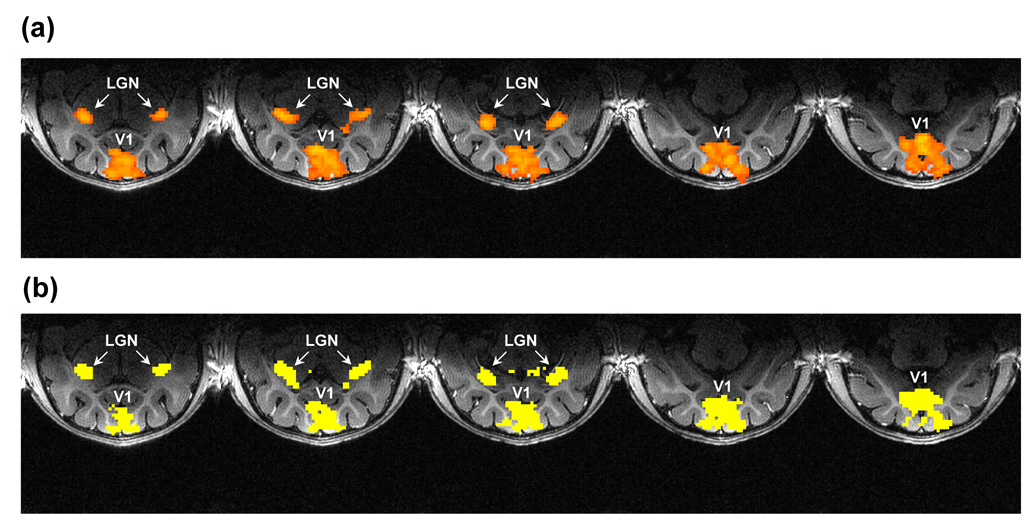
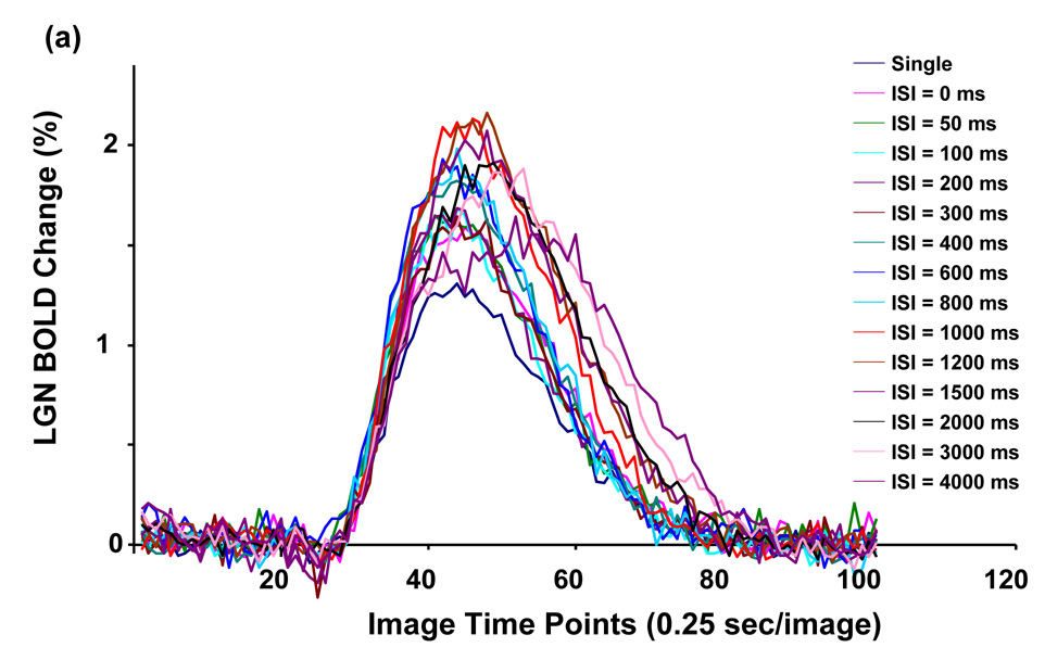
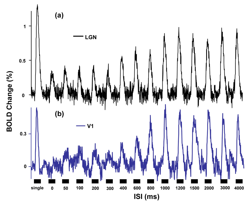
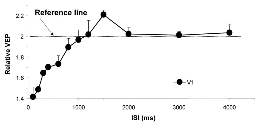
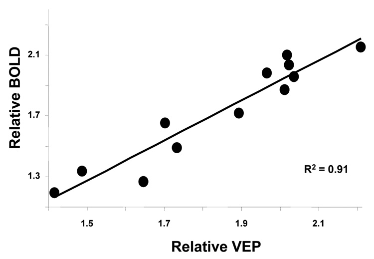
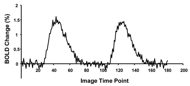
To date there is still no proper neuroimaging methods suitable for noninvasively providing both detailed spatial and temporal information of neural interaction across large-scale brain networks. This limitation has impeded the advance of neuroscience research. In an attempt to overcome this challenge, Ogawa et al. applied a paired-stimulus paradigm, which is composed of a pair of stimuli separated by a variable inter-stimulus interval (ISI), to decode temporal information of neural interaction from amplitude modulation of the blood-oxygenation-level-dependent (BOLD) responses elicited by the neural interaction pursued [Ogawa, S., Lee, T.-M., Stepnoski, R., Chen, W., Zhu, X.H., Ugurbil, K., 2000. An approach to probe neural systems interaction by functional MRI at neural time scale down to milliseconds. Proc. Natl. Acad Sci. U S A 97, 11026-11031.]. Although application of this paradigm has been demonstrated in a few publications, most of them only focused on investigating cortico-cortical interaction. Considering the vital roles that cortico-thalamic networks play in brain communication and function, extending the applicability of this method to studying cortico-thalamic neural interaction should be significant. In this study, we applied the paired-visual-stimulus paradigm to simultaneously measure the BOLD amplitude modulations as a function of ISI in the lateral geniculate nucleus (LGN) and primary visual cortex (V1) in the cat brain. The results reveal that both V1 and LGN BOLD responses were significantly suppressed when the visual system was within the refractory period at ISI<or=1 s and the suppression extent was gradually recovered when ISI became longer. Both BOLD and electrophysiological measurements show a facilitatory activity in V1 at ISI approximately 1.5 s, but not in LGN. Furthermore, there was additional and consistent reduction in the LGN BOLD response compared to V1 within the range of ISI below 4 s, which is likely controlled by inhibitory effects through the cortico-geniculate feedback. These findings together suggest that the dynamic fMRI approach applied in this study is sensitive to neuronal inhibitory and facilitatory interactions and it should be useful for noninvasively investigating large-scale cortico-thalamic neural networks.
Figures


References
-
- Arthurs OJ, Williams EJ, Carpenter TA, Pickard JD, Boniface SJ. Linear coupling between functional magnetic resonance imaging and evoked potential amplitude in human somatosensory cortex. Neuroscience. 2000;101:803–806. - PubMed
-
- Bachmann T. Time course of the subjective contrast enhancement for a second stimulus in successively paired above-threshold transient forms: perceptual retouch instead of forward masking. Vision Res. 1988;28:1255–1261. - PubMed
-
- Bandettini PA, Jesmanowicz A, Wong EC, Hyde JS. Processing strategies for time-course data sets in functional MRI of the human brain. Magn Reson Med. 1993;30:161–173. - PubMed
-
- Bartlett JR, Doty RW, Lee BB, Sr, Sakakura H. Influence of saccadic eye movements on geniculostriate excitability in normal monkeys. Exp Brain Res. 1976;25:487–509. - PubMed
-
- Breitmeyer BG. Unmasking visual masking: a look at the "why" behind the veil of the "how". Psychol Rev. 1980;87:52–69. - PubMed
Publication types
MeSH terms
Grants and funding
LinkOut - more resources
Full Text Sources
Medical
Miscellaneous
