. 2008 Jul 8;14(1):36-46.
doi: 10.1016/j.ccr.2008.05.003.

Menin critically links MLL proteins with LEDGF on cancer-associated target genes

Affiliations

Menin critically links MLL proteins with LEDGF on cancer-associated target genes

Akihiko Yokoyama et al. Cancer Cell. .

Abstract

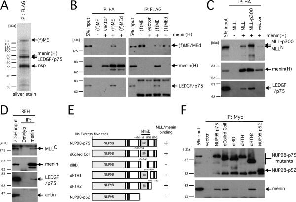
Menin displays the unique ability to either promote oncogenic function in the hematopoietic lineage or suppress tumorigenesis in the endocrine lineage; however, its molecular mechanism of action has not been defined. We demonstrate here that these discordant functions are unified by menin's ability to serve as a molecular adaptor that physically links the MLL (mixed-lineage leukemia) histone methyltransferase with LEDGF (lens epithelium-derived growth factor), a chromatin-associated protein previously implicated in leukemia, autoimmunity, and HIV-1 pathogenesis. LEDGF is required for both MLL-dependent transcription and leukemic transformation. Conversely, a subset of menin mutations in multiple endocrine neoplasia type 1 patients abrogate interaction with LEDGF while preserving MLL interaction but nevertheless compromise MLL/menin-dependent functions. Thus, LEDGF critically associates with MLL and menin at the nexus of transcriptional pathways that are recurrently targeted in diverse diseases.


Figures

Figure 1
Figure 1. LEDGF and NUP98-LEDGF associate with the MLL/menin histone methyltransferase complex
A. Silver stained image of an SDS-PAGE analysis shows presence of LEDGF in the purified MLL-ENL/menin immunoprecipitate. FLAG-tagged MLL-ENL [(f)ME] and HA-tagged menin [menin(H)] were transiently expressed in large scale culture of 293T cells. A nuclear extract was prepared from the cells and subjected to immunopurification using anti-FLAG antibody. Protein bands from SDS-PAGE analysis were analyzed by mass spectrometry and identified as indicated by arrows. The band at 52 kDa is a non-specific (nsp) product that was also observed in the control IP of a nuclear extract from non-transduced cells (not shown). Protein standards are shown on the left. B. Reciprocal immunoprecipitations of the MLL-ENL/menin complex. (f)ME, or a mutant [(f)MEd] lacking the high affinity menin binding motif, were transiently expressed in 293T cells with (+) or without (-) HA-tagged menin. Nuclear extracts from the transfected cells were subjected to IP with anti-FLAG or anti-HA antibodies followed by immunoblotting with anti-FLAG, anti-HA, and anti-LEDGF (p75) antibodies. C. MLL or MLL-p300 was transiently expressed with (+) or without (-) HA-tagged menin in 293T cells and subjected to IP with anti-HA antibody, followed by immunoblotting with anti-MLLN, anti-HA, and anti-LEDGF antibodies. D. To detect endogenous MLL/menin/LEDGF association, nuclear extract of REH cells was subjected to IP with anti-menin antibody followed by immunoblotting with anti-MLLC, anti-menin, anti-LEDGF, and anti-ACTIN antibodies. Anti-Drosophila Myb (DmMyb) antibody was used as a negative control. E. Schematic representations of the NUP98-LEDGF mutants are shown with a summary of their binding properties with MLL/menin on the right. The identified minimum MLL/menin binding domain (MmBD, residues 335-460) is indicated. F. His-Express-Myc-tagged NUP98-LEDGF proteins were expressed in 293T cells and subjected to IP with anti-MYC antibody followed by immunoblotting with anti-Express epitope and anti-menin antibodies.
Figure 2
Figure 2. MLL proteins co-localize with LEDGF on chromatin of cancer-associated genes
A. ChIP was performed on ML-2 cells using anti-MLLN, anti-menin and anti-LEDGF antibodies. MLL-AF6, menin and LEDGF occupancy was specifically observed at the HOXA7 and HOXA9 loci, but not on the MEIS1 and GAPDH loci. Negative and positive controls consisted of no antibody and anti-histone H3 antibody, respectively. B. ChIP followed by quantitative PCR was performed on ML-2 and U937 cells using anti-MLLN, anti-menin and anti-LEDGF antibodies. Values are expressed relative to the maximum value (arbitrarily set at 100%) in each group with error bars representing standard deviations for triplicate PCR analyses.
Figure 3
Figure 3. MLL oncoproteins associate with LEDGF to initiate myeloid transformation
A. Schematic structure of the MLL-ENL oncoprotein. Amino acid sequence of MLL encompassing the LEDGF binding domain (LBD, residues 109-153 denoted by red box) is aligned with that of fugu MLL. Arrow indicates the phenylalanine residue substituted to alanine. B. Various mutants of (f)ME were transiently expressed with (+) or without (-) HA-tagged menin in 293T cells and subjected to IP with anti-HA antibody followed by immunoblotting with anti-FLAG, anti-HA, and anti-LEDGF antibodies. C. The experimental scheme for myeloid progenitor transformation assay. The time point at which Hoxa9 expression was measured (end of 1st plating) is indicated. D. The colony forming units (CFU) per 104 plated cells are shown for each round of plating. Error bars represent standard deviations of three independent experiments. E. Relative expression levels of Hoxa9 are shown for first round colonies. Expression levels are normalized to β-actin and expressed relative to the ME value (arbitrarily set at 100%). Error bars represent standard deviations of triplicate PCR analyses. F. Subnuclear localizations of MLL fusion proteins in HeLa cells are shown as a merged image of signals for FITC (MLL) and DAPI (DNA). Scale bar, 10 μm.
Figure 4
Figure 4. Menin tethers LEDGF with MLL oncoproteins
A. Schematic structures of menin, MLL-ENL and LEDGF are shown at the top with sites of intermolecular interactions indicated by arrows. Binding properties and transforming abilities are summarized to the right of the respective MLL-ENL constructs. The numbers of 3rd round CFU in myeloid progenitor assays are shown with error bars representing the standard deviations of three independent experiments. B. Various mutants of pME were transiently expressed with (+) or without (-) HA-tagged menin in 293T cells and subjected to IP with anti-HA antibody followed by immunoblotting with anti-MLLN, anti-HA, and anti-LEDGF antibodies. Upper panel shows immunoblot of the nuclear extracts (NE) to determine protein inputs. Mutants lacking the hMBM failed to co-IP with menin and LEDGF. C. The relative expression levels are shown for Hoxa9 in first round colonies. Expression levels are normalized to β-Actin, and expressed relative to the ME value (arbitrarily set at 100%). Error bars represent standard deviations of triplicate PCR analyses. D. Subnuclear localization of the pME mutants in HeLa cells is shown as a merged image of signals for FITC (MLL) and DAPI (DNA). Scale bar, 10 μm. E. ChIP analysis of ME- or pME-transformed myeloid progenitors was performed using antibodies indicated at the top. Amplicons upstream of Hoxa9 and β-Actin genes were analyzed. F. Survival curves are shown for mice transplanted with the indicated transduced cells. Numbers of mice analyzed (n) are shown. G. Expression of ME and pME proteins in immortalized cells (IC) and leukemic cells (LC) from bone marrow (BM) and spleen (SP) were analyzed by immunoblotting with anti-MLLN and anti-SIN3A (control) antibodies. H. Morphology is shown for pME leukemic blasts (pME: right panel) and normal BM (wt: left) following May-Grunwald/Giemsa staining of cytospin preparations. Scale bar, 15 μm.
Figure 5
Figure 5. LEDGF is necessary for maintenance of MLL leukemic transformation
A. The experimental scheme for conditional inactivation of Men1 in MLL transformed cells. The time points at which CFU or gene expression were measured are indicated. B. The relative CFUs are shown for cells transformed by ME or pME in the absence (vector) or presence (Cre-ER) of Men1 inactivation (vector controls are arbitrarily set at 100%). Error bars represent the standard deviations of three independent analyses. C. Morphologies are shown for representative colonies from experiment in panel B. Scale bar, 150 μm. D. Western blot shows expression of pME, menin and SIN3A proteins after Men1 inactivation. E. Relative expression levels of Hoxa9 are shown for first round colonies after Cre-ER transduction. Expression levels were normalized to β-Actin, and expressed relative to the vector control values (arbitrarily set at 100%). Error bars represent standard deviations of triplicate PCR analyses. F. Experimental scheme for conditional inactivation of Ledgf by shRNA-mediated knockdown. The time points at which CFUs or gene expression were measured are indicated. G. The relative CFU activity of ME- or pME-transformed cells is shown with (sh-Ledgf#1 or sh-Ledgf#2) or without (vector) Ledgf knockdown (the vector controls are arbitrarily set at 100%). Error bars represent the standard deviations of three independent analyses. H. Relative expression levels of Hoxa9 and Ledgf are shown for first round colonies after shRNA vector transduction. Expression levels are normalized to β-actin, and expressed relative to the vector control values (arbitrarily set at 100%). Error bars represent the standard deviations of triplicate PCR analyses.
Figure 6
Figure 6. Menin functionally interacts with LEDGF
A. Schematic structures of menin and its mutants. The abilities of each protein to associate with MLL or LEDGF and to rescue MLL-dependent transcription are summarized on the right. *, indicates impaired association as opposed to complete loss of association. ND, not determined. B. Various HA-tagged menin proteins were transiently expressed with (+) or without (-) (f)ME in 293T cells and subjected to IP with anti-HA antibody, followed by immunoblotting with anti-FLAG, anti-HA, and anti-LEDGF antibodies. C. Experimental scheme for rescue of Men1 knockdown by wild type or mutant menin proteins. The time points at which CFUs or gene expression were measured are indicated. D. Western blot analysis shows expression of menin mutants in ME-transformed cells detected by anti-HA and anti-MLLN antibodies, respectively. E. Relative CFU are shown for ME-transformed cells transduced with various menin mutants with (+) or without (-) Men1 knock down (vector control was arbitrarily set at 100%). Error bars represent the standard deviations of three independent analyses. F. Relative expression levels of Hoxa9 and Men1 are shown for first round colonies after Men1 knockdown. Expression levels are normalized to Gapdh, and expressed relative to the ME/vector value (arbitrarily set at 100%). Error bars represent the standard deviations of triplicate PCR analyses. P values for differences compared to wild type menin rescue were determined by unpaired t test (*, P<0.0005; **, P<0.005).
Figure 7
Figure 7. Model for the role of LEDGF in the normal and neoplastic functions of the MLL/menin HMT complex
Upper: Specific association with LEDGF is required for transcriptional contributions by the MLL/menin HMT complex at its chromatin sites of action. Lower: Similarly, the constitutive transcriptional properties of MLL chimeric oncoproteins in complex with menin are also dependent on association with LEDGF, which may provide a molecular target for therapeutic intervention.

Comment in

  • A MENage à Trois in leukemia.
    Roudaia L, Speck NA. Roudaia L, et al. Cancer Cell. 2008 Jul 8;14(1):3-5. doi: 10.1016/j.ccr.2008.06.009. Cancer Cell. 2008. PMID: 18598937

Similar articles

Cited by

  • A dual role for PSIP1/LEDGF in T cell acute lymphoblastic leukemia.
    Demoen L, Matthijssens F, Reunes L, Palhais B, Lintermans B, T'Sas S, Fijalkowski I, Taminau J, Akele MZ, Van Belle S, Taghon T, Deforce D, Van Nieuwerburgh F, Berx G, Ntziachristos P, Debyser Z, Durinck K, Pieters T, Goossens S, Van Vlierberghe P. Demoen L, et al. Sci Adv. 2024 Nov;10(44):eado6765. doi: 10.1126/sciadv.ado6765. Epub 2024 Nov 1. Sci Adv. 2024. PMID: 39485844 Free PMC article.
  • Novel Targeted Therapeutics in Acute Myeloid Leukemia: an Embarrassment of Riches.
    Grieselhuber NR, Mims AS. Grieselhuber NR, et al. Curr Hematol Malig Rep. 2021 Apr;16(2):192-206. doi: 10.1007/s11899-021-00621-9. Epub 2021 Mar 18. Curr Hematol Malig Rep. 2021. PMID: 33738705 Free PMC article. Review.
  • Drug discovery toward antagonists of methyl-lysine binding proteins.
    Herold JM, Ingerman LA, Gao C, Frye SV. Herold JM, et al. Curr Chem Genomics. 2011;5:51-61. doi: 10.2174/1875397301005010051. Epub 2011 Aug 22. Curr Chem Genomics. 2011. PMID: 22145013 Free PMC article.
  • IMPDH inhibition activates TLR-VCAM1 pathway and suppresses the development of MLL-fusion leukemia.
    Liu X, Sato N, Yabushita T, Li J, Jia Y, Tamura M, Asada S, Fujino T, Fukushima T, Yonezawa T, Tanaka Y, Fukuyama T, Tsuchiya A, Shikata S, Iwamura H, Kinouchi C, Komatsu K, Yamasaki S, Shibata T, Sasaki AT, Schibler J, Wunderlich M, O'Brien E, Mizukawa B, Mulloy JC, Sugiura Y, Takizawa H, Shibata T, Miyake K, Kitamura T, Goyama S. Liu X, et al. EMBO Mol Med. 2023 Jan 11;15(1):e15631. doi: 10.15252/emmm.202115631. Epub 2022 Dec 1. EMBO Mol Med. 2023. PMID: 36453131 Free PMC article.
  • Epigenetic regulation of IL-12-dependent T cell proliferation.
    Schaller M, Ito T, Allen RM, Kroetz D, Kittan N, Ptaschinski C, Cavassani K, Carson WF 4th, Godessart N, Grembecka J, Cierpicki T, Dou Y, Kunkel SL. Schaller M, et al. J Leukoc Biol. 2015 Oct;98(4):601-13. doi: 10.1189/jlb.1A0814-375RR. Epub 2015 Jun 9. J Leukoc Biol. 2015. PMID: 26059830 Free PMC article.

References

    1. Ahuja HG, Hong J, Aplan PD, Tcheurekdjian L, Forman SJ, Slovak ML. t(9;11)(p22;p15) in acute myeloid leukemia results in a fusion between NUP98 and the gene encoding transcriptional coactivators p52 and p75-lens epithelium-derived growth factor (LEDGF) Cancer Res. 2000;60:6227–6229. - PubMed
    1. Alvarez-Venegas R, Avramova Z. Two Arabidopsis homologs of the animal trithorax genes: a new structural domain is a signature feature of the trithorax gene family. Gene. 2001;271:215–221. - PubMed
    1. Ayton PM, Chen EH, Cleary ML. Binding to nonmethylated CpG DNA is essential for target recognition, transactivation, and myeloid transformation by an MLL oncoprotein. Mol Cell Biol. 2004;24:10470–10478. - PMC - PubMed
    1. Ayton PM, Cleary ML. Transformation of myeloid progenitors by MLL oncoproteins is dependent on Hoxa7 and Hoxa9. Genes Dev. 2003;17:2298–2307. - PMC - PubMed
    1. Balogh K, Racz K, Patocs A, Hunyady L. Menin and its interacting proteins: elucidation of menin function. Trends Endocrinol Metab. 2006;17:357–364. - PubMed

Publication types

MeSH terms