. 2008 Jul 8;14(1):68-78.
doi: 10.1016/j.ccr.2008.05.017.

Mutations in Fbx4 inhibit dimerization of the SCF(Fbx4) ligase and contribute to cyclin D1 overexpression in human cancer

Affiliations

Mutations in Fbx4 inhibit dimerization of the SCF(Fbx4) ligase and contribute to cyclin D1 overexpression in human cancer

Olena Barbash et al. Cancer Cell. .

Abstract

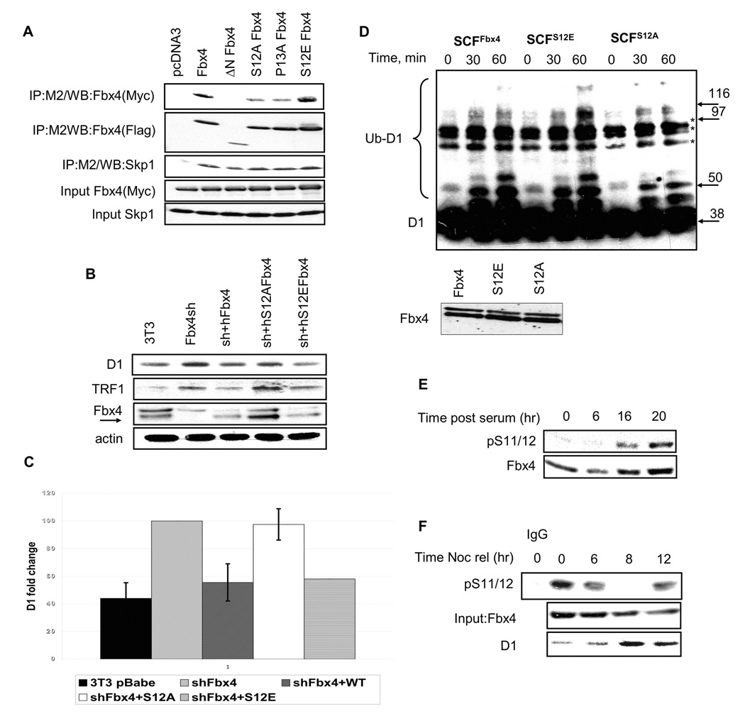
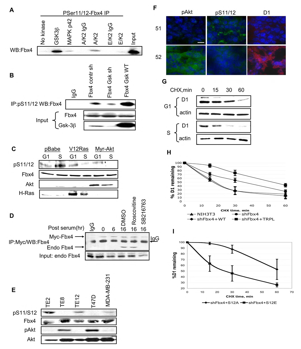
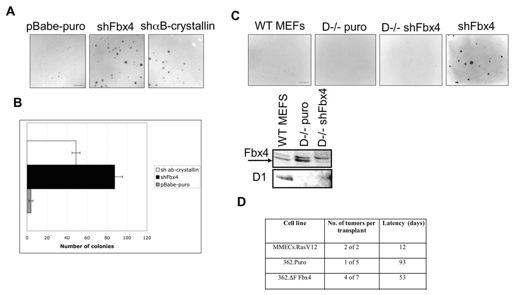
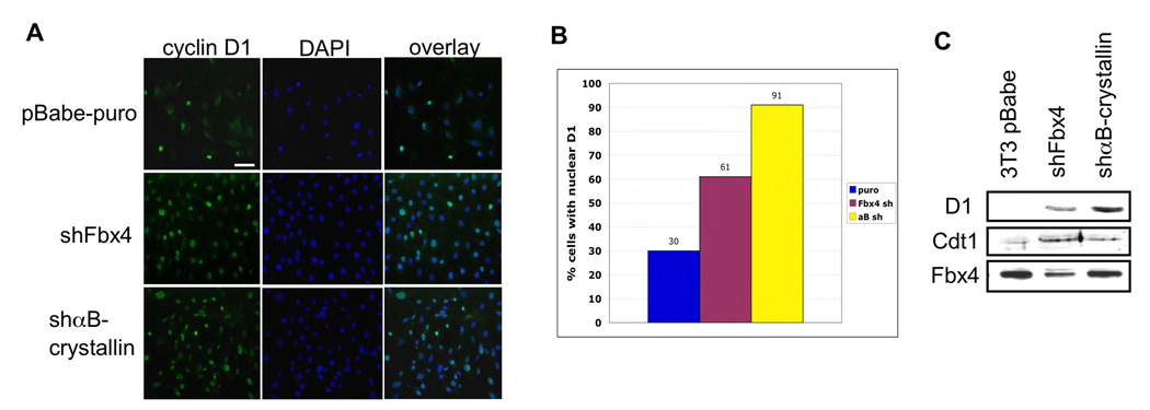
SCF(Fbx4) was recently identified as the E3 ligase for cyclin D1. We now describe cell-cycle-dependent phosphorylation and dimerization of Fbx4 that is regulated by GSK3beta and is defective in human cancer. We present data demonstrating that a pathway involving Ras-Akt-GSK3beta controls the temporal phosphorylation and dimerization of the SCF(Fbx4) E3 ligase. Inhibition of Fbx4 activity results in accumulation of nuclear cyclin D1 and oncogenic transformation. The importance of this regulatory pathway for normal cell growth is emphasized by the prevalence of mutations in Fbx4 in human cancer that impair dimerization. Collectively, these data reveal that inactivation of the cyclin D1 E3 ligase likely contributes to cyclin D1 overexpression in a significant fraction of human cancer.


Figures

Figure 1
Figure 1. Dimerization of Fbx4 occurs through its N-terminus
A and B. Lysates were prepared from 293T cells transfected with vectors encoding human wild type Myc-tagged Fbx4 and the indicated Flag-Fbx4 mutant constructs. Protein complexes were isolated by M2 affinity chromatography and detected by immunoblot with a Fbx4 antibody. C. 293T cells were co-transfected with Fbx4 constructs along with cyclin D1, CDK4 and αB-crystallin. 24hrs post-transfection cells were treated with MG132 for 6 hrs followed by cyclin D1 immunoprecipitation. Cyclin D1 was detected with the 13G11 monoclonal antibody.
Figure 2
Figure 2. Fbx4 serine 12 is required for Cyclin D1 ubiquitination and proteolysis
A. 293T cells were transfected with plasmids encoding Flag-tagged Fbx4 mutants and wild type Myc-tagged Fbx4. Complexes were isolated by affinity chromatography using the M2 conjugated agarose and individual components detected by immunoblot with Fbx4 and Skp1 antibodies. B. NIH-3T3 cells, wherein Fbx4 had previously been knocked down by shRNA, were transfected with wt, S12A or S12E Fbx4 pcDNA3 followed by G418 selection of stably expressing clones. Asynchronous cells were harvested and subjected to Western blotting with cyclin D1, TRF1 and Fbx4 antibodies. C. Quantification of B. Error bars indicate +/−SD. D. Ligase complexes (purified protein shown in bottom panel) were purified from stable cell lines expressing wt, S12E and S12A Fbx4 (described in B). In vitro ubiquitination reactions were performed using GST-tagged purified cyclin D1. Ubiquitin-conjugated cyclin D1 was detected by immunoblot. Non-specific complexes are denoted by asterisk. E. NIH3T3 cells were serum starved for 48 hrs and released into 10% FBS containing media for the indicated intervals. Immunoblot analysis was performed using pS11/12-Fbx4 and a total Fbx4 antibody. F. Cells synchronized via a nocodazole block were harvested at the indicated intervals following nocodazole release. Fbx4 phosphorylation was detected by precipitation with the pS11/12 antibody and immunoblot with the total Fbx4 antibody. Total cyclin D1 and Fbx4 levels were assessed by direct western.
Figure 3
Figure 3. GSK3β phosphorylates S11/12 of Fbx4 in S phase of cell cycle
A. In vitro kinase assay was performed using purified GST-tagged Fbx4 and indicated kinases. Following in vitro phosphorylation, samples were precipitated with the pS11/12–Fbx4 antibody and subjected to immunoblot with a Fbx4 antibody. B. 293T cells were transfected with wild type Fbx4 along with control shGFP vector, a GSK3β shRNA vector, or a plasmid encoding wild type GSK3β Fbx4 was immunoprecipitated from cellular extracts using pS11/12-Fbx4 antibody 48hrs post–transfection and detected by immunoblot with the total Fbx4 antibody. C. NIH3T3 cells were transfected with an empty vector, or RasV12 or Myr Akt expressing constructs. Cells were synchronized by serum starvation and released into complete medium for 6hrs (G1) or 16hrs(S). Protein extracts were processed for immunoblot using pS11/12-Fbx4, total Fbx4, total Akt and H-Ras antibodies. D. NIH3T3 cells were transfected with vector encoding Myc-tagged Fbx4 and 24 hrs later were synchronized in G0. Cells were released into cell cycle for the indicated intervals (cells were treated with kinase inhibitors for 6 hrs before harvesting samples at 16 hrs time point). Myc-Fbx4 pull down was performed using Myc-agarose. Myc-Fbx4 associated endogenous Fbx4 was detected by immunoblot with total Fbx4 antibody. E. Total cell extracts from esophageal carcinoma cell lines (TE2, TE8 and TE12) and breast carcinoma cell lines (T47D and MDA-MB-231) were subjected to precipitaion with the pS11/12 antibody followed by immunoblot with the Fbx4 antibody. Direct Western blotting was performed using pAkt (Ser473) and total Akt antibodies. F. Frozen sections from MMTV-Neu breast tumors were analyzed by IHC using following antibodies: pS11/12-Fbx4, pAkt (Ser473) and cyclin D1 (bar, 8µM). G. NIH3T3 cells were arrested by serum starvation following by replating in complete media. Cycloheximide was added for the indicated intervals during G1 (6 hrs post-release) and S phases of cell cycle (16 hrs post-release). Cyclin D1 turnover was analyzed by Western analysis using cyclin D1 antibody. H. NIH3T3 shFbx4 stable cell line was transfected with either wild type or Fbx4 trpl. Cycloheximide chase was performed in asynchronous cells 48 hrs after transfection. Quantification of 3 independent experiments is provided, error bars represent +/− SD. I. Fbx4S12A or Fbx4S12E were individually re-introduced into Fbx4 knockdown cells and cyclin D1 turnover was assessed in S phase cells; quantification of 3 independent experiments is provided, error bars represent +/− SD.
Figure 4
Figure 4. Inhibition of SCFFbx4/αB-crystallin activity leads to neoplastic growth
A. Soft agar colony formation assay was performed using pBabe-puro, shFbx4 and shαB-crystallin knockdown cell lines. Cells were plated in soft agar and grown for 21 days (bar, 250µM). B. Quantification of A, error bars represent +/− SD. C. Same as A using wt and cyclin D1−/− MEFs infected with control (puro) or shFbx4 retrovirus (bar, 250µM). Levels of Fbx4 and cyclin D1 proteins are provided in the bottom panel. D. 106 cells were transplanted into cleared mammary fat pads of 3 week-old NOD-SCID mice. Mice were monitored for palpable tumor formation bi-weekly.
Figure 5
Figure 5. Disruption of SCFFbx4/αB-crystallin activity leads to nuclear accumulation of cyclin D1
A. Immunofluorescence was performed on the indicated cell lines using the 13G11 cyclin D1 monoclonal antibody and counterstained with DAPI (bar, 25 µM). B. Quantification of A. C. Protein extracts from stable NIH3T3 cell lines expressing shFbx4 or shαB-crystallin were analyzed by Western Blot using Cdt1 and cyclin D1 antibodies.
Figure 6
Figure 6. Analysis of Fbx4 mutations in human esophageal tumors
A. and B. Immunohistochemical analysis of cyclin D1 or TRF1 expression in esophageal tumors: S8R Fbx4, P76T Fbx4, S12L and wtFbx4, normal esophagus (bars, 50 (A) and 25 (B) µM). C. shFbx4 NIH3T3 lines were transfected with indicated Fbx4 expression vectors (immunoblot of Fbx4 levels is presented in the bottom panel) were assessed for growth in soft agar (21 days, bar, 250 µM). D. Quantification of C, error bars represent +/− SD.

References

    1. Aggarwal P, Lessie MD, Lin DI, Pontano L, Gladden AB, Nuskey B, Goradia A, Wasik MA, Klein-Szanto AJ, Rustgi AK, et al. Nuclear accumulation of cyclin D1 during S phase inhibits Cul4-dependent Cdt1 proteolysis and triggers p53-dependent DNA rereplication. Genes Dev. 2007;21:2908–2922. - PMC - PubMed
    1. Alt JR, Cleveland JL, Hannink M, Diehl JA. Phosphorylation-dependent regulation of cyclin D1 nuclear export and cyclin D1-dependent cellular transformation. Genes Dev. 2000;14:3102–3114. - PMC - PubMed
    1. Beausoleil SA, Jedrychowski M, Schwartz D, Elias JE, Villen J, Li J, Cohn MA, Cantley LC, Gygi SP. Large-scale characterization of HeLa cell nuclear phosphoproteins. Proc Natl Acad Sci U S A. 2004;101:12130–12135. - PMC - PubMed
    1. Benzeno S, L F, Guo M, Barbash O, Zhang F, Herman JG, Klein PS, Rustgi A, Diehl JA. Identification of mutations that disrupt phosphorylation-dependent nuclear export of cyclin D1. Oncogene. 2006 - PubMed
    1. Chew EH, Poobalasingam T, Hawkey CJ, Hagen T. Characterization of cullin-based E3 ubiquitin ligases in intact mammalian cells--evidence for cullin dimerization. Cell Signal. 2007;19:1071–1080. - PubMed

Publication types

MeSH terms