Review
. 2008 Jul;4(3):235-45.
doi: 10.1016/j.hfc.2008.02.007.

Embryogenesis of the heart muscle

Affiliations
Review

Embryogenesis of the heart muscle

David Sedmera et al. Heart Fail Clin. 2008 Jul.

Abstract

This article concerns the development of myocardial architecture--crucial for contractile performance of the heart and its conduction system, essential for generation and coordinated spread of electrical activity. Topics discussed include molecular determination of cardiac phenotype (contractile and conducting), remodeling of ventricular wall architecture and its blood supply, and relation of trabecular compaction to noncompaction cardiomyopathy. Illustrated are the structure and function of the tubular heart, time course of trabecular compaction, and development of multilayered spiral systems of the compact layer.


Figures
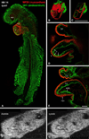
Figure 1
Figure 1
Structure of a tubular heart. A-E: whole mount immunostaining for myocardial marker myosin heavy chain (red) and endothelial QH1 marker (green) shows beautifully the shape of the heart (S-loop). Individual confocal sections (D, E) show the diminished amount of cardiac jelly in the ventricular apex, resulting in close contact between these two layers. Endocardial ruffles (arrowheads) show the site of prospective trabecular formation. Three-dimensional reconstructions (B, C) show the whole heart and an oblique section illustrating the variable extent of the cardiac jelly. Scale bars 500 µm (B, C) and 100 µm (D, E). Echocardiographic views (F, G) show the heart loop in two different phases of cardiac cycle. Note the echo-free acellular layer of cardiac jelly (arrows) along the inner curvature during systole. CJ, cardiac jelly, endo, endocardium, myo, myocardium, NT, neural tube, OT, outflow tract, V, ventricle. Scale bar 100 µm smallest division.
Figure 2
Figure 2
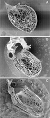
Time course of ventricular compaction in the human left ventricle. Note increasing proportion and thickness of the outer compact layer. A. Numerous fine trabeculae are present at 6 weeks. B. The trabeculae start to compact at their basal portion, contributing to added thickness of the compact layer at 12 weeks when ventricular septation is completed. C. The compact layer forms the bulk of the myocardial mass after completion of compaction in the early fetal period. Scale bars 100 microns (A, B), 1 mm (C). A. and C. were originally published in Sedmera et al. .
Figure 3
Figure 3
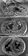
Development of spiral architecture in the compact layer. A, pre-compaction stage (E13.5 embryonic mouse heart in transverse section) showing thin, avascular compact layer with circumferential arrangement of myocytes. B, post-compaction, vascularized compact layer at E15.5 shows spiral arrangement in both compact myocardium (Co) and trabeculae (Tr). C, in the adult mouse heart, the typical three-layered structure with inner predominantly longitudinal (l), middle circular (c) and outer oblique (o) orientation can be seen in the left ventricle (although recent quantitative studies show that the change in orientation occurs in continuum 29). LV, left ventricle, RV, right ventricle, mp, papillary muscles. Ventricular midportion slabs, viewed from the apex towards the outflow. The top two specimens were prepared by late Dr. Si Minh Pham at University of Lausanne.

Similar articles

Cited by

References

    1. Anderson RH, Ho SY. The architecture of the sinus node, the atrioventricular conduction axis, and the internodal atrial myocardium. J Cardiovasc Electrophysiol. 1998;9:1233. - PubMed
    1. Ben-Shachar G, Arcilla RA, Lucas RV, et al. Ventricular trabeculations in the chick embryo heart and their contribution to ventricular and muscular septal development. Circ Res. 1985;57:759. - PubMed
    1. Blausen BE, Johannes RS, Hutchins GM. Computer-based reconstructions of the cardiac ventricles of human embryos. Am J Cardiovasc Pathol. 1990;3:37. - PubMed
    1. Butcher JT, McQuinn TC, Sedmera D, et al. Transitions in early embryonic atrioventricular valvular function correspond with changes in cushion biomechanics that are predictable by tissue composition. Circ Res. 2007;100:1503. - PubMed
    1. Challice CE, Viragh S. The embryonic development of the mammalian heart. In: Challice CE, Viragh S, editors. Ultrastructure of the mammalian heart. New York: Academic Press; 1973. p. 91.

Publication types