Fluorescent LYVE-1 antibody to image dynamically lymphatic trafficking of cancer cells in vivo
- PMID: 18599080
- PMCID: PMC2628480
- DOI: 10.1016/j.jss.2007.12.769
Fluorescent LYVE-1 antibody to image dynamically lymphatic trafficking of cancer cells in vivo
Abstract
Background: The lymphatic system is a major route for cancer cell dissemination, and a potential target for antitumor therapy. Despite ongoing interest in this area of research, the real-time behavior of cancer cells trafficking in the lymphatic system is poorly understood due to lack of appropriate tools to image this process.
Materials and methods: We have used monoclonal-antibody and fluorescence technology to color-code lymphatic vessels and the cancer cells inside them in a living animal. Monoclonal anti-mouse LYVE-1 antibody was conjugated to a green fluorophore and delivered to the lymphatic system of a nude mouse, allowing imaging of mouse lymphatics. Tumor cells engineered to express red fluorescent protein were then imaged traveling within the labeled lymphatics in real time.
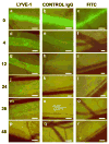
Results: AlexaFluor-labeled monoclonal anti-mouse LYVE-1 created a durable signal with clear delineation of lymphatic architecture. The duration of fluorescent signal after conjugated LYVE-1 delivery was far superior to that of fluorescein isothiocyanate-dextran or control fluorophore-conjugated IgG. Tumor cells engineered to express red fluorescent protein delivered to the inguinal lymph node enabled real-time tracking of tumor cell movement within the green fluorescent-labeled lymphatic vessels.
Conclusions: This technology offers a powerful tool for the in vivo study of real-time trafficking of tumor cells within lymphatic vessels, for the deposition of the tumor cells in lymph nodes, as well as for screening of potential antitumor lymphatic therapies.
Figures


Similar articles
-
Real-time imaging of tumor-cell shedding and trafficking in lymphatic channels.Cancer Res. 2007 Sep 1;67(17):8223-8. doi: 10.1158/0008-5472.CAN-07-1237. Cancer Res. 2007. PMID: 17804736
-
Inhibition of tumor formation and metastasis by a monoclonal antibody against lymphatic vessel endothelial hyaluronan receptor 1.Cancer Sci. 2018 Oct;109(10):3171-3182. doi: 10.1111/cas.13755. Epub 2018 Aug 26. Cancer Sci. 2018. PMID: 30058195 Free PMC article.
-
Imaging tumor-induced sentinel lymph node lymphangiogenesis with LyP-1 peptide.Amino Acids. 2012 Jun;42(6):2343-51. doi: 10.1007/s00726-011-0976-1. Epub 2011 Jul 19. Amino Acids. 2012. PMID: 21769497 Free PMC article.
-
Tumor imaging with multicolor fluorescent protein expression.Int J Clin Oncol. 2011 Apr;16(2):84-91. doi: 10.1007/s10147-011-0201-y. Epub 2011 Feb 25. Int J Clin Oncol. 2011. PMID: 21347627 Review.
-
AlexaFluor 488-conjugated antibody against lymphatic vessel endothelial hyaluronan receptor-1 (LYVE-1).2011 Mar 24 [updated 2011 Jul 6]. In: Molecular Imaging and Contrast Agent Database (MICAD) [Internet]. Bethesda (MD): National Center for Biotechnology Information (US); 2004–2013. 2011 Mar 24 [updated 2011 Jul 6]. In: Molecular Imaging and Contrast Agent Database (MICAD) [Internet]. Bethesda (MD): National Center for Biotechnology Information (US); 2004–2013. PMID: 21755634 Free Books & Documents. Review.
Cited by
-
Noninvasive detection of breast cancer lymph node metastasis using carbonic anhydrases IX and XII targeted imaging probes.Clin Cancer Res. 2012 Jan 1;18(1):207-19. doi: 10.1158/1078-0432.CCR-11-0238. Epub 2011 Oct 20. Clin Cancer Res. 2012. PMID: 22016510 Free PMC article.
-
Involvement of insulin-like growth factor-binding protein-3 in the effects of histone deacetylase inhibitor MS-275 in hepatoma cells.J Biol Chem. 2011 Aug 26;286(34):29540-7. doi: 10.1074/jbc.M111.263111. Epub 2011 Jul 7. J Biol Chem. 2011. PMID: 21737444 Free PMC article.
-
Current status of optical imaging for evaluating lymph nodes and lymphatic system.Korean J Radiol. 2015 Jan-Feb;16(1):21-31. doi: 10.3348/kjr.2015.16.1.21. Epub 2015 Jan 9. Korean J Radiol. 2015. PMID: 25598672 Free PMC article. Review.
-
Emerging lymphatic imaging technologies for mouse and man.J Clin Invest. 2014 Mar;124(3):905-14. doi: 10.1172/JCI71612. Epub 2014 Mar 3. J Clin Invest. 2014. PMID: 24590275 Free PMC article. Review.
-
Imaging of the interaction of cancer cells and the lymphatic system.Adv Drug Deliv Rev. 2011 Sep 10;63(10-11):886-9. doi: 10.1016/j.addr.2011.05.015. Epub 2011 Jun 28. Adv Drug Deliv Rev. 2011. PMID: 21718727 Free PMC article. Review.
References
-
- Rizk N, Venkatraman E, Park B, Flores R, Bains MS, Rusch V. The prognostic importance of the number of involved lymph nodes in esophageal cancer: implications for revisions of the American Joint Committee on Cancer staging system. J Thorac Cardiovasc Surg. 2006;132:1374–1381. - PubMed
-
- Singletary SE, Connolly JL. Breast cancer staging: working with the sixth edition of the AJCC Cancer Staging Manual. CA Cancer J Clin. 2006;56:37–47. quiz 50-31. - PubMed
-
- Stacker SA, Achen MG, Jussila L, Baldwin ME, Alitalo K. Lymphangiogenesis and cancer metastasis. Nat Rev. 2002;2:573–583. - PubMed
-
- Boardman KC, Swartz MA. Interstitial flow as a guide for lymphangiogenesis. Circ Res. 2003;92:801–808. - PubMed
-
- Hoshida T, Isaka N, Hagendoorn J, di Tomaso E, Chen YL, Pytowski B, Fukumura D, Padera TP, Jain RK. Imaging steps of lymphatic metastasis reveals that vascular endothelial growth factor-C increases metastasis by increasing delivery of cancer cells to lymph nodes: therapeutic implications. Cancer Res. 2006;66:8065–8075. - PubMed
Publication types
MeSH terms
Substances
Grants and funding
LinkOut - more resources
Full Text Sources
Other Literature Sources
Medical
Miscellaneous

