. 2009 Mar;152(1):54-60.
doi: 10.1016/j.jss.2007.12.784. Epub 2008 Jan 28.

Steatosis reversibly increases hepatocyte sensitivity to hypoxia-reoxygenation injury

Affiliations

Steatosis reversibly increases hepatocyte sensitivity to hypoxia-reoxygenation injury

François Berthiaume et al. J Surg Res. 2009 Mar.

Abstract

Background: Steatosis decreases survival of liver grafts after transplantation due to poorly understood mechanisms. We examined the effect of steatosis on the survival of liver grafts in a rat liver transplantation model and the viability of cultured rat hepatocytes after hypoxia and reoxygenation.

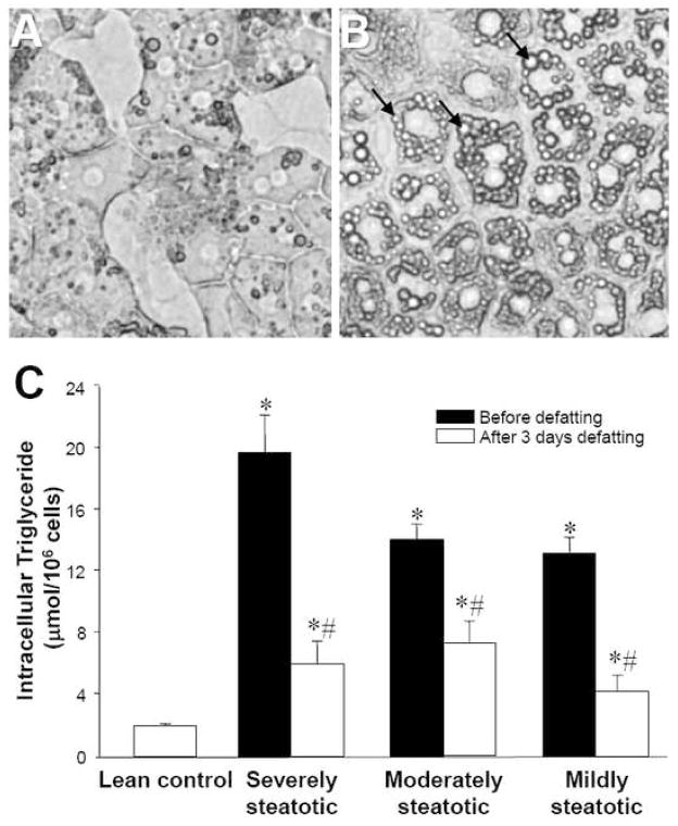
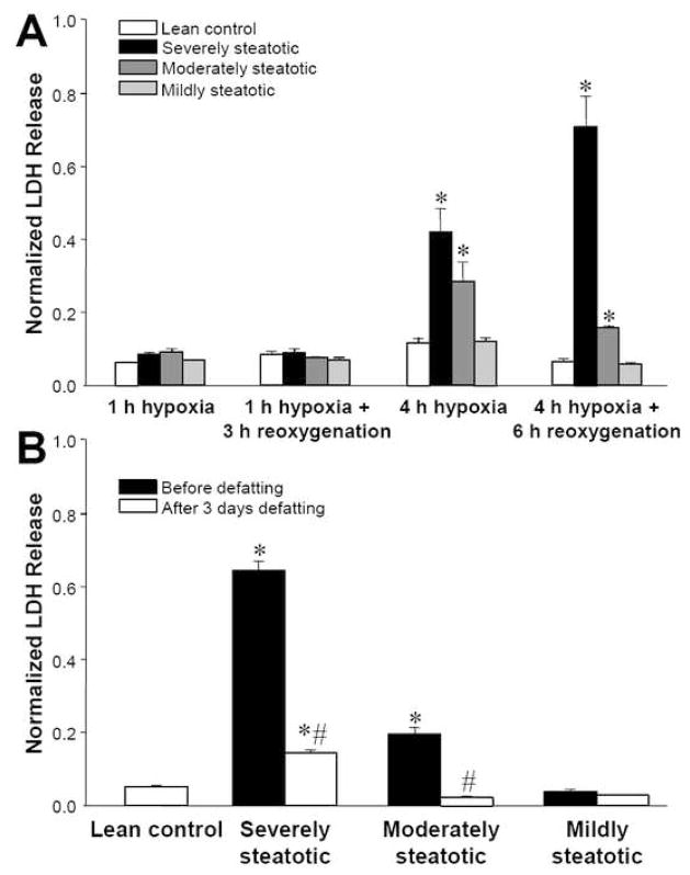
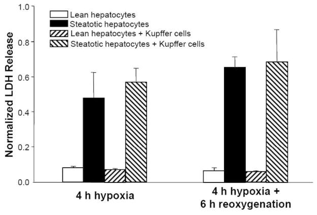
Materials and methods: Rats were fed a choline and methionine-deficient diet to induce hepatic steatosis, and the livers were transplanted into recipient rats after 6 h of cold storage. Cultured hepatocytes were made steatotic by incubation for 3 d in fatty acid-supplemented medium. Hypoxia and reoxygenation were induced by placing the cultures in a 90% N(2)/10% CO(2) atmosphere for 4 h, followed by return to normoxic conditions for 6 h. Hepatocyte viability was assessed by lactate dehydrogenase release and mitochondrial potential staining.

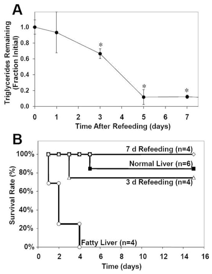
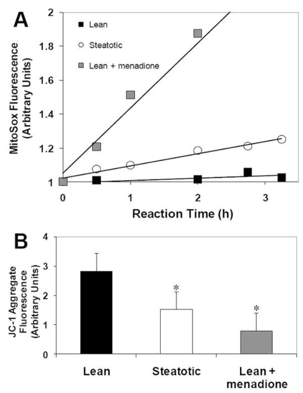
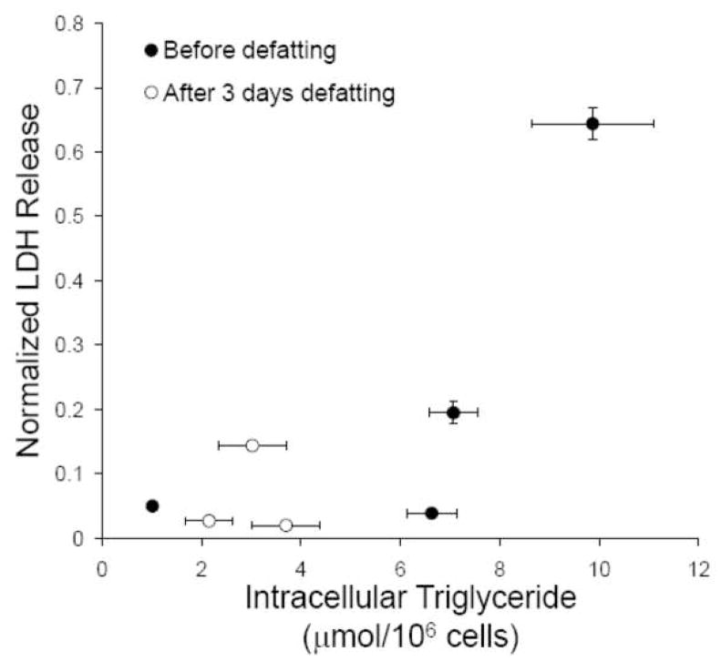
Results: Transplanted steatotic livers exhibited 0% viability compared with 90% for lean liver controls. When donor choline and methionine-deficient diet rats were returned to a normal diet, hepatic fat content decreased while viability of the grafts after transplantation increased. Cultured steatotic hepatocytes generated more mitochondrial superoxide, exhibited a lowered mitochondrial membrane potential, and released significantly more lactate dehydrogenase after hypoxia and reoxygenation than lean hepatocyte controls. When steatotic hepatocytes were defatted by incubating in fatty acid-free medium, they became less sensitive to hypoxia and reoxygenation as the remaining intracellular triglyceride content decreased.

Conclusions: Hepatic steatosis reversibly decreases viability of hepatocytes after hypoxia and reoxygenation in vitro. The decreased viability of steatotic livers after transplantation may be due to a direct effect of hypoxia and reoxygenation on hepatocytes, and can be reversed by defatting.


Figures

Figure 1
Figure 1
Correlation between liver triglyceride content and survival rate after transplantation in a fatty liver rat model. Fatty liver was induced by feeding a choline and methionine-deficient diet for 6 weeks, after which the rats were returned to a normal diet. (A) Effect of refeeding time on liver triglyceride content. Initial content was 220±20 mg/g liver. *: significantly different (p<0.05) compared to initial triglyceride content by Student’s t-test. (B) Effect of refeeding time on survival curves of rats. Fatty liver curve is significantly different (p<0.05) from the other groups.
Figure 2
Figure 2
Effect of fatty medium on lipid content in cultured hepatocytes. (A, B) Bright-field microscopy of primary rat hepatocytes cultured in standard hepatocyte culture medium, and after 2 days of culture in “fatty medium,” respectively. Arrows point to lipid droplets. (C) Hepatocytes were cultured for two days in media containing different proportions of fatty medium and subsequently defatted in regular medium for 3 days. Data shown are triglyceride content of hepatocytes as a function of proportion of fatty medium and defatting, expressed as averages ± SD of triplicate samples. *: significantly different (p<0.05) compared to lean control; #: significantly different compared to initial triglyceride content in same group by Student’s t-test. Each experiment was repeated at least twice with a different cell batch with similar findings.
Figure 3
Figure 3
Effect of steatosis on LDH released in the culture medium by hepatocytes after hypoxia and reoxygenation. Prior to the experiment, hepatocytes were made steatotic by culture for 2 days in fatty medium (severely steatotic), a 1:1 mixture of fatty and regular medium (moderately steatotic), a 1:7 mixture of fatty and regular medium (mildly steatotic). Lean controls were placed in fresh regular medium. Hypoxia was induced by incubation in a 90% N2/10% CO2 atmosphere. For reoxygenation, the medium was replaced with fresh medium and the cultures incubated in a standard 90% air/10% CO2 atmosphere. (A) LDH release as a function of severity of steatosis and protocol of hypoxia and reoxygenation. *: significantly different (p<0.05) compared to lean control by Student’s t-test. (B) Comparison of LDH release after exposure to 4 h hypoxia followed by 6 h reoxygenation for fatty hepatocytes before and after defatting in regular medium for 3 days. *: significantly different (p<0.05) compared to lean control; #: significantly different compared to before defatting in same group by Student’s t-test. Data shown are expressed as averages ± SD of triplicate samples from one experiment. Each experiment was repeated at least twice with a different cell batch with similar findings.
Figure 4
Figure 4
Effect of hepatocyte co-culture with Kupffer cells on LDH release after 4 h hypoxia followed by 6 h reoxygenation. Data shown are averages ± SD of triplicate samples from one experiment. Each experiment was repeated at least twice with a different cell batch with similar findings.
Figure 5
Figure 5
Effect of steatosis on mitochondrial membrane potential and mitochondrial superoxide generation. Hepatocytes were made steatotic by culture for 3 days in fatty medium, while lean controls remained in regular medium. (A) Effect of steatosis on superoxide generation as detected by the Mitosox dye. Menadione was used as a positive control. (B) JC-1 aggregate fluorescence indicating mitochondrial membrane potential. Data shown are averages ± SD of triplicate samples from one experiment. *: significantly different (p<0.05) compared to lean control by Student’s t-test. Each experiment was repeated at least twice with a different cell batch with similar findings.
Figure 6
Figure 6
Correlation between triglyceride content and LDH release after 4 h hypoxia followed by 6 h reoxygenation. Data from Figures 2C and 3B were combined.

References

    1. Busuttil RW, Tanaka K. The utility of marginal donors in liver transplantation. Liver Transpl. 2003;9:651–663. - PubMed
    1. Adam R, Reynes M, Johann M, Morino M, Astarcioglu I, Kafetzis I, Castaing D, Bismuth H. The outcome of steatotic grafts in liver transplantation. Transplant Proc. 1991;23:1538–1540. - PubMed
    1. Todo S, Demetris AJ, Makowka L, Teperman L, Podesta L, Shaver T, Tzakis A, Starzl TE. Primary nonfunction of hepatic allografts with preexisting fatty infiltration. Transplantation. 1989;47:903–905. - PMC - PubMed
    1. Chui AK, Shi LW, Rao AR, Verran DJ, Painter D, Koorey D, McCaughan GW, Sheil AG. Donor fatty (steatotic) liver allografts in orthotopic liver transplantation: a revisit. Transplant Proc. 2000;32:2101–2102. - PubMed
    1. Nakamuta M, Morizono S, Soejima Y, Yoshizumi T, Aishima S, Takasugi S, Yoshimitsu K, Enjoji M, Kotoh K, Taketomi A, Uchiyama H, Shimada M, Nawata H, Maehara Y. Short-term intensive treatment for donors with hepatic steatosis in living-donor liver transplantation. Transplantation. 2005;80:608–612. - PubMed

Publication types