. 2008 Jul 30;249(2-3):194-203.
doi: 10.1016/j.tox.2008.05.009. Epub 2008 May 24.

Toxicological analysis of low-nicotine and nicotine-free cigarettes

Affiliations

Toxicological analysis of low-nicotine and nicotine-free cigarettes

Jinguo Chen et al. Toxicology. .

Abstract

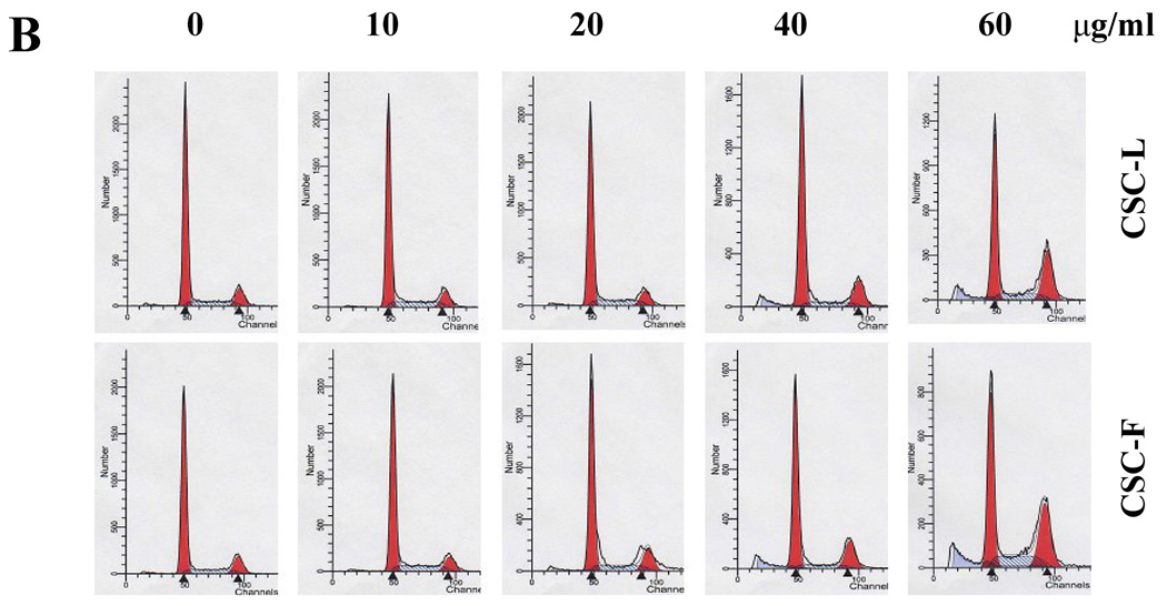
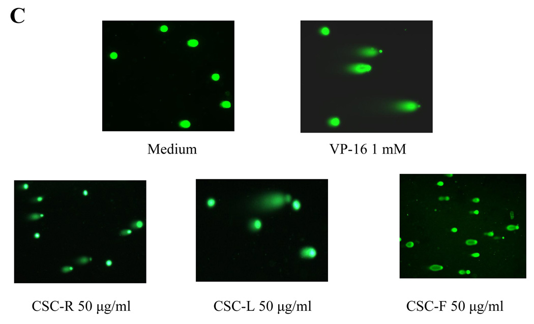
Low-nicotine and nicotine-free cigarettes are commercially available under the brand-name Quest. Some consumers may believe that these are safer cigarettes, and they may smoke more cigarettes or inhale more smoke to compensate for low nicotine yields. Thus, we have studied the toxicological effects of these two cigarettes and compared them with the Kentucky reference cigarette 2R4F. Also, the availability of nicotine-free cigarettes allows for the assessing the role of nicotine in cigarette smoke. In addition to nicotine, some tobacco-specific nitrosamines, aldehydes, and volatile organic compounds were also reduced in the Quest cigarettes compared to the 2R4F. However, aromatic amines were higher in the nicotine-free compared with low nicotine cigarettes. The Ames test revealed that cigarette smoke condensates from the nicotine-free (CSC-F), low nicotine (CSC-L) and 2R4F (CSC-R) cigarettes had a similar mutagenic potency. Exposure to any CSC caused a similar dose-dependent LDH leakage from normal human bronchial epithelial cells. However, CSC-F had more inhibitory effects on the cell growth than CSC-L and CSC-R. Adding nicotine to the CSC-F attenuated this inhibition. Both Quest CSCs decreased gap junction intercellular communication and caused cell cycle arrest. CSC exposure increased cytoplasmic nucleosomes, sub-G1/G0 population and apoptotic comet tails. Proapoptotic protein Bax increased independent of p53 induction after exposure to CSC-F. In conclusion, these studies are not consistent with a perception that low-nicotine or nicotine-free cigarettes may have less toxicity in human cells. Nicotine, as it exists in CSC, attenuates cytotoxicity possibly in part through inhibition of apoptotic pathways.


Figures

Fig. 1
Fig. 1
Mutagenic activity of different CSCs by Ames test. Evaluation was performed in bacterial strain TA98 in the presence of S9 solution. Mutagenicity was expressed as revertants/per plate per mg CSC. All testing was conducted with triplicate plates at each concentration. Ames test in TA100 (data not shown here) revealed a similar pattern. Shown is one example of three or more independent experiments, all with similar results.
Fig. 2
Fig. 2
Cytotoxic and anti-proliferative effects of CSC and nicotine in normal human bronchial epithelial (NHBE) cells. NHBE cells were exposed to the indicated concentration of CSC from Kentucky 2R4F reference cigarettes (CSC-R), or Quest® low-nicotine cigarette (CSC-L) or nicotine-free cigarette (CSC-F) in the absence or presence of nicotine (10 µM): (A) the level of LDH released from cytoplasm into the culture media after exposure for 3 hours was determined with Sigma TOX-7 kit; (B) after overnight exposure, cell viability was assayed with a CCK-8 kit, which measures mitochondrial dehydrogenase activities of viable cells by reducing WST-8 to produce a yellow color formazan dye; (C) cell number after exposure for 3 days. The inhibition of cell growth was expressed as percentage of untreated control wells. All tests were performed in triplicate. Shown is one example of three or more independent experiments, all with similar results.
Fig. 3
Fig. 3
Effects of CSC on gap junction intercellular communication (GJIC) analyzed by fluorescent dye transfer between NHBE cells. Double-stained (Calcein AM and PKH26) normal or CSC treated donor cells were mixed with non-dyed recipient cells at a ratio of 1:10. Calcein transfer occurred after contact between donor and recipient cells. The number of coupled recipient (calcein-dyed) cells per donor cell (coupling ratio Cr) was determined by flow cytometry after co-culture for 0 h, 1 h and 2 h. Shown is one example of two or more independent experiments, all with similar results.
Fig. 4
Fig. 4
Effects of CSC and nicotine on the induction of apoptosis. For all assays for apoptosis, NHBE cells were placed in 6-well plate and treated with different dose of CSC and/or nicotine or apoptosis-inducers at 37°C overnight. The attached and floating cells were then harvested, washed and resuspended. For DNA fragmentation, the amount of cytoplasmic nucleosome (A) were determined with Cell Death Detection ELISA kit. For DNA content (B), the cells were collected and fixed with 75% ethanol. After stained with propidium iodine, cell cycle analysis was performed by flow cytometry. Sub-diploid DNA peaks were considered apoptotic cells. For neutral Comet assay (C), the cells were mixed with molten LMAgarose and placed in CometSlide. Cells in the slides were then lyzed and subjected to electrophoresis in TBE buffer horizontally at 1 Volt/cm for 15 min. After fixing with 70% ethanol and staining with SYBR Green I, cells were observed and photographed under an epifluorescence microscopy. The comet was scored from 0 ~ 4 according to Collins criteria by measuring tail length, head size, tail intensity and head intensity. (D) Effects of CSC and nicotine on the induction of apoptosis-related protein expression. NHBE cells were exposed to CSC-F and/or nicotine (10 µM), or apoptosis-inducer VP-16 (0.5 mM) at 37°C overnight. Cell lysates (30 µg) were then immunoblotted with indicated antibodies.
Fig. 4
Fig. 4
Effects of CSC and nicotine on the induction of apoptosis. For all assays for apoptosis, NHBE cells were placed in 6-well plate and treated with different dose of CSC and/or nicotine or apoptosis-inducers at 37°C overnight. The attached and floating cells were then harvested, washed and resuspended. For DNA fragmentation, the amount of cytoplasmic nucleosome (A) were determined with Cell Death Detection ELISA kit. For DNA content (B), the cells were collected and fixed with 75% ethanol. After stained with propidium iodine, cell cycle analysis was performed by flow cytometry. Sub-diploid DNA peaks were considered apoptotic cells. For neutral Comet assay (C), the cells were mixed with molten LMAgarose and placed in CometSlide. Cells in the slides were then lyzed and subjected to electrophoresis in TBE buffer horizontally at 1 Volt/cm for 15 min. After fixing with 70% ethanol and staining with SYBR Green I, cells were observed and photographed under an epifluorescence microscopy. The comet was scored from 0 ~ 4 according to Collins criteria by measuring tail length, head size, tail intensity and head intensity. (D) Effects of CSC and nicotine on the induction of apoptosis-related protein expression. NHBE cells were exposed to CSC-F and/or nicotine (10 µM), or apoptosis-inducer VP-16 (0.5 mM) at 37°C overnight. Cell lysates (30 µg) were then immunoblotted with indicated antibodies.

Similar articles

Cited by

References

    1. Blouquit S, Morel H, Hinnrasky J, Naline E, Puchelle E, Chinet T. Characterization of ion and fluid transport in human bronchioles. Am. J. Respir. Cell Mol. Biol. 2002;27(4):503–510. - PubMed
    1. Carlisle DL, Hopkins TM, Gaither-Davis A, Silhanek MJ, Luketich JD, Christie NA, Siegfried JM. Nicotine signals through muscle-type and neuronal nicotinic acetylcholine receptors in both human bronchial epithelial cells and airway fibroblasts. Respir. Res. 2004;5(1):27. - PMC - PubMed
    1. Chen J, Xu XM, Underhill CB, Yang S, Wang L, Chen Y, Hong S, Creswell K, Zhang L. Tachyplesin activates the classic complement pathway to kill tumor cells. Cancer Res. 2005;65(11):4614–4622. - PubMed
    1. Chu M, Guo J, Chen CY. Long-term exposure to nicotine, via ras pathway, induces cyclin D1 to stimulate G1 cell cycle transition. J. Biol. Chem. 2005;280(8):6369–6379. - PubMed
    1. Collins AR. The comet assay. Principles, applications, and limitations. Methods Mol. Biol. 2002;203:163–177. - PubMed

Publication types

MeSH terms

Substances