. 2008 Nov;22(8):1178-89.
doi: 10.1016/j.bbi.2008.05.004. Epub 2008 Jul 2.

Proinflammatory cytokines oppose opioid-induced acute and chronic analgesia

Affiliations

Proinflammatory cytokines oppose opioid-induced acute and chronic analgesia

Mark R Hutchinson et al. Brain Behav Immun. 2008 Nov.

Abstract

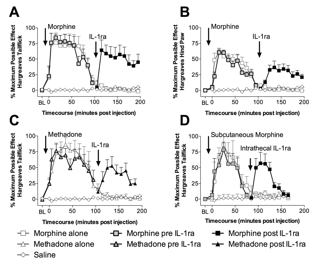
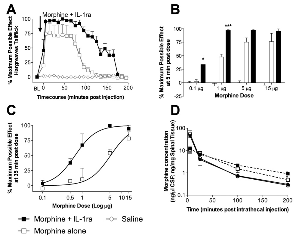
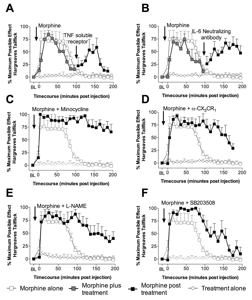
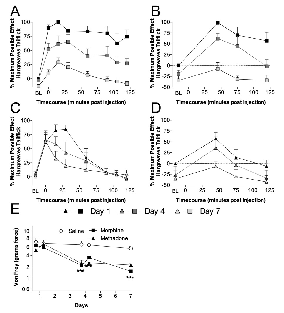
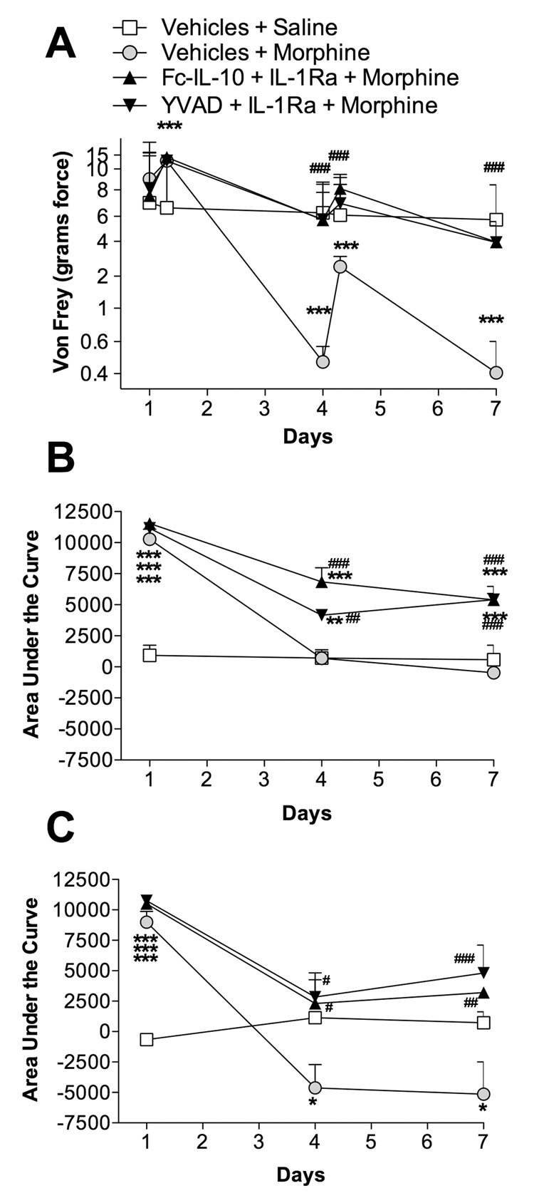
Spinal proinflammatory cytokines are powerful pain-enhancing signals that contribute to pain following peripheral nerve injury (neuropathic pain). Recently, one proinflammatory cytokine, interleukin-1, was also implicated in the loss of analgesia upon repeated morphine exposure (tolerance). In contrast to prior literature, we demonstrate that the action of several spinal proinflammatory cytokines oppose systemic and intrathecal opioid analgesia, causing reduced pain suppression. In vitro morphine exposure of lumbar dorsal spinal cord caused significant increases in proinflammatory cytokine and chemokine release. Opposition of analgesia by proinflammatory cytokines is rapid, occurring < or =5 min after intrathecal (perispinal) opioid administration. We document that opposition of analgesia by proinflammatory cytokines cannot be accounted for by an alteration in spinal morphine concentrations. The acute anti-analgesic effects of proinflammatory cytokines occur in a p38 mitogen-activated protein kinase and nitric oxide dependent fashion. Chronic intrathecal morphine or methadone significantly increased spinal glial activation (toll-like receptor 4 mRNA and protein) and the expression of multiple chemokines and cytokines, combined with development of analgesic tolerance and pain enhancement (hyperalgesia, allodynia). Statistical analysis demonstrated that a cluster of cytokines and chemokines was linked with pain-related behavioral changes. Moreover, blockade of spinal proinflammatory cytokines during a stringent morphine regimen previously associated with altered neuronal function also attenuated enhanced pain, supportive that proinflammatory cytokines are importantly involved in tolerance induced by such regimens. These data implicate multiple opioid-induced spinal proinflammatory cytokines in opposing both acute and chronic opioid analgesia, and provide a novel mechanism for the opposition of acute opioid analgesia.


Figures

Figure 1
Figure 1. Intrathecal interleukin-1 receptor antagonist "unmasks" both intrathecal and systemic opioid analgesia
A: Intrathecal injection of 15 µg of morphine (in 1 µl with a 10 µl flush; black square with gray center) produces significant increases in tailflick latencies (5 min to 85 min compared to vehicle treated animals or saline plus IL-1ra treated animals [gray diamond with white center]; P < 0.05) which dissipated by ~100 min, following which intrathecal IL-1ra (100 µg in 1 µl with a 10 µl flush; black square) unmasked continuing analgesia (115 min to 175 min compared to morphine followed by vehicle treated animals [gray square with white center] and vehicle followed by IL-1ra [gray diamond with white center]; P < 0.05). B: Intrathecal injection of 15 µg of morphine (in 1 µl with a 10 µl flush; black square with gray center) produces significant increases in hind paw withdraw latencies (15 min to 85 min compared to vehicle treated animals or saline plus IL-1ra treated animals [gray diamond with white center]; P < 0.05) which dissipated by ~100 min, following which intrathecal IL-1ra (100 µg in 1 µl with a 10 µl flush; black square) unmasked continuing analgesia (125 min to 165 min compared to morphine followed by vehicle treated animals [gray square with white center] and vehicle followed by IL-1ra [gray diamond with white center]; P < 0.05). C: Intrathecal injection of 15 µg of methadone (in 1 µl with a 10 µl flush; black triangle with gray center) produces significant analgesia (5 min to 85 min compared to vehicle treated animals or saline plus IL-1ra treated animals [gray diamond with white center]; P < 0.01) which dissipates by ~100 min, following which intrathecal IL-1ra (100 µg in 1 µl with a 10 µl flush; black triangle) unmasked continuing analgesia (125 min to 165 min; P < 0.05 compared to methadone followed by vehicle treated animals [gray triangle with white center] and vehicle followed by IL-1ra [gray diamond with white center]) in a similar fashion to the IL-1ra unmasking of morphine analgesia. D: Subcutaneous morphine (4 mg/kg in a dose volume of 1 ml/kg; black square with gray center) produced significant analgesia (5 min to 55 min compared to vehicle treated animals or saline plus IL-1ra treated animals [gray diamond with white center]; P < 0.05) dissipating by ~75 min. Intrathecal administration of IL-1ra (100 µg in 1 µl with a 10 µl flush; black square) unmasked significant continuing analgesia (95 min to 125 min compared to morphine followed by vehicle treated animals [gray square with white center] and vehicle followed by IL-1ra [gray diamond with white center]; P < 0.01) that lasted for a further 50 min.
Figure 2
Figure 2. Intrathecal interleukin-1 receptor antagonist potentiates morphine analgesia, independent of a change in pharmacokinetics
A: Intrathecal co-administration of morphine and intrathecal IL-1ra (15 µg morphine in 1 µl and IL-1ra 100 µg in 1 µl with 10 µl flush; black square) produced significant analgesia lasting for 175 min (0 min to 165 min compared to vehicle plus IL-1ra treated animals [gray diamond with white center]; P < 0.001) which was significantly greater than morphine alone (85 to 145 min; gray square with white center; P < 0.05). B: Responses to the Hargreaves test were assessed 5 min after intrathecal administration of morphine alone (0.1 µg, 0.5 µg, 1 µg, 5 µg and 15 µg in 1 µl with a 10 µl flush; open bars), morphine co-administered with IL-1ra (0.1 µg, 0.5 µg, 1 µg, 5 µg and 15 µg morphine in 1 µl plus 100 µg IL-1ra in 1 µl followed by a 10 µl flush; filled bars) and vehicle treated animals (gray bars). Co-administration of IL-1ra with morphine significantly potentiated 0.1 µg and 1 µg morphine analgesia 5 min following drug administration, demonstrating the speed of onset of IL-1 opposition of morphine analgesia. C: Responses to the Hargreaves test were assessed 35 min after intrathecal administration of morphine alone (0.1 µg, 0.5 µg, 1 µg, 5 µg and 15 µg in 1 µl with a 10 µl flush; black square with white center) and morphine co-administered with IL-1ra (0.1 µg, 0.5 µg, 1 µg, 5 µg and 15 µg morphine in 1 µl plus 100 µg IL-1ra in 1 µl followed by a 10 µl flush; black square). Morphine alone produced an dose response curve with an EC50 of 4.9 µM, while morphine co-administered with IL-1ra caused a leftward shift in the dose response function with an EC50 of 0.66 µM. D: CSF concentrations of morphine (ng morphine per µl CSF) over time following an intrathecal injection of morphine alone (15 µg in 1 µl with a 10 µl flush; black circle with white center) and morphine co-administered with IL-1ra (15 µg morphine in 1 µl plus 100 µg IL-1ra in 1 µl with a 10 µl flush; black circle) were quantified. Dorsal spinal cord concentrations of morphine over time following an intrathecal injection of morphine alone (15 µg in 1 µl with a 10 µl flush; black square with white center) and morphine co-administered with IL-1ra (15 µg morphine in 1 µl plus 100 µg IL-1ra in 1 µl with a 10 µl flush; black square) were also quantified. No significant differences in morphine CSF or dorsal spinal cord tissue concentration were observed at any time point (P > 0.05).
Figure 3
Figure 3. Characterization of potential mediators opposing acute morphine analgesia
A: Intrathecal injection of morphine (15 µg in 1 µl with a 10 µl flush; black square with gray center) produces analgesia that dissipated by ~100 min, following which intrathecal TNF-α soluble receptor (300 µg in 5 µl as previously published by Milligan et al. (2001) with a 10 µl flush; black square) unmasked significant continuing analgesia (135 min and 155 min compared to morphine control gray square with white center; P < 0.05), whilst IgG control or TNF-α soluble receptor alone had no effect (controls average, gray diamond with white center). B: Intrathecal injection of morphine (15 µg in 1 µl with a 10 µl flush; black square with gray center) produces analgesia dissipating by ~100 min, following which intrathecal IL-6 neutralizing antibody (0.325 µg; in 5 µl as previously published by Milligan et al. (2005; with a 10 µl flush; black square) unmasked significant continuing analgesia (125 min to 185 min compared to morphine control gray square with white center; P < 0.05), whilst IgG or IL-6 neutralizing antibody alone had no effect (controls average, gray diamond with white center). C: Intrathecal co-administration of morphine (15 µg in 1 µl with a 10 µl flush; black square) and minocycline (100 µg in 3 µl as previously published by Ledeboer et al. (2005) at time of catheter implant and 33.3 µg in 1 µl with 15 µg morphine in 1 µl with a 10 µl flush) produced significantly potentiated analgesia compared to morphine alone (gray square with white center; 85 min to 245 min; P < 0.05), whilst minocycline alone had no effect (gray diamond with white center). D: Intrathecal co-administration of morphine (15 µg in 1 µl with a 10 µl flush; black square) and fractalkine receptor antibody (α-CX3CR1 10 µg in 1 µl as previously published by Johnston et al. (2004) at time of catheter implant and then again co-administered with 15 µg morphine in 1 µl with a 10 µl flush) produced significantly potentiated analgesia compared to morphine alone (gray square with white center; 95 min to 155 min; P < 0.05), whilst fractalkine receptor antibody alone had no effect (gray diamond with white center). E: Intrathecal co-administration of morphine (15 µg in 1 µl with a 10 µl flush; black square) and L-NAME (5 µg in 1 µl as previously published by Holguin et al. (2004) with 15 µg morphine in 1 µl with a 10 µl flush) produced significantly potentiated analgesia compared to morphine alone (gray square with white center; 95 min to 155 min; P < 0.05), whilst L-NAME administration alone had no significant effect (gray diamond with white center). F: Intrathecal co-administration of morphine (15 µg in 1 µl with a 10 µl flush; black square) and SB203508 (10 µg in 1 µl as previously published by Wu et al. (2006) with 15 µg morphine in 1 µl with a 10 µl flush) produced significantly potentiated analgesia compared to morphine alone (gray square with white center; 85 min to 155 min; P < 0.05), whilst SB203508 alone had no effect (gray diamond with white center).
Figure 4
Figure 4. Chronic intrathecal morphine and methadone administration produce significant tolerance, hyperalgesia, allodynia, and alterations in mRNA and/or protein levels of cytokines, chemokines and a glial activation marker
Animals were administered once daily 15 µg morphine (A,B), 15 µg methadone (C,D) or saline (in 1 µl with a 25 µl flush) intrathecally for 7 days via an indwelling catheter. On days 1 (black square and triangle), 4 (black square and triangle with dark gray center) and 7 (black square and triangle with light gray center) animals were tested for short baseline latency Hargreaves stimuli (A,C) to assess analgesia and long baseline latency Hargreaves stimuli (B,D) to assess hyperalgesia over a 2 hr timecourse following drug administration. Significant reductions in analgesia were observed across the multiple days dosing, indicating the development of tolerance (A,C) and hyperalgesia (B,D). Mechanical allodynia (E) was assessed using von Frey filaments 60 min prior and 2 hr after administration on day 1 and day 4 and only 60 min prior to drug administration on day 7 for morphine (black square), methadone (black triangle) or saline (black circle with white center). *** P < 0.001
Figure 5
Figure 5. Chronic intrathecal co-administration of anti-inflammatory treatments blunts the development of morphine tolerance, hyperalgesia and allodynia
Animals received 20 µg morphine (in 1 µl with a 25 µl flush) twice daily via an indwelling intrathecal catheter, co-administered with IL-1ra (100 µg in 1 µl) plus Fc-IL-10 (250 ng in 5 µl; black triangle, point up), IL-1ra (100 µg in 1 µl) plus YVAD (500 ng in 5 µl; black triangle, point down), morphine co-administered with vehicles (black circle with gray center), or saline co-administered with vehicles (black square with white center). Fc-IL-10 is a stabilized human IL-10 which has an extended half-life. YVAD is a caspase-1 inhibitor, the enzyme that cleaves IL-1 into its mature form. YVAD will reduce the levels of mature active IL-1. The aim of Fc-IL-10 and YVAD treatments were to reduce the production of proinflammatory cytokines by producing an anti-inflammatory environment (Fc-IL-10) and by blocking the production of mature IL-1 (YVAD). IL-1ra was included to provide an immediate block of IL-1 actions. Animals were tested on days 1, 4 and 7 prior to, during and after the morning drug administration. The development of allodynia (von Frey; A), tolerance (short baseline latency Hargreaves stimuli; B) and hyperalgesia (long baseline latency Hargreaves stimuli; C) were measured. A two-way ANOVA with Bonferroni post hoc test was conducted to assess statistical significance between saline co-administered with vehicles versus the animals receiving morphine treatments (# P < 0.05, ##, P < 0.01 and ### P < 0.001), as well as the difference between morphine plus vehicles versus other treatments (* P < 0.05, ** P < 0.01 and *** P < 0.001).

Similar articles

Cited by

References

    1. Carmody J. Avoiding fallacies in nociceptive measurements. Pain. 1995;63:136. - PubMed
    1. Chaplan SR, Bach FW, Pogrel JW, Chung JM, Yaksh TL. Quantitative assessment of tactile allodynia in the rat paw. J Neurosci Methods. 1994;53:55–63. - PubMed
    1. Clark AK, Yip PK, Grist J, Gentry C, Staniland AA, Marchand F, Dehvari M, Wotherspoon G, Winter J, Ullah J, Bevan S, Malcangio M. Inhibition of spinal microglial cathepsin S for the reversal of neuropathic pain. Proc Natl Acad Sci U S A. 2007;104:10655–10660. - PMC - PubMed
    1. Cui Y, Chen Y, Zhi JL, Guo RX, Feng JQ, Chen PX. Activation of p38 mitogen-activated protein kinase in spinal microglia mediates morphine antinociceptive tolerance. Brain Res. 2006;1069:235–243. - PubMed
    1. Doverty M, Somogyi AA, White JM, Bochner F, Beare CH, Menelaou A, Ling W. Methadone maintenance patients are cross-tolerant to the antinociceptive effects of morphine. Pain. 2001;93:155–163. - PubMed

Publication types

MeSH terms