Comparative Study
. 2008 Sep 10;16(9):1333-44.
doi: 10.1016/j.str.2008.06.005. Epub 2008 Jul 3.

Structure of IL-22 bound to its high-affinity IL-22R1 chain

Affiliations
Comparative Study

Structure of IL-22 bound to its high-affinity IL-22R1 chain

Brandi C Jones et al. Structure. .

Abstract

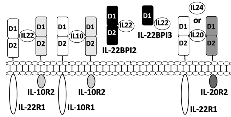
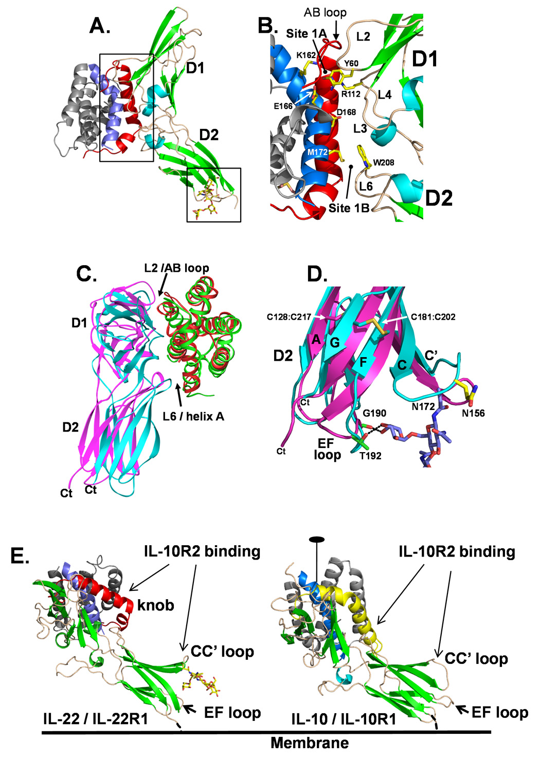
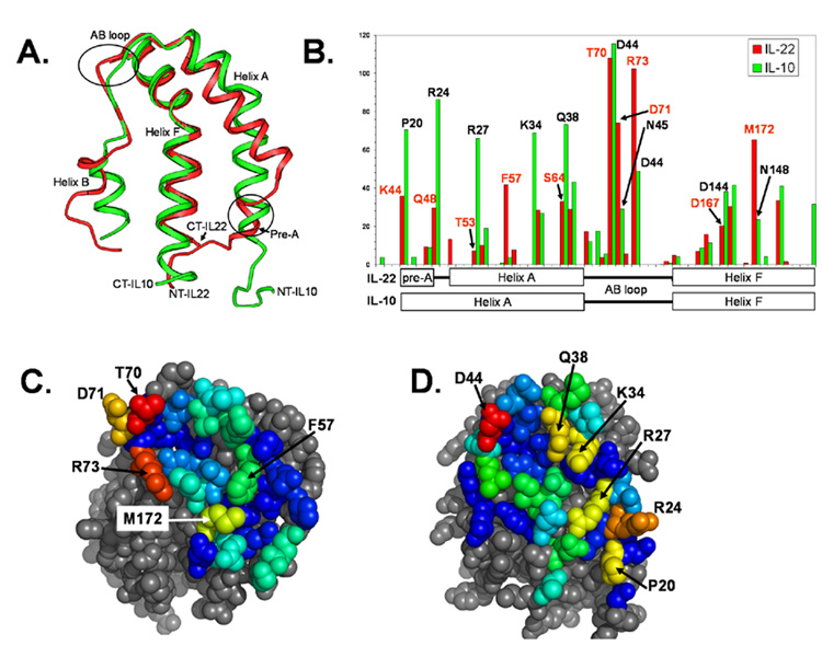
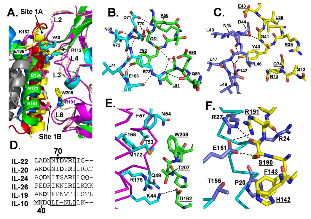
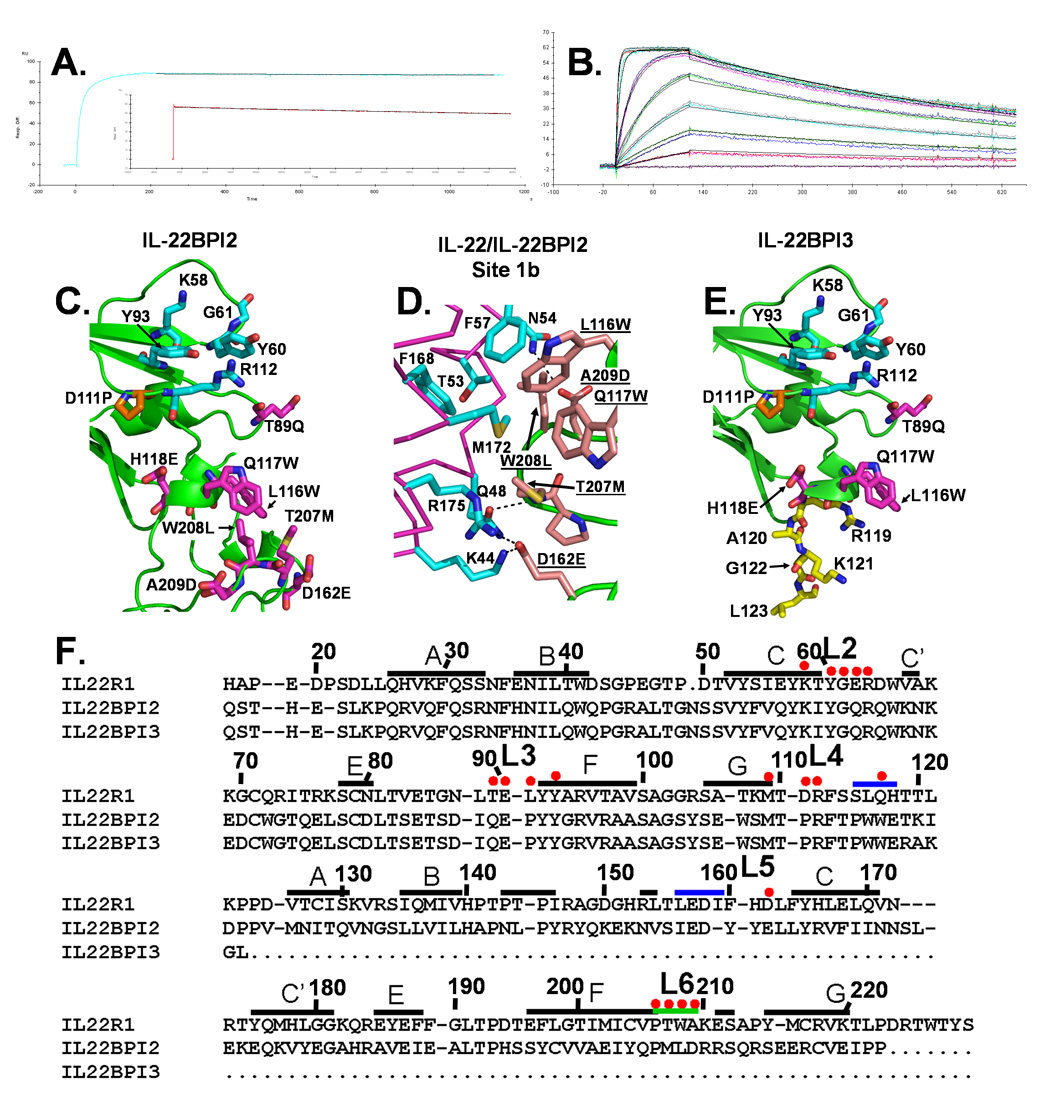
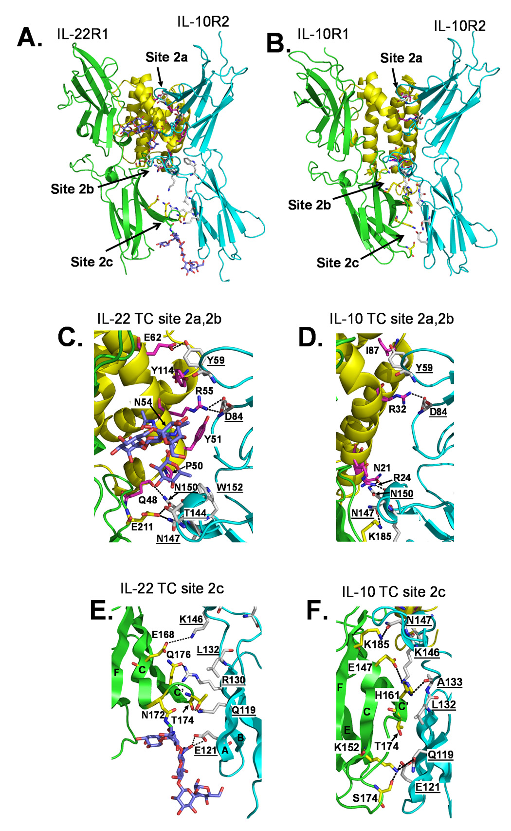
IL-22 is an IL-10 family cytokine that initiates innate immune responses against bacterial pathogens and contributes to immune disease. IL-22 biological activity is initiated by binding to a cell-surface complex composed of IL-22R1 and IL-10R2 receptor chains and further regulated by interactions with a soluble binding protein, IL-22BP, which shares sequence similarity with an extracellular region of IL-22R1 (sIL-22R1). IL-22R1 also pairs with the IL-20R2 chain to induce IL-20 and IL-24 signaling. To define the molecular basis of these diverse interactions, we have determined the structure of the IL-22/sIL-22R1 complex. The structure, combined with homology modeling and surface plasmon resonance studies, defines the molecular basis for the distinct affinities and specificities of IL-22 and IL-10 receptor chains that regulate cellular targeting and signal transduction to elicit effective immune responses.


Figures

Figure 1
Figure 1. Schematic diagram of protein complexes formed by IL-22 receptors
Although not shown, IL-10R2 also forms IL-20R1/IL-10R2 and IL-28R1/IL-10R2 cell surface complexes that activate IL-26, IL-28, and IL-29 signaling.
Figure 2
Figure 2. Structure of the IL-22/IL-22R1 complex
(A) Ribbon diagram of IL-22/IL-22R1 complex. Helix A/AB loop and helix F that form the sIL-22R1 binding site are colored red and blue, respectively. Boxes show the location of Figures 2B and 2E, respectively. (B) IL-22/sIL-22R1 site 1 interface. (C) Superpositionof IL-22/sIL-22R1 and one half of the IL-10/sIL-10R1 dimer structure. IL-22 is colored red, sIL-22R1 is magenta, IL-10 is colored green, sIL-10R1 is cyan. (D) Structural differences, including the role of carbohydrate, between sIL-22R1 and sIL-10R1 D2 domains. (E) Putative orientation of IL-22/IL-22R1 and IL-10/IL-10R1 cell surface complexes. The IL-10/IL-10R1 complex was orientated so that the IL-10 dimer twofold axis was perpendicular to the membrane. The helix A/AB loop and helix F residues of IL-10, which form the IL-10R1 binding site are colored yellow and blue.
Figure 3
Figure 3. IL-22 and IL-10 exhibit distinct site 1 structures and binding surfaces
(A) Superposition of IL-22 and IL-10 site 1 binding epitopes colored as described in Figure 2C. (B) Bar graph of IL-22 and IL-10 surface area buried in the site 1 interfaces of IL-22/sIL-22R1 and IL-10/sIL-10R1. Space filling models colored according to the amount of surface area buried (red greatest, dark blue least) at each residue position in IL-22 (C) and IL-10 (D) site 1 interfaces.
Figure 4
Figure 4. Comparison of IL-22/sIL-22R1 and IL-10/sIL-10R1 interfaces
(A) Enlarged picture of IL-22 and IL-10 site 1 interfaces, as shown in Figure 2A, except the D1 domains of the receptors are superimposed. Important AB loop hydrogen bonds formed in IL-22/sIL-22R1 site 1a (B) and IL-10/sIL-10R1 site 1a (C). (D) Sequence alignment of IL-10 family cytokine AB loops. Interactions in IL-22/sIL-22R1 site 1b (E) and IL-10/sIL-10R1 site 1b (F) are also shown. Receptor residue labels are distinguished from the ligand labels by underlining.
Figure 5
Figure 5. Binding and structure of IL-22BP isoforms
(A) Short (7 minute) and long (22 hour, inset) period sensorgrams used to derive the affinity of the IL-22/IL-22BPI2 interaction (See Table 1). (B) Sensorgram performed using IL-22BPI3-his6 captured to anti-his Ab surfaces, followed by the injection of IL-22. Larger versions of the sensorgram are shown in Figure S1. (C) Homology model of IL-22BPI2 colored to show the location of residue differences and similarities with sIL-22R1. Residues conserved between IL-22BPI2 and sIL-22R1 are colored cyan, and differences are colored magenta. The orange residue, Pro-111IL-22BP highlights the one amino change in D1 that alters contacts with IL-22. (D) Site 1b interface observed in the IL-22/IL-22BPI2 homology model. (E) Model of the IL-22BPI3 colored as described in C. Unique C-terminal residues of IL-22BPI3 are shown in yellow. (F) Sequence alignment of sIL-22R1 and IL-22BP isoforms. sIL-22R1 residues that bind IL-22 are labeled with red dots.
Figure 6
Figure 6. Models of the IL-22/IL-22R1/IL-10R2 and IL-10/IL-10R1/IL-10R2 ternary complexes
Overall IL-22 (A) and IL-10 (B) ternary complex structures. Enlarged pictures of the IL-22 (C) and IL-10 (D) site 2a/2b and IL-22 (E) and IL-10 (F) site 2c are also shown.

References

    1. Aujla SJ, Chan YR, Zheng M, Fei M, Askew DJ, Pociask DA, Reinhart TA, McAllister F, Edeal J, Gaus K, et al. IL-22 mediates mucosal host defense against Gram-negative bacterial pneumonia. Nat Med. 2008;14:275–281. - PMC - PubMed
    1. Blumberg H, Conklin D, Xu WF, Grossmann A, Brender T, Carollo S, Eagan M, Foster D, Haldeman BA, Hammond A, et al. Interleukin 20: discovery, receptor identification, and role in epidermal function. Cell. 2001;104:9–19. - PubMed
    1. Boniface K, Guignouard E, Pedretti N, Garcia M, Delwail A, Bernard FX, Nau F, Guillet G, Dagregorio G, Yssel H, et al. A role for T cell-derived interleukin 22 in psoriatic skin inflammation. Clin Exp Immunol. 2007;150:407–415. - PMC - PubMed
    1. Brown RJ, Adams JJ, Pelekanos RA, Wan Y, McKinstry WJ, Palethorpe K, Seeber RM, Monks TA, Eidne KA, Parker MW, Waters MJ. Model for growth hormone receptor activation based on subunit rotation within a receptor dimer. Nat Struct Mol Biol. 2005;12:814–821. - PubMed
    1. Brunger AT, Adams PD, Clore GM, DeLano WL, Gros P, Grosse-Kunstleve RW, Jiang JS, Kuszewski J, Nilges M, Pannu NS, et al. Crystallography & NMR system: A new software suite for macromolecular structure determination. Acta Crystallogr D Biol Crystallogr. 1998;54(Pt 5):905–921. - PubMed

Publication types

MeSH terms