. 2008 Jul 8;105(27):9337-42.
doi: 10.1073/pnas.0801717105. Epub 2008 Jul 1.

Dependence of antibody-mediated presentation of antigen on FcRn

Affiliations

Dependence of antibody-mediated presentation of antigen on FcRn

Shuo-Wang Qiao et al. Proc Natl Acad Sci U S A. .

Abstract

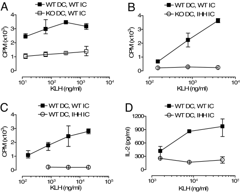
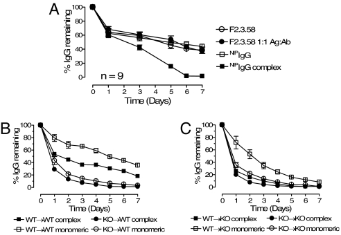
The neonatal Fc receptor for IgG (FcRn) is a distant member of the MHC class I protein family. It binds IgG and albumin in a pH-dependent manner and protects these from catabolism by diverting them from a degradative fate in lysosomes. In addition, FcRn-mediated IgG transport across epithelial barriers is responsible for the transmission of IgG from mother to infant and can also enhance IgG-mediated antigen uptake across mucosal epithelia. We now show a previously undescribed role for FcRn in mediating the presentation of antigens by dendritic cells when antigens are present as a complex with antibody by uniquely directing multimeric immune complexes, but not monomeric IgG, to lysosomes.


Conflict of interest statement

The authors declare no conflict of interest.

Figures

Fig. 1.
Fig. 1.
The IHH mutation abrogates FcRn binding but does not affect binding to FcγR. (A) Human monocyte-derived DC lysates were subjected to sequential binding with WT or IHH-mutated NIPIgG and protein G beads at pH 6 or 8. FcRn protein was detected as a 40-kDa band in a human FcRn-specific immunoblot. The light chain of NIPIgG was also detected (arrowhead). (B) WT (square) or IHH-mutated (triangle) NIPIgG were tested in an ELISA for binding affinity to recombinant GST-tagged human FcRn at pH 5.5, (C) to human FcγRI, and (D) to human FcγRIIa at pH 7.4.
Fig. 2.
Fig. 2.
Enhanced in vitro presentation of FcRn-binding OVA-ICs by FcRn-expressing DC. Spleen DC from WT mice (filled symbols) or FcRn-deficient (KO) mice (open symbols) were used to present NIP-OVA (triangles), ICs of WT NIPIgG and NIP-OVA (squares), or non-FcRn-binding ICs of IHH-mutated NIPIgG and NIP-OVA (circles) to an OVA-reactive OT-II T cell line. Error bars indicate the observed range within triplicates in one representative experiment of four. WT DC presenting WT ICs was statistically significantly different from all other APC and Ag combinations (P < 0.01) and FcRn-deficient DC presenting WT ICs was significantly different from FcRn-deficient DC-presenting soluble NIP-OVA (P < 0.05), whereas all of the other Ag conditions did not display statistically significant differences from each other in one-way ANOVA with Bonferroni post test (P > 0.05).
Fig. 3.
Fig. 3.
Enhanced in vivo presentation of FcRn-binding KLH-ICs by in vitro Ag-loaded DC. Spleen DC from WT (filled symbols) or FcRn-deficient (KO, open symbols, A and B) mice loaded in vitro overnight with Ags were injected into hind footpads of WT B6 mice. Ags used were ICs of WT NIPIgG + NIP-KLH (filled symbols) or IHH-mutated NIPIgG + NIP-KLH (open symbols, B–D). Five days after injection, draining popliteal lymph nodes were assayed in vitro for T cell recall response to KLH. Error bars indicate the observed range within triplicates in one representative experiment of three. All pairs of closed and open graphs were significantly different in A–D (Mann–Whitney test, P < 0.05).
Fig. 4.
Fig. 4.
Enhanced in vivo Ag presentation and T cell proliferation of FcRn-binding ICs in WT mice. The proliferation of CFSE-labeled DO11.10 CD4 T cells was assessed in draining popliteal lymph nodes of WT and FcRn-deficient (KO) Balb/C mice 3–5 days after transfer and Ag immunization in hind footpads. Ags used were ICs of WT NIPIgG + NIP-OVA on the left side (L) or IHH-mutated NIPIgG + NIP-OVA on the right side (R). The mitosis per progenitor cell (M/P) ratio was calculated, and a Wilcoxon paired test was performed for each group of mice.
Fig. 5.
Fig. 5.
Selective FcRn-mediated presentation enhancement of multimeric ICs in human in vitro T cell proliferation assay. Monocyte-derived DC from an HLA-DQ2 donor were used to present monomeric IgG-Ag molecules (A) or multimeric ICs (B) to a CD4 T cell clone reactive to the IQPEPQPAQL peptide presented on HLA-DQ2 molecules. NIP-peptide (NIP-CLRMKLGIIQPEQPAQL) or NIP-gliadin was used alone (filled triangle) or in complex with 200 μg/ml WT NIPIgG (filled square) or IHH-mutated NIPIgG (filled circle). Error bars indicate the observed range within triplicates in one representative experiment of three. NIP-peptide alone was statistically significantly different from both NIPIgG + NIP-peptide conditions (A) (P < 0.05) and WT NIPIgG + NIP-gliadin was statistically significant from the other two Ags tested in B (P < 0.05). (C) A gliadin-derived peptide conjugated with two NIP molecules NIP-LRMKLQPFPQPELPYPQPELPYLRMK(-NIP)L was used to form multimeric ICs in the presence of WT (filled symbols) or IHH-mutated (open symbols) NIPIgG. The Ags were presented by FcRn-positive DC (squares) or FcRn-negative, EBV transformed B cells (circles).
Fig. 6.
Fig. 6.
Trafficking of FcRn to LAMP1-positive lysosomes after IC stimulation in human monocyte derived DC. Human DC were incubated with Ags in HBSS, pH 7.4, at 37°C, fixed, permeabilized, and stained for LAMP1 encoded in green with rabbit polyclonal anti-LAMP1 and endogenously expressed FcRn encoded in red with biotinylated mouse IgG2b anti-human FcRn. (A) Medium. (B) Alexa647-conjugated FcRn-binding multimeric ICs encoded in blue, removed after 20 min by washing and further incubated in HBSS for 5 min. (C) Monomeric FcRn ligands, removed after 20 min and further incubated in HBSS for 30 min. (D) Alexa647-ICs encoded in blue, removed after 20 min and further incubated in HBSS for 30 min, and (E) Alexa647-conjugated FcRn nonbinding IHH-mutated ICs encoded in blue, removed after 20 min, and further incubated in HBSS for 30 min. Arrowheads in D indicate white areas of triple colocalized FcRn, IgG, and LAMP1, a phenomenon seen in ≈50% of cells investigated, and the arrow indicates yellow area of FcRn colocalized with LAMP1.
Fig. 7.
Fig. 7.
IgG in multimeric ICs is cleared faster from the circulation than free IgG and monomeric IgG-Ag molecules with clearance dependent on FcRn expression in hematopoietic cells. (A) Two groups of WT BALB/c mice were injected i.v. with either WT NIPIgG (open square) mixed with monoclonal mouse anti-OVA F2.3.58 IgG1 (open circle), or mixtures of NIPIgG in multimeric ICs (NIPIgG + NIP-BSA, filled square) and F2.3.58 in monomeric complex (F2.3.58 + OVA, filled circle) in PBS. The remaining levels of both transferred antibodies were measured by specific ELISA and are shown as percentage of the starting serum IgG level in the same animal 2 h after injection. The NIPIgG complex group is significantly different from each of the other three groups (P < 0.01). (B) WT B6 Thy1.1 recipient mice were lethally irradiated and reconstituted with BM from WT (squares, WT→WT, n = 3) or FcRn-deficient Thy1.2 mice (circles, KO→W,T n = 3). Nine weeks after reconstitution, the mice were injected i.v. with NIPIgG + NIP-BSA ICs or monomeric F2.3.58-OVA complex. The serum levels of NIPIgG (filled symbols) and F2.3.58 (open symbols) were measured daily and are expressed as percentage of the starting level 2 h after injection. Pairwise comparisons with one-way ANOVA showed that all groups were statistically different from each other (P < 0.001 after Bonferroni correction) except complex and monomeric IgG in the KO→WT group (filled circle vs. open circle) that were not statistically different (P > 0.05). (C) Lethally irradiated FcRn-deficient mice were reconstituted with WT (squares, WT→KO, n = 5) or FcRn-deficient BM (circles, KO→KO n = 4). Nine weeks later, the mice received the same IgG transfer as in B. Monomeric IgG degradation in the WT→KO group (open square) was statistically different from each of the other three graphs (P < 0.01), whereas these three groups were not statistically different from each other. Error bars indicate standard error of the mean.

References

    1. Liu Y, et al. Regulated expression of FcγR in human dendritic cells controls cross-presentation of antigen-antibody complexes. J Immunol. 2006;177:8440–8447. - PubMed
    1. Regnault A, et al. Fcγ receptor-mediated induction of dendritic cell maturation and major histocompatibility complex class I-restricted antigen presentation after immune complex internalization. J Exp Med. 1999;189:371–380. - PMC - PubMed
    1. Ravetch JV, Bolland S. IgG Fc receptors. Annu Rev Immunol. 2001;19:275–290. - PubMed
    1. Hirano M, et al. IgEb immune complexes activate macrophages through FcγRIV binding. Nat Immunol. 2007;8:762–771. - PubMed
    1. Simister NE, Mostov KE. An Fc receptor structurally related to MHC class I antigens. Nature. 1989;337:184–187. - PubMed

Publication types

MeSH terms

LinkOut - more resources