. 2008 Jul 15;105(28):9522-7.
doi: 10.1073/pnas.0801866105. Epub 2008 Jul 3.

Directed assembly of cell-laden microgels for fabrication of 3D tissue constructs

Affiliations

Directed assembly of cell-laden microgels for fabrication of 3D tissue constructs

Yanan Du et al. Proc Natl Acad Sci U S A. .

Abstract

We present a bottom-up approach to direct the assembly of cell-laden microgels to generate tissue constructs with tunable microarchitecture and complexity. This assembly process is driven by the tendency of multiphase liquid-liquid systems to minimize the surface area and the resulting surface free energy between the phases. We demonstrate that shape-controlled microgels spontaneously assemble within multiphase reactor systems into predetermined geometric configurations. Furthermore, we characterize the parameters that influence the assembly process, such as external energy input, surface tension, and microgel dimensions. Finally, we show that multicomponent cell-laden constructs could be generated by assembling microgel building blocks and performing a secondary cross-linking reaction. This bottom-up approach for the directed assembly of cell-laden microgels provides a powerful and highly scalable approach to form biomimetic 3D tissue constructs and opens a paradigm for directing the assembly of mesoscale materials.


Conflict of interest statement

The authors declare no conflict of interest.

Figures

Fig. 1.
Fig. 1.
Schematic diagram of microgel assembly process. Microgel units were synthesized by photolithography, transferred into a dish containing mineral oil, and subjected to mechanical agitation applied by manually manipulating a pipette-tip in a back-and-forth manner. Four structural types of microgel assemblies were observed: linear, branched, random, and offset. Secondary cross-linking was achieved by exposing the microgel assemblies to UV light. (Scale bars, 200 μm.)
Fig. 2.
Fig. 2.
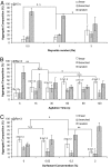
Optimization of the microgel assembly. Effects of (A) agitation rate (fast, medium, and slow), (B) agitation time, and (C) the addition of surfactant on microgel assembly. Compositions of linear, branched, or random microgel assemblies were compared. Average chain length of linear assemblies with SD was also labeled in B and C. Data are means ± SD, n = 3. *, P < 0.05; **, P < 0.01. N.S., not significant.
Fig. 3.
Fig. 3.
Effects of the microgel dimensions on microgel assembly. (A) Assembly composition and (B) average chain length of linear, branched, or random microgel assemblies containing microgel units with different aspect ratios were compared (phase image in B). Data are means ± SD, n = 3. *, P < 0.05. N.S., not significant.
Fig. 4.
Fig. 4.
Secondary cross-linking of the microgel assembles. (A) Dissociated microgel assemblies after replacing mineral oil with culture medium without secondary cross-linking. (B) Stabilized microgel assemblies in culture medium after secondary cross-linking. (C) Dissociated microgel assemblies after secondary cross-linking with removed residual prepolymer. (D–F) Diffusion of dyes through the hydrogel. (D) Free diffusion of rhodamine-dextran (Mr = 10 kDa) out of the hydrogel into PBS. (E) Two groups of microgels labeled with rhodamine-dextran or FITC-dextran (Mr = 2,000 kDa) were assembled, and the rhodamine-dextran diffused throughout the entire hydrogel assembly. (F) Microgel assemblies with FITC-dextran- or Nile red-stained microgels. (G–I) Microbead-containing and plain microgels were used to model microgels containing two types of cells by varying the initial mixing ratios of these two types of microgels (1:1, 2:1, and 4:1, respectively). (Scale bars, 200 μm.)
Fig. 5.
Fig. 5.
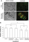
Cell-laden microgel assemblies. (A) Phase-contrast and fluorescence images of cell-laden (NIH 3T3) microgel assemblies after first and secondary cross-linking, respectively, to show the morphology. (B) Quantified cell viability after each step in the procedure to form hydrogel aggregates. (Scale bars, 100 μm.)
Fig. 6.
Fig. 6.
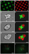
Directed assembly of lock-and-key-shaped microgels. (A) Fluorescence images of cross-shaped microgels stained with FITC-dextran. (B) Rod-shaped microgels stained with Nile red. (C–H) Phase-contrast and fluorescence images of lock-and-key assemblies with one to three rods per cross. (I and J) Fluorescence images of microgel assembly composed of cross-shaped microgels containing red-stained cells, and rod-shaped microgels containing green-stained cells. (Scale bars, 200 μm.)

References

    1. Costanzo L. Physiology. 3rd Ed. Philadelphia: Saunders; 2006.
    1. Langer R, Vacanti JP. Tissue engineering. Science. 1993;260:920–926. - PubMed
    1. Griffith LG, Swartz MA. Capturing complex 3D tissue physiology in vitro. Nat Rev Mol Cell Biol. 2006;7:211–224. - PubMed
    1. Levenberg S, et al. Engineering vascularized skeletal muscle tissue. Nat Biotechnol. 2005;23:879–884. - PubMed
    1. Khademhosseini A, Langer R. Microengineered hydrogels for tissue engineering. Biomaterials. 2007;28:5087–5092. - PubMed

Publication types

LinkOut - more resources