Golgi alpha-mannosidase II cleaves two sugars sequentially in the same catalytic site
- PMID: 18599462
- PMCID: PMC2474516
- DOI: 10.1073/pnas.0802206105
Golgi alpha-mannosidase II cleaves two sugars sequentially in the same catalytic site
Abstract
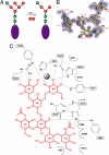
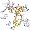
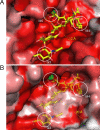
Golgi alpha-mannosidase II (GMII) is a key glycosyl hydrolase in the N-linked glycosylation pathway. It catalyzes the removal of two different mannosyl linkages of GlcNAcMan(5)GlcNAc(2), which is the committed step in complex N-glycan synthesis. Inhibition of this enzyme has shown promise in certain cancers in both laboratory and clinical settings. Here we present the high-resolution crystal structure of a nucleophile mutant of Drosophila melanogaster GMII (dGMII) bound to its natural oligosaccharide substrate and an oligosaccharide precursor as well as the structure of the unliganded mutant. These structures allow us to identify three sugar-binding subsites within the larger active site cleft. Our results allow for the formulation of the complete catalytic process of dGMII, which involves a specific order of bond cleavage, and a major substrate rearrangement in the active site. This process is likely conserved for all GMII enzymes-but not in the structurally related lysosomal mannosidase-and will form the basis for the design of specific inhibitors against GMII.
Conflict of interest statement
The authors declare no conflict of interest.
Figures


Similar articles
-
The molecular basis of inhibition of Golgi alpha-mannosidase II by mannostatin A.Chembiochem. 2009 Jan 26;10(2):268-77. doi: 10.1002/cbic.200800538. Chembiochem. 2009. PMID: 19101978 Free PMC article.
-
Probing the substrate specificity of Golgi alpha-mannosidase II by use of synthetic oligosaccharides and a catalytic nucleophile mutant.J Am Chem Soc. 2008 Jul 16;130(28):8975-83. doi: 10.1021/ja711248y. Epub 2008 Jun 18. J Am Chem Soc. 2008. PMID: 18558690 Free PMC article.
-
1,4-Dideoxy-1,4-imino-D- and L-lyxitol-based inhibitors bind to Golgi α-mannosidase II in different protonation forms.Org Biomol Chem. 2022 Nov 23;20(45):8932-8943. doi: 10.1039/d2ob01545e. Org Biomol Chem. 2022. PMID: 36322142 Free PMC article.
-
Structure, mechanism and inhibition of Golgi α-mannosidase II.Curr Opin Struct Biol. 2012 Oct;22(5):558-62. doi: 10.1016/j.sbi.2012.06.005. Epub 2012 Jul 20. Curr Opin Struct Biol. 2012. PMID: 22819743 Review.
-
Golgi alpha-mannosidase II deficiency in vertebrate systems: implications for asparagine-linked oligosaccharide processing in mammals.Biochim Biophys Acta. 2002 Dec 19;1573(3):225-35. doi: 10.1016/s0304-4165(02)00388-4. Biochim Biophys Acta. 2002. PMID: 12417404 Review.
Cited by
-
The molecular basis of inhibition of Golgi alpha-mannosidase II by mannostatin A.Chembiochem. 2009 Jan 26;10(2):268-77. doi: 10.1002/cbic.200800538. Chembiochem. 2009. PMID: 19101978 Free PMC article.
-
A long-wavelength fluorescent substrate for continuous fluorometric determination of alpha-mannosidase activity: resorufin alpha-D-mannopyranoside.Anal Biochem. 2010 Apr 1;399(1):7-12. doi: 10.1016/j.ab.2009.11.039. Epub 2009 Dec 21. Anal Biochem. 2010. PMID: 20026005 Free PMC article.
-
Characterisation of class I and II α-mannosidases from Drosophila melanogaster.Glycoconj J. 2013 Dec;30(9):899-909. doi: 10.1007/s10719-013-9495-5. Epub 2013 Aug 25. Glycoconj J. 2013. PMID: 23979800
-
Engineering β1,4-galactosyltransferase I to reduce secretion and enhance N-glycan elongation in insect cells.J Biotechnol. 2015 Jan 10;193:52-65. doi: 10.1016/j.jbiotec.2014.11.013. Epub 2014 Nov 25. J Biotechnol. 2015. PMID: 25462875 Free PMC article.
-
Glycan-protein interactions determine kinetics of N-glycan remodeling.RSC Chem Biol. 2021 Apr 16;2(3):917-931. doi: 10.1039/d1cb00019e. RSC Chem Biol. 2021. PMID: 34212152 Free PMC article.
References
-
- Roth J. Protein N-glycosylation along the secretory pathway: Relationship to organelle topography and function, protein quality control, and cell interactions. Chem Rev. 2002;102:285–303. - PubMed
-
- Moremen KW. Golgi α-mannosidase II deficiency in vertebrate systems: Implications for asparagine-linked oligosaccharide processing in mammals. Biochim Biophys Acta. 2002;1573:225–235. - PubMed
-
- Chui D, et al. Alpha-mannosidase-II deficiency results in dyserythropoiesis and unveils an alternate pathway in oligosaccharide biosynthesis. Cell. 1997;90:157–167. - PubMed
Publication types
MeSH terms
Substances
Associated data
- Actions
- Actions
- Actions
Grants and funding
LinkOut - more resources
Full Text Sources
Molecular Biology Databases

