. 2008 Sep 5;283(36):24571-83.
doi: 10.1074/jbc.M802673200. Epub 2008 Jul 3.

The Poly(ADP-ribose) polymerase PARP-1 is required for oxidative stress-induced TRPM2 activation in lymphocytes

Affiliations

The Poly(ADP-ribose) polymerase PARP-1 is required for oxidative stress-induced TRPM2 activation in lymphocytes

Ben Buelow et al. J Biol Chem. .

Abstract

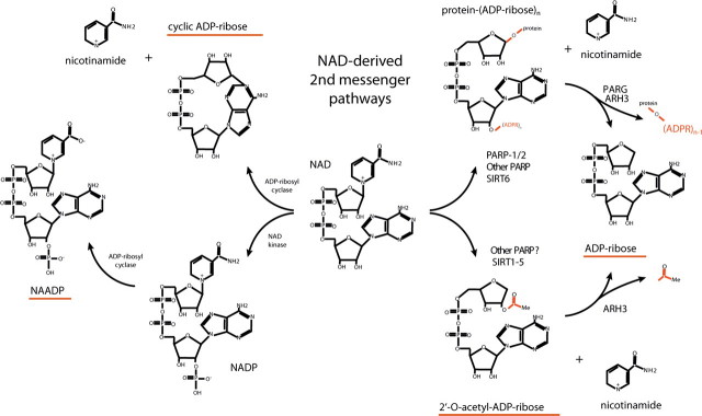
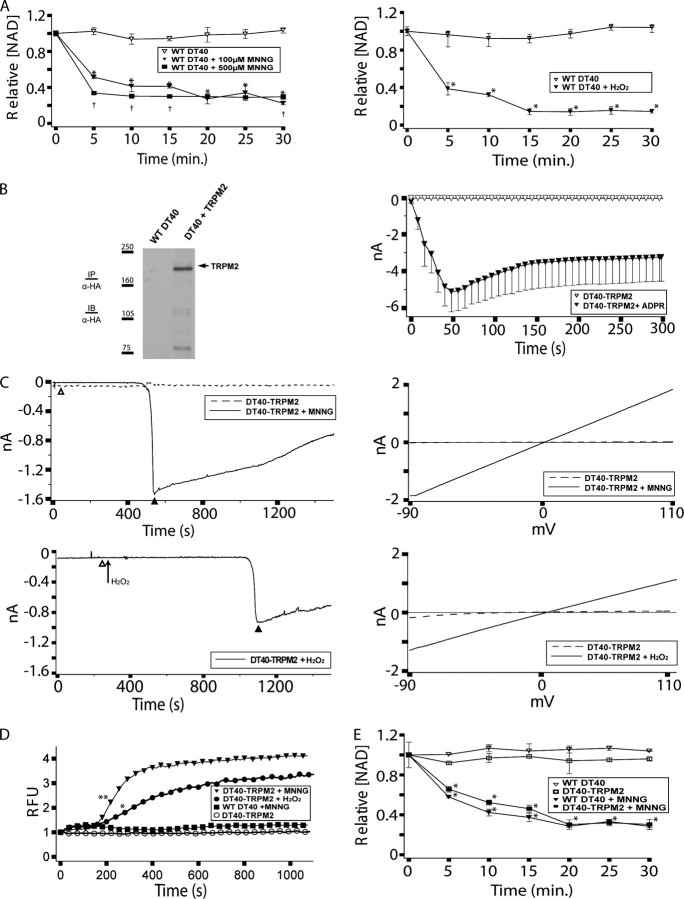
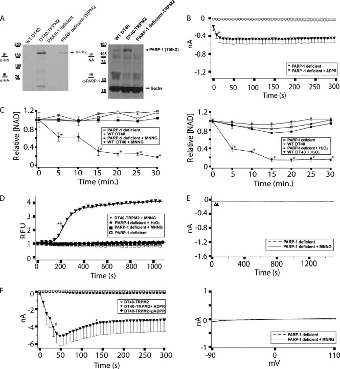
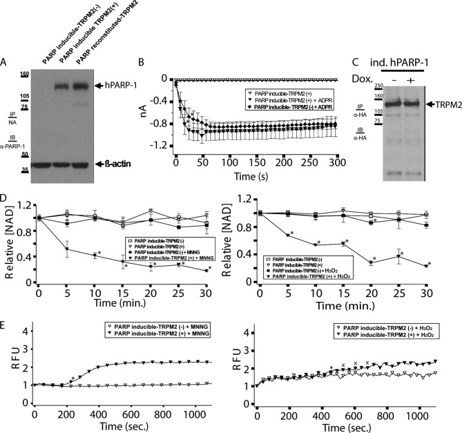
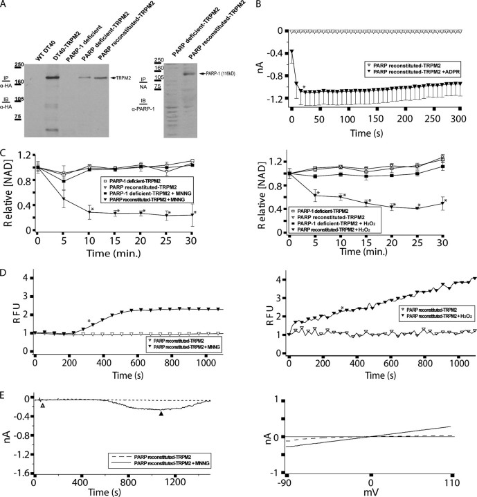
TRPM2 cation channels are widely expressed in the immune system and are thought to play a role in immune cell responses to oxidative stress. Patch clamp analyses suggest that TRPM2 channel activation can occur through a direct action of oxidants on TRPM2 channels or indirectly through the actions of a related group of adenine nucleotide 2nd messengers. However, the contribution of each gating mechanism to oxidative stress-induced TRPM2 activation in lymphocytes remains undefined. To better understand the molecular events leading to TRPM2 activation in lymphocytes, we analyzed oxidative stress-induced turnover of intracellular NAD, the metabolic precursor of adenine nucleotide 2nd messengers implicated in TRPM2 gating, and oxidative stress-induced TRPM2-mediated currents and Ca2+ transients in DT40 B cells. TRPM2-dependent Ca2+ entry did not influence the extent or time course of oxidative stress-induced turnover of NAD. Furthermore, expression of oxidative stress-activated poly(ADP-ribose) polymerases (PARPs) was required for oxidative stress-induced NAD turnover, TRPM2 currents, and TRPM2-dependent Ca2+ transients; no oxidant-induced activation of TRPM2 channels could be detected in PARP-deficient cells. Together, our results suggest that during conditions of oxidative stress in lymphocytes, TRPM2 acts as a downstream effector of the PARP/poly(ADP-ribose) glycohydrolase pathway through PARP-dependent formation of ADP-ribose.


Figures

FIGURE 1.
FIGURE 1.
Schematic of pathways leading to adenine nucleotide 2nd messenger production. Metabolic labeling studies indicate that NAD is turned over solely through hydrolysis of the glycosidic nicotinamide-ribose bond. Two major intracellular mechanisms are known to involve hydrolysis of the nicotinamide moiety from NAD as follows: protein ADP-ribosylation, which is mediated by members of the PARP family as well as several members of the sirtuin (SIRT) protein family, and NAD-dependent protein deacetylation, which at present is thought to be mediated solely by members of the SIRT protein family. Following hydrolysis of the nicotinamide moiety from NAD, both mechanisms require an additional enzymatic step to form ADPR. In the former case, ADPR adducts are released as ADPR in deribosylation reactions catalyzed by PARG or ARH3 (ADP-ribose protein hydrolases 3); in the latter case, 2′-O-acetylated ADPR may be deacetylated by ARH3 or other as yet undefined ADPR deacetylases (75, 76). NAD may also be turned over in a similar manner by CD38 and its close homologue BST-1, resulting in the production of ADPR or cADPR, which is subsequently thought to be metabolized to ADPR. Finally, phosphorylation of NAD to NADP allows the possibility for formation of NAADP by CD38/BST-1 at low pH (77). Accumulating information implicates ADP-ribosylation/deribosylation and protein deacetylation in myriad cell physiological processes (38, 52, 66, 78, 79). PARP family ADP-ribosyltransferases have been implicated in DNA repair and transcription (PARP-1/PARP-2 (reviewed in Refs. 37, 38, 52)), spindle structure formation, and telomere maintenance (tankyrase-1 and tankyrase-2 (–84)), STAT6-dependent transcriptional regulation (CoaSt6 (85, 86)), and transcriptional regulation of as yet undefined genes (BAL-2/3 (87)). In addition, six SIRT family proteins with documented ADPR transferase or protein deacetylase enzymatic activities have been implicated in protein deacetylation linked to metabolic and receptor-mediated signals (SIRT1 and SIRT2 (–93)), regulation of mitochondrial metabolism, and nuclear-mitochondrial signaling (SIRT3–5, with the role of SIRT4 in regulation of mitochondrial metabolism being secondarily linked to insulin secretion in pancreatic beta cells (–104)), and chromosomal instability and aging (SIRT6 (105, 106)).
FIGURE 2.
FIGURE 2.
Wild type DT40 cells support oxidative stress-dependent NAD turnover and TRPM2-dependent Ca2+ transients and currents. A, NAD turnover in WT DT40 cells following application of either 500 μm MNNG or 100 μm MNNG or H2O2. WT DT40 cells were analyzed for NAD content before and after application of 100 or 500 μm MNNG, as indicated. Asterisks indicate a p value of ≤0.001 as compared with the untreated controls. Daggers indicate a p value of ≤0.03 as compared with WT DT40 + 100 μm MNNG at the same time point. B, TRPM2 expression in DT40-TRPM2 cells. Left panel, WT DT40 cells stably expressing TRPM2 (DT40-TRPM2 cells) were generated as described under “Experimental Procedures.” HA-tagged TRPM2 was immunoprecipitated (IP) with mouse anti-HA antibody from 500 μg of DT40-TRPM2 protein extract, and the immunoprecipitates were analyzed by Western immunoblotting (IB) using anti-HA antibody. Right panel, DT40-TRPM2 cells show ADPR-dependent currents by whole cell patch clamp. DT40-TRPM2 cells were patched in the whole cell configuration; the pipette contained IC solution with or without 100 μm ADPR, as indicated. No current was detected in the absence of ADPR, but when the pipette solution contained 100 μm ADPR, ∼5 nA of current developed within 50 s, followed by an extended plateau. I/V curves were linear, as is characteristic of TRPM2. C, DT40-TRPM2 cells show oxidative stress-induced linear currents. Following establishment of the perforated patch configuration, control recordings were taken from a patched cell (the dashed line shows a representative trace) for 1000 sweeps (∼30 min). Subsequently, MNNG (top panels) or H2O2 (bottom panels) was added to the bath solution to a final concentration of 500 μm, and recordings were taken for another 1000 sweeps (the solid line shows a representative trace). In the H2O2 trace shown, the control and treatment I/V curves were taken from the same series of sweeps, before and after application of H2O2, respectively. I/V curves from sweeps recorded during the control series (open triangle) and the treatment series (closed triangle) are shown in the right panels. D, DT40-TRPM2 cells exhibit oxidative stress-induced Ca2+ transients. Intracellular Ca2+ was analyzed by Fluo-4 in DT40-TRPM2 cells without and with application of 500 μm MNNG or H2O2, as indicated. The asterisk indicates a p value of ≤0.05 from WT DT40 + MNNG. The double asterisk indicates a p value of ≤0.003 from WT DT40 + MNNG. For both MNNG and H2O2 all subsequent points have a p value satisfying these criteria. E, TRPM2 expression does not affect NAD turnover. NAD turnover in WT DT40 and DT40-TRPM2 cells was analyzed as in A in the absence or presence of 100 μm MNNG, as indicated. Asterisks indicate a p value of ≤0.001 as compared with the untreated controls.
FIGURE 3.
FIGURE 3.
PARP-deficient DT40 cells do not support oxidative stress-induced NAD turnover or TRPM2-dependent Ca2+ transients and currents. A, expression of TRPM2 and PARP-1 in DT40-TRPM2 and PARP-deficient TRPM2 cells. Left panel, HA-tagged TRPM2 was immunoprecipitated (IP) with mouse anti-HA antibody from 500 μg of protein extracted from each of the indicated cell types. The immunoprecipitates were then analyzed by Western immunoblotting (IB) with the same antibody. Note: 1st two lanes of this gel are the same as in Fig. 2A and are provided for comparison purposes. Right panel, WT and PARP-deficient clones were lysed, and 50 μg of protein was run in each lane of an SDS-polyacrylamide gel. Rabbit anti-human PARP-1 polyclonal antibody was used for immunoblotting of PARP-1. NA, not applicable. B, PARP-deficient TRPM2 cells exhibit ADPR-dependent currents by whole cell patch clamp. PARP-deficient TRPM2 cells were patched in the whole cell configuration; when the pipette solution contained 100 μm ADPR, ∼0.5 nA of current developed within 50 s, followed by an extended plateau characteristic of TRPM2, as indicated. The asterisk indicates a p value of ≤0.05 from the ADPR-free control recording. All subsequent points also differ from the control with a p value of ≤0.05. C, NAD turnover is eliminated in PARP-deficient DT40 cells. NAD turnover in the indicated cell lines was assessed as in Fig. 2, left panel, MNNG treatment. Fig. 2, right panel, H2O2 treatment. Asterisks indicate a p value of ≤0.001 as compared with the untreated controls. Note that the data for DT40 cells in this figure is the same as presented in Fig. 2A. D, PARP-deficient TRPM2 cells exhibit no oxidative stress-induced Ca2+ transients, as indicated. PARP-deficient TRPM2 cells were left untreated, or were treated with 500 μm MNNG or H2O2, and intracellular Ca2+ was monitored using Fluo-4. Asterisks indicate a p value of ≤0.05 from DT40-TRPM2. Double asterisks indicate a p value of ≤0.001 from DT40-TRPM2. For DT40-TRPM2 + MNNG, all subsequent points have a p value of ≤0.001. E, PARP-deficient TRPM2 cells do not exhibit oxidative stress-induced TRPM2 gating. The experiment was performed as described in Fig. 2C, except using PARP-deficient TRPM2 cells. Top panel, no MNNG application, dashed line; with MNNG application, solid line. Sweeps recorded during the control series (open triangle) and the treatment series (closed triangle) are shown in the bottom panel. F, DT40-TRPM2 cells show monomeric ADPR-dependent currents by whole cell patch clamp. DT40-TRPM2 cells were patched in the whole cell configuration; the pipette contained IC solution with or without 100 μm ADPR or pADPR, as indicated. No current was detected in the absence of ADPR, but when the pipette solution contained 100 μm ADPR, ∼5 nA of current developed within 50 s, followed by an extended plateau. I/V curves were linear, as is characteristic of TRPM2. All points between the asterisks differ from the ADPR-free control recording with a p value of ≤0.05. Very small or no current was detected following application of 100 μm pADPR.
FIGURE 4.
FIGURE 4.
Inducible reconstitution of PARP-deficient DT40 cells with human PARP-1 restores oxidative stress-dependent NAD turnover and TRPM2-dependent Ca2+ transients. A, PARP inducible-TRPM2 cells were either left untreated (-) or treated with 1 μg/ml doxycycline (+) to induce hPARP-1 expression. 50 μg of protein from the untreated and treated cells were separated by SDS-PAGE, and analyzed by Western immunoblotting (IB) along with PARP reconstituted-TRPM2 cells with rabbit anti-human PARP-1 polyclonal antibody. IP, immunoprecipitated. Note that the antibody we used cross-reacts with chicken PARP, allowing us to make comparisons between cell lines expressing chicken PARP (Fig. 3A), or between cell lines expressing human PARP (this figure), but not across these two groups. B, PARP inducible-TRPM2 cells show similar ADPR-dependent currents by whole cell patch clamp in the presence or absence of hPARP-1 expression. Cells were patched in the whole cell configuration; when the pipette solution contained 100 μm ADPR, ∼1 nA of current developed within 50 s in both untreated and doxycycline-treated (1 μg/ml) cells, followed by an extended plateau characteristic of TRPM2, as indicated. Asterisks indicate a p value of ≤0.05 from the ADPR-free control. All subsequent points differ from the control with a p value of ≤0.05. Following application of ADPR, the uninduced and induced cells produced TRPM2-dependent whole cell currents that were statistically indistinguishable from one another. C, PARP inducible-TRPM2 cells were either left untreated or treated with 1 μg/ml doxycycline. HA-tagged TRPM2 was immunoprecipitated with anti-HA antibody from 500 μg of protein and analyzed by Western immunoblotting. D, NAD turnover in PARP-inducible TRPM2 cells requires induction of hPARP1 expression, as indicated. Asterisks indicate a p value of ≤0.001 as compared with the untreated controls. E, PARP-inducible TRPM2 cells show oxidative stress-induced Ca2+ transients, as indicated. PARP-inducible TRPM2 cells were either left untreated or treated with 1 μg/ml doxycycline, and were subsequently treated with 500 μm MNNG or H2O2 or left untreated, and intracellular Ca2+ was monitored using Fluo-4. Asterisks indicate a p value of ≤0.05 from untreated PARP-inducible TRPM2 (-). All subsequent points differ from the uninduced trace with a p value of ≤0.05 except where indicated with an X.
FIGURE 5.
FIGURE 5.
Reconstitution of PARP-deficient DT40 cells with human PARP-1 restores oxidative stress-dependent NAD turnover and TRPM2-dependent Ca2+ transients and currents. A, left panel, expression of TRPM2 in DT40-TRPM2, PAR-deficient TRPM2 and PARP-reconstituted TRPM2 cells. HA-tagged TRPM2 was immunoprecipitated (IP) from 500 μg of protein extracted from each cell type with mouse anti-HA, run on an SDS-polyacrylamide gel, and immunoblotted (IB) with the same antibody. Note: the first 4 lanes of this gel are the same as in Fig. 3A and are provided for comparison purposes. Right panel: PARP-deficient TRPM2 and PARP-reconstituted TRPM2 cells were lysed, and 50 μg of protein was run in each lane of an SDS-polyacrylamide gel. Rabbit anti-human PARP-1 polyclonal antibody was used for immunoblotting of PARP-1. B, PARP-reconstituted TRPM2 cells show ADPR-dependent currents by whole cell patch clamp. PARP-reconstituted TRPM2 cells were patched in the whole cell configuration; when the pipette solution contained 100 μm ADPR, ∼1 nA of current developed within 50 s, followed by an extended plateau characteristic of TRPM2, as indicated. Asterisks indicate a p value of ≤0.05 from the ADPR-free control recording. All subsequent points differ from the control with a p value of ≤0.05. C, NAD turnover in PARP-reconstituted TRPM2 cells. NAD turnover was assayed in PARP-deficient TRPM2 cells and PARP-reconstituted TRPM2 cells without and with application of 100 μm MNNG or H2O2, as indicated. Asterisks indicate a p value of ≤0.001 as compared with the untreated controls. D, PARP-reconstituted TRPM2 cells show oxidative stress-induced Ca2+ transients. PARP-reconstituted TRPM2 cells were left untreated or were treated with 500 μm MNNG or H2O2, and intracellular Ca2+ was monitored using Fluo-4, as indicated. Asterisks indicate a p value of ≤0.05 from the untreated control. All subsequent points also differ from the control with a p value of ≤0.05. E, PARP-reconstituted TRPM2 cells show oxidative stress-induced currents. PARP-reconstituted TRPM2 cells were analyzed for MNNG-induced currents using the perforated patch method as described in Fig. 2E and under “Experimental Procedures.” I/V relationships recorded during the control series (open triangle), and the treatment series (closed triangle) are shown in the right panel as dashed or solid lines, respectively.

References

    1. Inada, H., Iida, T., and Tominaga, M. (2006) Biochem. Biophys. Res. Commun. 350 762-767 - PubMed
    1. Gasser, A., Bruhn, S., and Guse, A. H. (2006) J. Biol. Chem. 281 16906-16913 - PubMed
    1. Beck, A., Kolisek, M., Bagley, L. A., Fleig, A., and Penner, R. (2006) FASEB J. 20 962-964 - PubMed
    1. Gasser, A., Glassmeier, G., Fliegert, R., Langhorst, M. F., Meinke, S., Hein, D., Kruger, S., Weber, K., Heiner, I., Oppenheimer, N., Schwarz, J. R., and Guse, A. H. (2006) J. Biol. Chem. 281 2489-2496 - PubMed
    1. Kraft, R., Grimm, C., Grosse, K., Hoffmann, A., Sauerbruch, S., Kettenmann, H., Schultz, G., and Harteneck, C. (2004) Am. J. Physiol. 286 C129-C137 - PubMed

MeSH terms