. 2008 Sep 5;283(36):24506-13.
doi: 10.1074/jbc.M802415200. Epub 2008 Jul 3.

Supramolecular interactions in the dermo-epidermal junction zone: anchoring fibril-collagen VII tightly binds to banded collagen fibrils

Affiliations

Supramolecular interactions in the dermo-epidermal junction zone: anchoring fibril-collagen VII tightly binds to banded collagen fibrils

Daniela Villone et al. J Biol Chem. .

Abstract

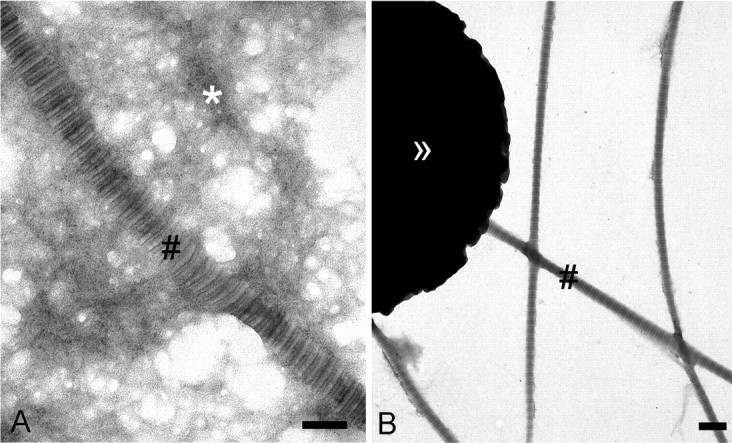
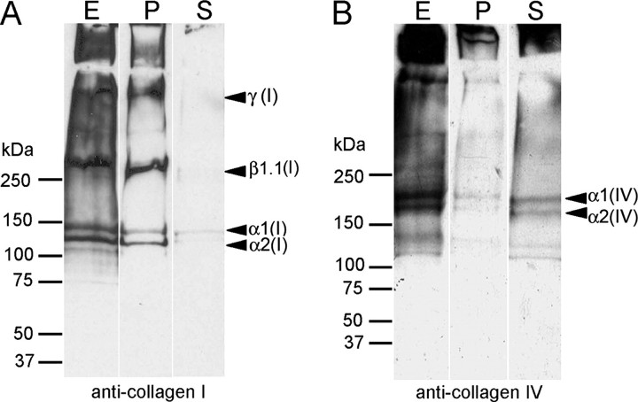
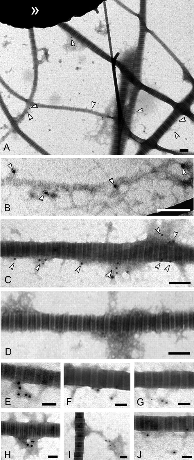
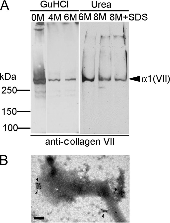
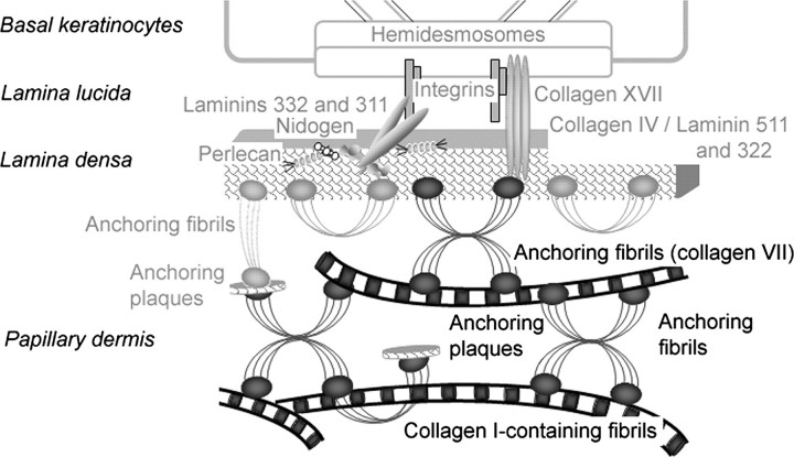
The dermis and the epidermis of normal human skin are functionally separated by a basement membrane but, together, form a stable structural continuum. Anchoring fibrils reinforce this connection by insertion into the basement membrane and by intercalation with banded collagen fibrils of the papillary dermis. Structural abnormalities in collagen VII, the major molecular constituent of anchoring fibrils, lead to a congenital skin fragility condition, dystrophic epidermolysis bullosa, associated with skin blistering. Here, we characterized the molecular basis of the interactions between anchoring fibrils and banded collagen fibrils. Suprastructural fragments of the dermo-epidermal junction zone were generated by mechanical disruption and by separation with magnetic Immunobeads. Anchoring fibrils were tightly attached to banded collagen fibrils. In vitro binding studies demonstrated that a von Willebrand factor A-like motif in collagen VII was essential for binding of anchoring fibrils to reconstituted collagen I fibrils. Since collagen I and VII molecules reportedly undergo only weak interactions, the attachment of anchoring fibrils to collagen fibrils depends on supramolecular organization of their constituents. This complex is stabilized in situ and resists dissociation by strong denaturants.


Figures

FIGURE 1.
FIGURE 1.
Collagen I-containing fibrils are mechanically separable from basement membrane networks. A, ultrastructural analysis by transmission electron microscopy of a crude skin extract obtained after tissue homogenization and differential centrifugation. The extract contains banded fibrils (#) and electron-dense network-like material. B, the crude extract was fractionated by immunomagnetic beads (>>) coated with antibodies to collagen I. D-periodically banded collagen I-containing fibrils (#) are separated from most of the network/like material. Bars, 200 nm.
FIGURE 2.
FIGURE 2.
Collagen I-containing fibrils are mechanically separable from basement membrane networks. Immunoblots are shown of the fraction containing banded fibrils attached to immunomagnetic beads coated with antibodies to collagen I and of unbound material in the supernatants. The blots were reacted with antibodies to collagen I, the major component of banded fibrils (A) or basement membrane collagen IV (B). Lanes E, crude extract before separation; lanes P, material attached to immunomagnetic beads; and lanes S, supernatants after reaction with immunomagnetic beads.
FIGURE 3.
FIGURE 3.
Anchoring fibrils are co-localized with D-periodically banded fibrils. Indirect immunogold electron microscopy of D-periodically banded fibrils sequentially treated with antibodies to collagen VII and gold-conjugated antibodies to immunoglobulins (white arrowheads) is shown. A, overview of the isolated collagen I-containing fibril fraction. Anchoring fibrils are associated with both small diameter (B) and large diameter (C) fibrils. D, negative control without treatment with antibodies to collagen VII. Bars, 200 nm. A gallery of immunogold-labeled structures is shown at higher magnification (EJ). Bars, 100 nm.
FIGURE 4.
FIGURE 4.
D-periodically banded fibrils and anchoring fibrils interact with high affinity. A, fibril fractions isolated with anti-collagen I-immunomagnetic beads were treated and washed with denaturants as indicated, and the extracts were analyzed by immunoblotting with antibodies to collagen VII. Proteins were separated by SDS-PAGE on a 4.5% gel under reducing conditions. Note: Treatment of collagen I-containing fibrils with guanidinium chloride and urea at high, denaturing concentrations does not eliminate collagen VII (α1(VII): 290 kDa). B, immunogold electron microscopy using antibodies to collagen VII (black arrowheads) reveals suprastructures containing collagen VII connected to banded fibrils after the partial denaturation with 2 m guanidinium chloride. Black asterisk, intact fibril section; white asterisk, fibril section disintegrated by the denaturant. Bar, 100 nm.
FIGURE 5.
FIGURE 5.
Schematic representation of the domain structures of recombinantly expressed full-length collagen VII and truncated collagen VII fusion constructs. The non-collagenous domains NC-1 and NC-2 are the amino- and the carboxyl-terminal globular domains, respectively, and separated by an interrupted triple helical domain. The NC-1 domain contains two von Willebrand factor A-like domains (vWFA) that are separated by nine fibronectin III-like repeats. The cleavage site for BMP-1 is located in the NC-2 domain. A, in the recombinant protein, a FLAG tag has been introduced at the amino terminus. B, mini-collagen VII results from the fusion between the amino acids 1042–1284 and 2684–2944. BPTI, bovine pancreatic trypsin inhibitor. C, mini-collagen VII short comprises the amino acids 2522–2944. Both fragments contain an octahistidine tag at their carboxyl terminus, which is used for purification.
FIGURE 6.
FIGURE 6.
Procollagen VII and mini-collagen VII but not mini-collagen VII short-bind to reconstituted collagen I fibrils. In vitro fibrillogenesis was performed with solutions of purified collagen I. Newly formed fibrils were reacted with variants of recombinant collagen VII, and immunogold labeling was performed analogously to the experiments shown in Fig. 3. Collagen I fibrils reacted with full-length procollagen VII (A, overview; B–D, gallery of filamentous structures in detail). Black and white arrowheads, double labeling with antibodies against the NC-1(VII) domain (12-nm gold particles, black arrowheads) and the carboxyl terminus of collagen VII (18-nm gold particles, white arrowheads). Reconstituted collagen I fibrils were exposed to mini-collagen VII (E) and mini-collagen VII short form (F). If present, these proteins were identified with antibodies to the carboxyl terminus (white arrowheads). * highlights collagen I fibrils. Note the absence of gold labeling in panel F. Bars: 200 nm in A, E, and F; 100 nm in B, C, and D.
FIGURE 7.
FIGURE 7.
Procollagen VII and mini-collagen VII but not mini-collagen VII short-bind to reconstituted collagen I fibrils. The graphs show the quantification of the binding of procollagen VII and procollagen VII fragments constructs to reconstituted collagen I fibrils. Procollagen VII (A) and the procollagen VII fragments (B) were incubated at different concentrations with collagen I fibrils and labeled with antibodies to the carboxyl terminus of collagen VII (NC-2-10). Binding was quantified by counting gold particles per μm2 of collagen I fibril in 10 arbitrarily chosen EM fields. Control: collagen VII ligand omitted.
FIGURE 8.
FIGURE 8.
Procollagen VII and mini-collagen VII but not mini-collagen VII short are covalently cross-linked to collagen I fibrils in vitro. Full-length procollagen VII (A), mini-collagen VII (B), and mini-collagen VII short form (C) were coupled to Sulfo-SBED and incubated with reconstituted collagen I fibrils. After the irradiation with UV light, the reaction products were reduced, submitted to SDS-PAGE, and electrotransferred to nitrocellulose membrane (lanes 1). For controls, collagen VII ligands (lanes 2) or collagen I fibrils (lanes 3) were omitted from reaction mixtures. The biotin transfer was detected by ExtrAvidin coupled to horseradish peroxidase. Note: Biotin was transferred to collagen I only in complete reaction mixtures (lanes 1) and only from procollagen VII (A) and mini-collagen VII (B). For further controls (right panels), immunoblotting was conducted with antibodies to procollagen VII.
FIGURE 9.
FIGURE 9.
Schematic representation of the organization of the anchoring complex within the DEJZ. The novel, covalently stabilized interaction between collagen I-containing fibrils and anchoring fibrils is highlighted in black, whereas interactions previously described are kept in gray. Inset: molecular organization of anchoring fibrils; antiparallel dimers of collagen VII molecules subsequently form lateral aggregates.

References

    1. Ellison, J., and Garrod, D. R. (1984) J. Cell Sci. 72 163–172 - PubMed
    1. Palade, G. E., and Farquhar, M. G. (1965) J. Cell Biol. 27 215–224 - PMC - PubMed
    1. Burgeson, R. E., Lunstrum, G. P., Rokosova, B., Rimberg, C. S., Rosenbaum, L. M., and Keene, D. R. (1990) Ann. N. Y. Acad. Sci. 580 32–43 - PubMed
    1. Shimizu, H., Ishiko, A., Masunaga, T., Kurihara, Y., Sato, M., Bruckner-Tuderman, L., and Nishikawa, T. (1997) Lab. Investig. 76 753–763 - PubMed
    1. Keene, D. R., Sakai, L. Y., Lunstrum, G. P., Morris, N. P., and Burgeson, R. E. (1987) J. Cell Biol. 104 611–621 - PMC - PubMed

Publication types

MeSH terms

LinkOut - more resources