

ABT-737 induces expression of the death receptor 5 and sensitizes human cancer cells to TRAIL-induced apoptosis
- PMID: 18599488
- PMCID: PMC2529128
- DOI: 10.1074/jbc.M802511200
ABT-737 induces expression of the death receptor 5 and sensitizes human cancer cells to TRAIL-induced apoptosis
Abstract
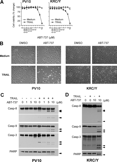
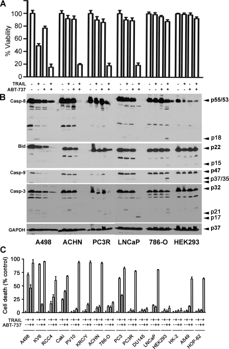
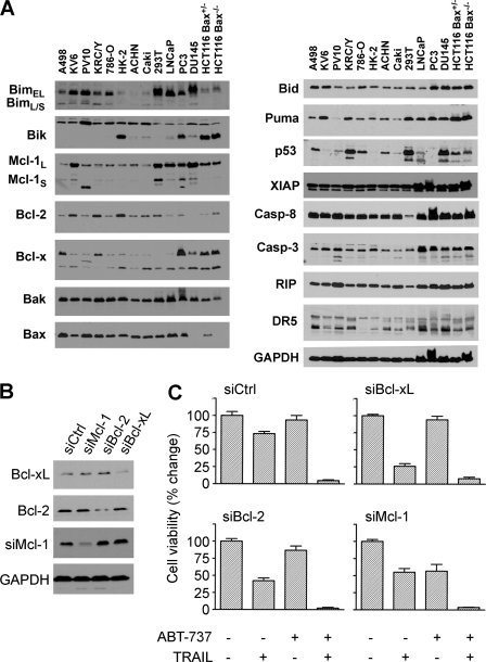
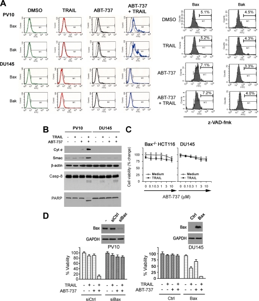
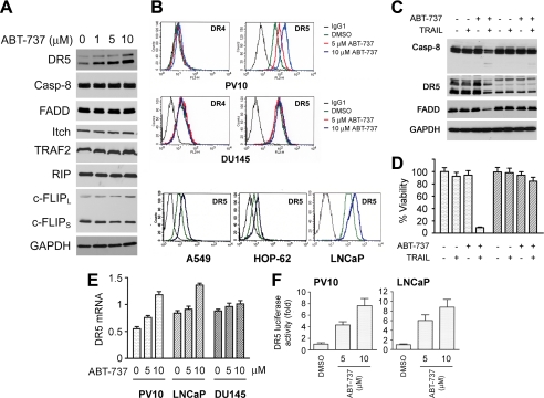
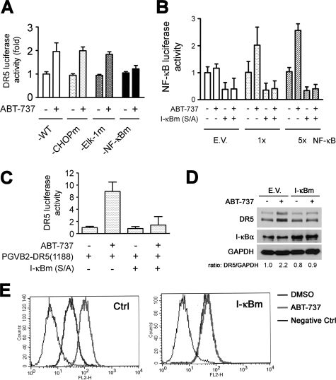
Because Bcl-2 family members inhibit the ability of tumor necrosis factor-related apoptosis-inducing ligand (TRAIL) to induce apoptosis, we investigated whether ABT-737, a small molecule Bcl-2 inhibitor, enhances TRAIL killing. We demonstrate that a combination of ABT-737 and TRAIL induced significant cell death in multiple cancer types, including renal, prostate, and lung cancers, although each agent individually had little activity in these tumor cells. All of these cell lines expressed the Mcl-1 protein that is known to block the activity of ABT-737 and TRAIL but did not block the synergy between these agents. However, Bax-deficient cell lines, including DU145 and HCT116 cells and those cell lines expressing low levels of TRAIL receptor, were resistant to apoptosis induced by these agents. To understand how ABT-737 functions to markedly increase TRAIL sensitivity, the levels of specific death-inducing signaling complex components were evaluated. Treatment with ABT-737 did not change the levels of c-FLIP, FADD, and caspase-8 but up-regulated the levels of the TRAIL receptor DR5. DR5 up-regulation induced by ABT-737 treatment occurred through a transcriptional mechanism, and mutagenesis studies demonstrated that the NF-kappaB site found in the DR5 promoter was essential for the ability of ABT-737 to increase the levels of this mRNA. Using luciferase reporter plasmids, ABT-737 was shown to stimulate NF-kappaB activity. Together, these results demonstrate that the ability of ABT-737 and TRAIL to induce apoptosis is mediated through activation of both the extrinsic and intrinsic pathways. Combinations of ABT-737 and TRAIL can be exploited therapeutically where antiapoptotic Bcl-2 family members drive tumor cell resistance to current anticancer therapies.
Figures
Similar articles
-
Bcl-2 family proteins contribute to apoptotic resistance in lung cancer multicellular spheroids.Am J Respir Cell Mol Biol. 2009 Jul;41(1):14-23. doi: 10.1165/rcmb.2008-0320OC. Epub 2008 Dec 18. Am J Respir Cell Mol Biol. 2009. PMID: 19097992 Free PMC article.
-
Bcl-xL inhibition by molecular-targeting drugs sensitizes human pancreatic cancer cells to TRAIL.Oncotarget. 2015 Dec 8;6(39):41902-15. doi: 10.18632/oncotarget.5881. Oncotarget. 2015. PMID: 26506422 Free PMC article.
-
BH3 mimetic ABT-737 potentiates TRAIL-mediated apoptotic signaling by unsequestering Bim and Bak in human pancreatic cancer cells.Cancer Res. 2008 Apr 15;68(8):2944-51. doi: 10.1158/0008-5472.CAN-07-2508. Cancer Res. 2008. PMID: 18413764 Free PMC article.
-
Down-regulation of intracellular anti-apoptotic proteins, particularly c-FLIP by therapeutic agents; the novel view to overcome resistance to TRAIL.J Cell Physiol. 2018 Oct;233(10):6470-6485. doi: 10.1002/jcp.26585. Epub 2018 May 9. J Cell Physiol. 2018. PMID: 29741767 Review.
-
BH3 mimetics to improve cancer therapy; mechanisms and examples.Drug Resist Updat. 2007 Dec;10(6):207-17. doi: 10.1016/j.drup.2007.08.002. Epub 2007 Oct 24. Drug Resist Updat. 2007. PMID: 17921043 Free PMC article. Review.
Cited by
-
Generation of TRAIL-resistant cell line models reveals distinct adaptive mechanisms for acquired resistance and re-sensitization.Oncogene. 2021 May;40(18):3201-3216. doi: 10.1038/s41388-021-01697-6. Epub 2021 Mar 25. Oncogene. 2021. PMID: 33767436
-
Ceramide synthase-6 confers resistance to chemotherapy by binding to CD95/Fas in T-cell acute lymphoblastic leukemia.Cell Death Dis. 2018 Sep 11;9(9):925. doi: 10.1038/s41419-018-0964-4. Cell Death Dis. 2018. PMID: 30206207 Free PMC article.
-
Harnessing TRAIL-Induced Apoptosis Pathway for Cancer Immunotherapy and Associated Challenges.Front Immunol. 2021 Aug 20;12:699746. doi: 10.3389/fimmu.2021.699746. eCollection 2021. Front Immunol. 2021. Retraction in: Front Immunol. 2023 Sep 04;14:1285126. doi: 10.3389/fimmu.2023.1285126. PMID: 34489946 Free PMC article. Retracted. Review.
-
Combination of TRAIL and actinomycin D liposomes enhances antitumor effect in non-small cell lung cancer.Int J Nanomedicine. 2012;7:1449-60. doi: 10.2147/IJN.S24711. Epub 2012 Mar 19. Int J Nanomedicine. 2012. PMID: 22619505 Free PMC article.
-
Real-time imaging of the dynamics of death receptors and therapeutics that overcome TRAIL resistance in tumors.Oncogene. 2013 Jun 6;32(23):2818-27. doi: 10.1038/onc.2012.304. Epub 2012 Jul 23. Oncogene. 2013. PMID: 22824792 Free PMC article.
References
-
- Walczak, H., Miller, R. E., Ariail, K., Gliniak, B., Griffith, T. S., Kubin, M., Chin, W., Jones, J., Woodward, A., Le, T., Smith, C., Smolak, P., Goodwin, R. G., Rauch, C. T., Schuh, J. C., and Lynch, D. H. (1999) Nat. Med. 5 157-163 - PubMed
-
- Hao, C., Song, J. H., Hsi, B., Lewis, J., Song, D. K., Petruk, K. C., Tyrrell, D. L., and Kneteman, N. M. (2004) Cancer Res. 64 8502-8506 - PubMed
-
- Ichikawa, K., Liu, W., Zhao, L., Wang, Z., Liu, D., Ohtsuka, T., Zhang, H., Mountz, J. D., Koopman, W. J., Kimberly, R. P., and Zhou, T. (2001) Nat. Med. 7 954-960 - PubMed
-
- Walczak, H., and Krammer, P. H. (2000) Exp. Cell Res. 256 58-66 - PubMed
Publication types
MeSH terms
Substances
Grants and funding
LinkOut - more resources
Full Text Sources
Other Literature Sources
Molecular Biology Databases
Research Materials
Miscellaneous
