

Review
doi: 10.1038/eye.2008.206.
Epub 2008 Jul 4.
Macrophages in neovascular age-related macular degeneration: friends or foes?
Affiliations
- PMID: 18600240
- PMCID: PMC8204908
- DOI: 10.1038/eye.2008.206
Item in Clipboard
Review
Macrophages in neovascular age-related macular degeneration: friends or foes?
Eye (Lond).
2009 Apr.
Abstract
The events that lead to choroidal neovascularization in eyes with age-related macular degeneration are poorly understood. One possibility that has been explored in a number of studies is that macrophages can promote neovascular changes. In this paper, we summarize the evidence for inflammation in general and macrophages in particular in pathologic neovascularization, and discuss how the diverse functions of these cells may promote or inhibit macular disease. We also discuss some of the conflicting findings regarding the role of macrophages in experimental choroidal neovascularization in mouse models, and suggest areas for future research.
Figures
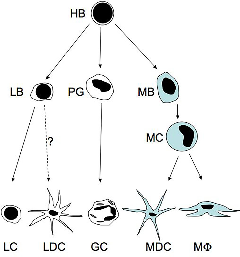
Bone marrow hemocytoblasts (HB), which may already be specialized in some cases, give rise to progenitor cells including lymphoblasts (LB), progranulocytes (PG), and monoblasts (MB). These cells then differentiate into lymphocytes (LC) or arguably lymphoid dendritic cells (LDC), different classes of granulocytes (GC), and monocytes (MC), respectively. Circulating monocytes leave the vasculature and differentiate into myeloid dendritic cells (MDC) and macrophages (MΦ), based on microenvironmental cues.
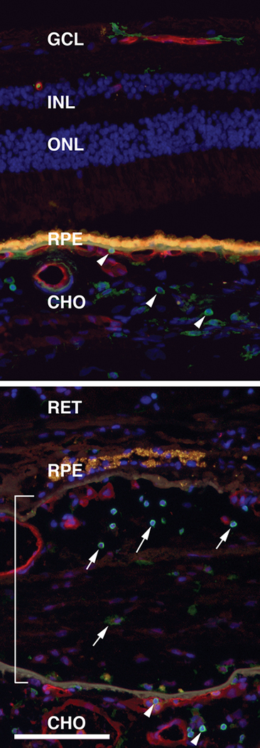
Leukocytes localized in the normal human choroid of an 84 year old donor (top panel) and in an eye from an 80 year old donor with choroidal neovascularization (lower panel). Sections were labeled with antibodies directed against leukocyte common antigen, a protein expressed in all classes of leukocytes (CD45; green labeling) and with the lectin Ulex europeaus agglutinin-I to visualize the vasculature (red labeling)(85). Nuclei are counterstained blue with DAPI. Yellow-orange fluorescence is due to RPE lipofuscin. Choroidal leukocytes are indicated by arrowheads, leukocytes within the CNVM are indicated by arrows, and the CNVM is indicated by the bracket. GCL, ganglion cell layer; INL, inner nuclear layer; ONL, outer nuclear layer; CHO, choroid. Scale bar = 100μm.
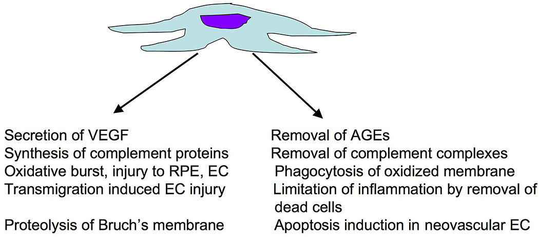
Potential harmful (left) and beneficial (right) roles of macrophages in the progression of neovascular AMD.
References
-
- Klein R, Klein B, Cruickshanks K. The prevalence of age-related maculopathy by geographic region and ethnicity. Prog Retin Eye Res. 1999;18:371–89. - PubMed
-
- Buch H, Vinding T, Nielsen N. Prevalence and causes of visual impairment according to World Health Organization and United States criteria in an aged, urban Scandinavian population: the Copenhagen City Eye Study. Ophthalmology. 2001;108:2347–57. - PubMed
-
- Smith W, Assink J, Klein R, Mitchell P, Klaver C, Klein B, et al. Risk factors for age-related macular degeneration: Pooled findings from three continents. Ophthalmology. 2001;108:697–704. - PubMed
-
- Friedman D, O'Colmain B, Munoz B, Tomany S, McCarty C, deJong P, et al. Prevalence of age-related macular degeneration in the United States. Arch Ophthalmol. 2004;122:564–72. - PubMed
-
- VanNewkirk M, Nanjan M, Wang J, Mitchell P, Taylor H, McCarty C. The prevalence of age-related maculopathy: the visual impairment project. Ophthalmology. 2000;107:1593–600. - PubMed
Publication types
MeSH terms
Grants and funding
LinkOut - more resources
Full Text Sources
Medical
