Review
. 2008 Oct;32(8):1451-84.
doi: 10.1016/j.neubiorev.2008.05.023. Epub 2008 May 24.

Cetacean sleep: an unusual form of mammalian sleep

Affiliations
Review

Cetacean sleep: an unusual form of mammalian sleep

Oleg I Lyamin et al. Neurosci Biobehav Rev. 2008 Oct.

Abstract

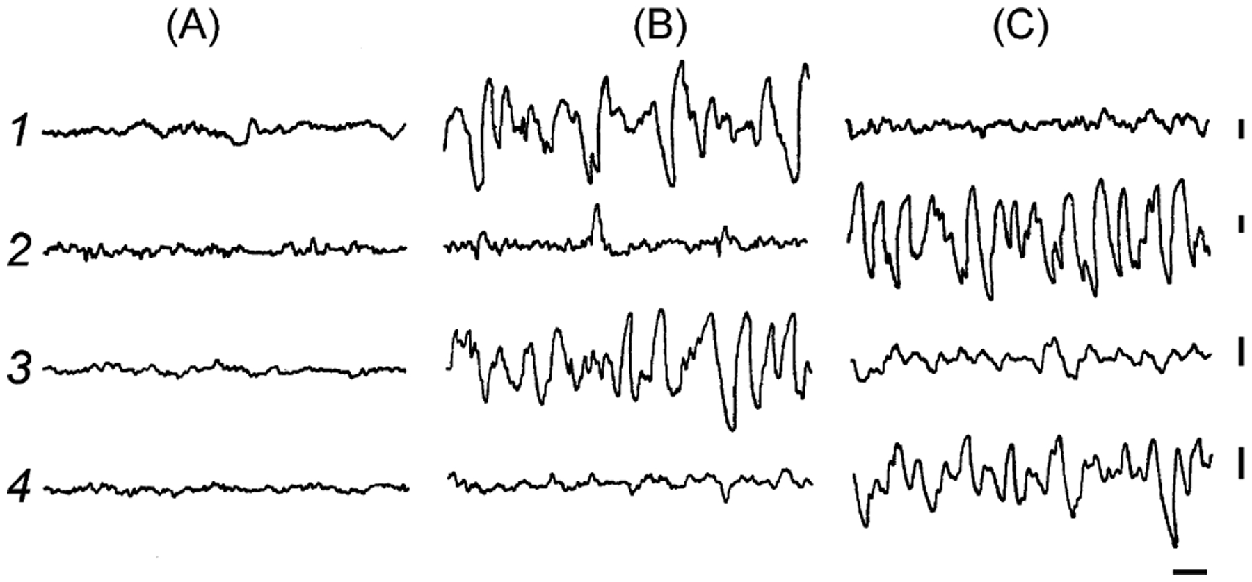
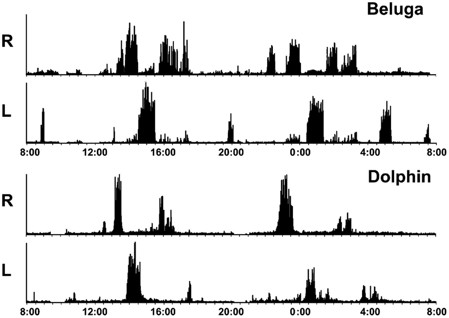
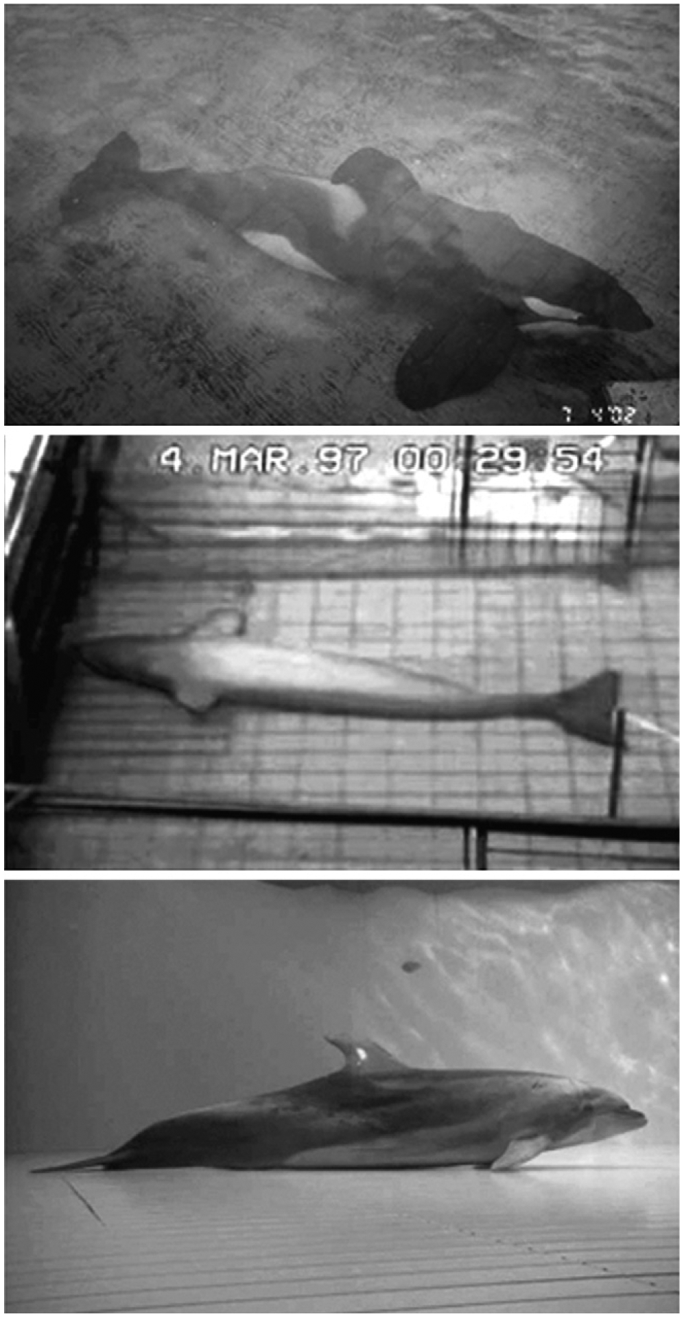
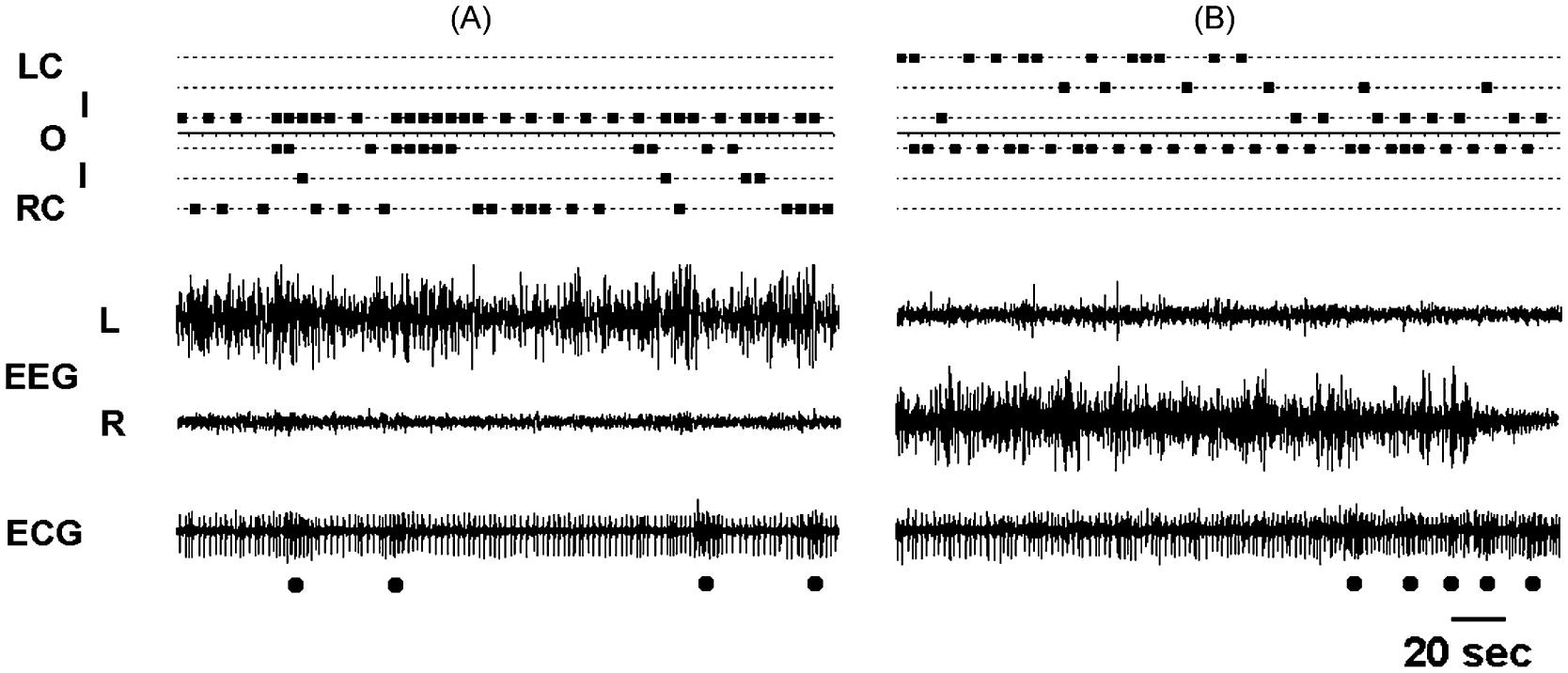
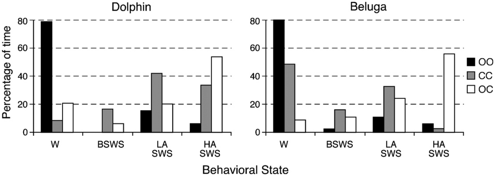
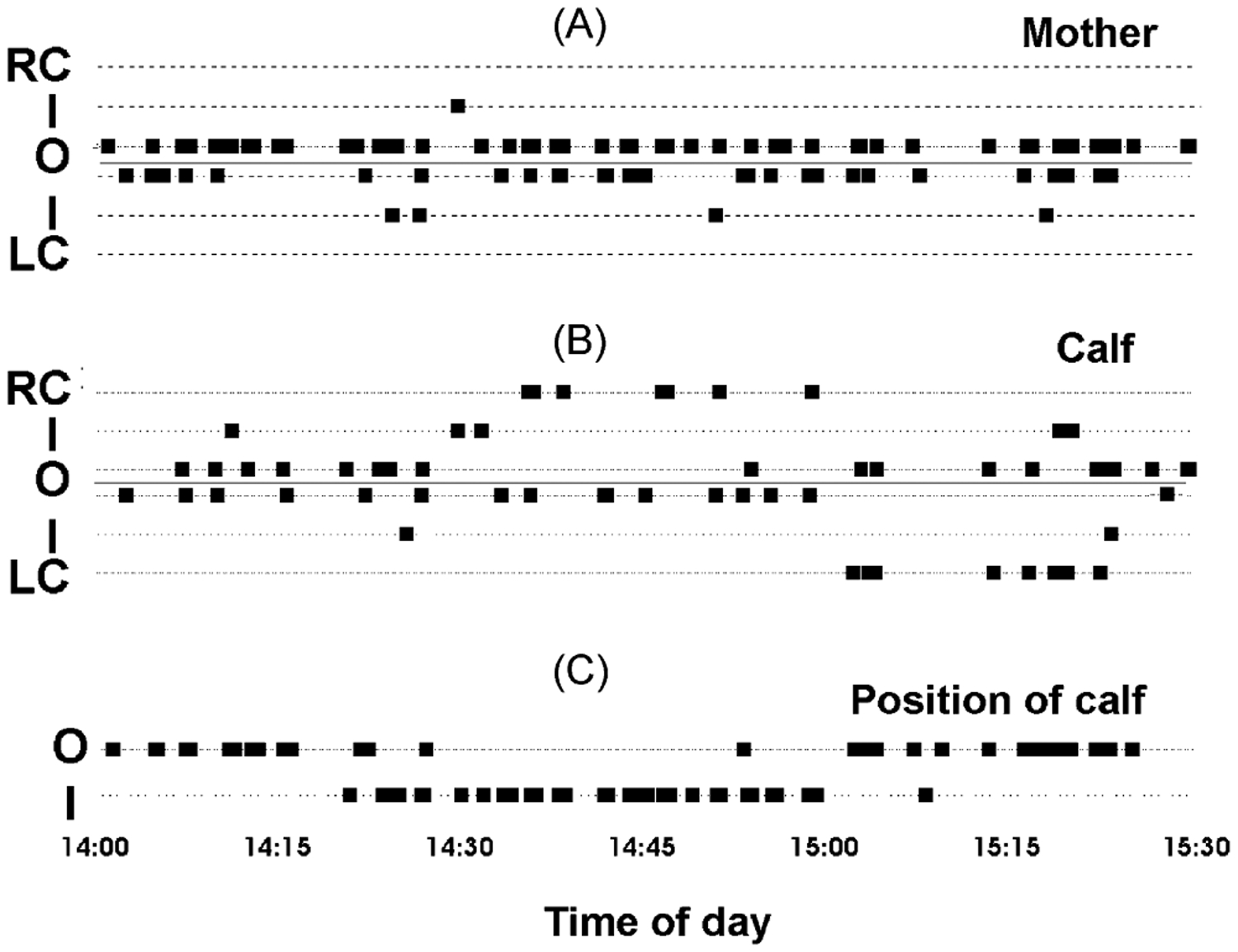
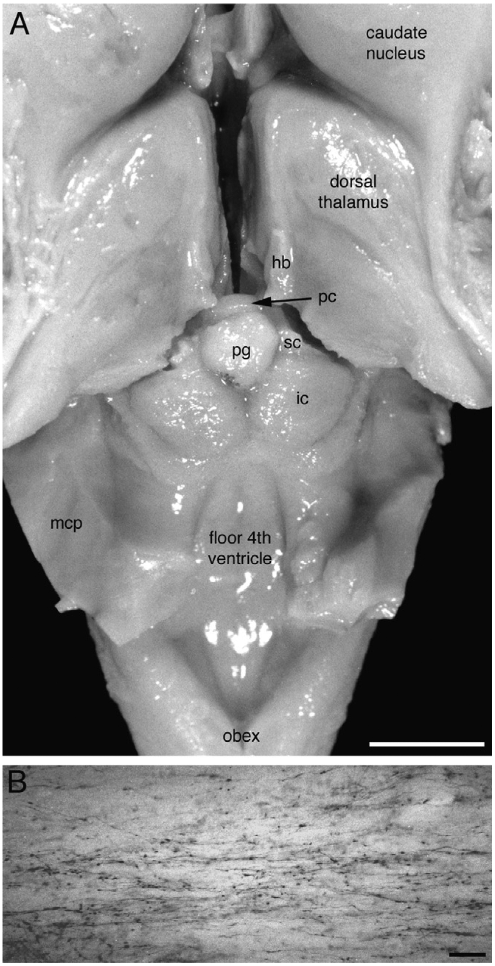
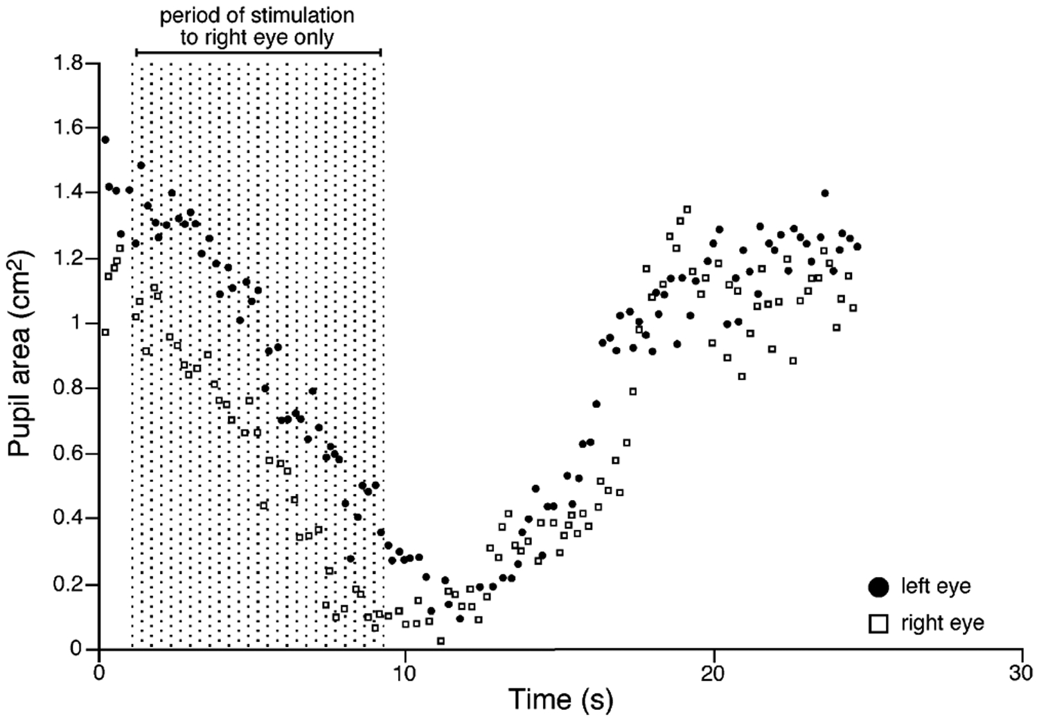
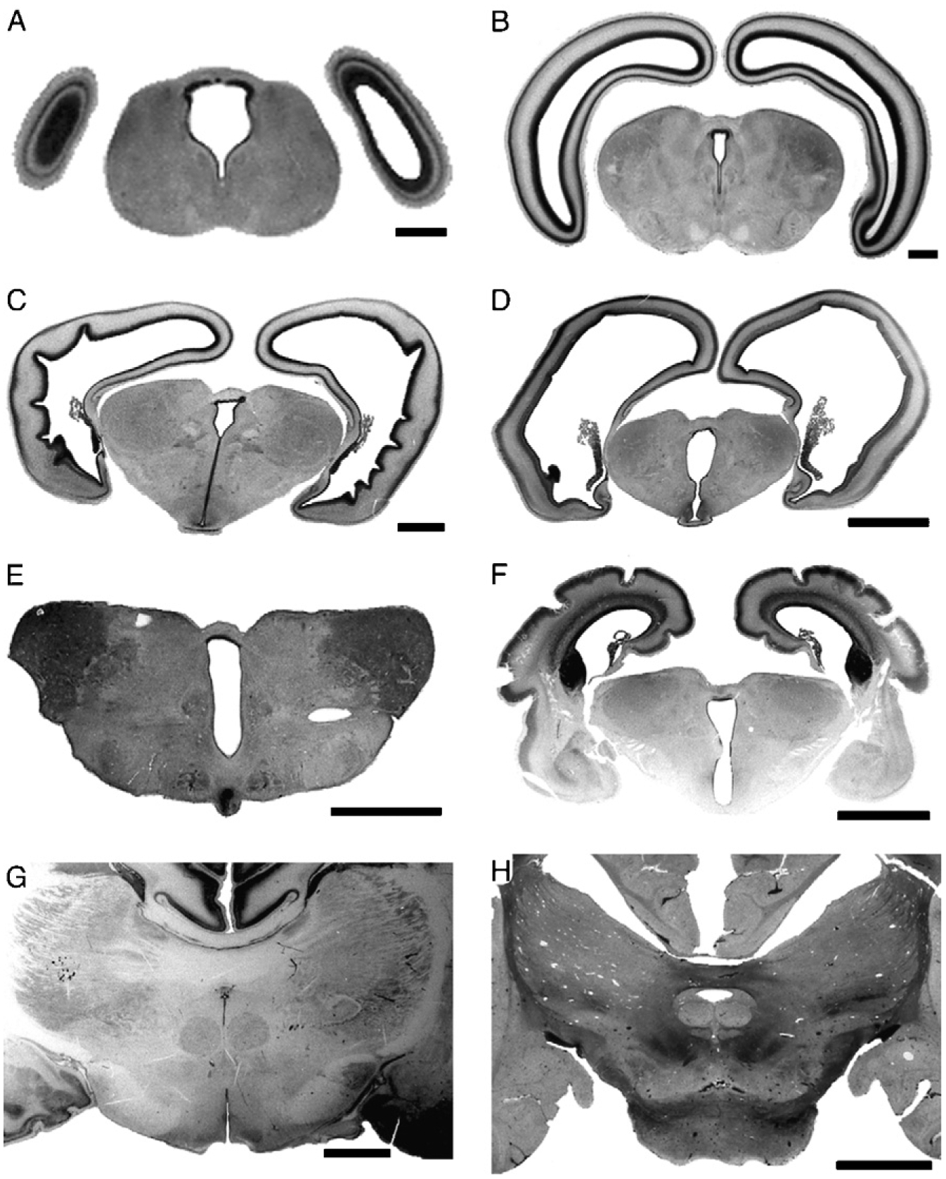
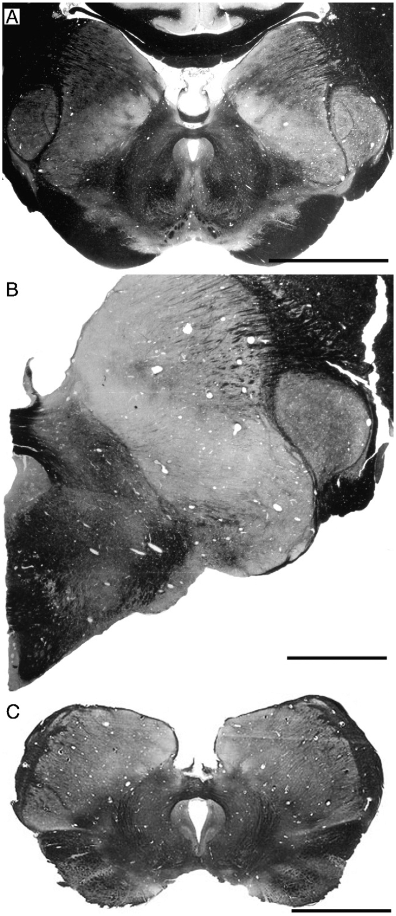
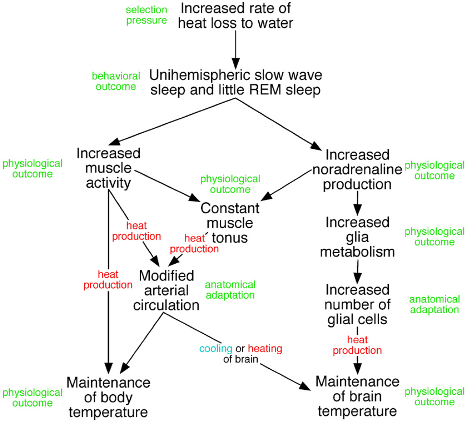
Our knowledge of the form of lateralized sleep behavior, known as unihemispheric slow wave sleep (USWS), seen in all members of the order Cetacea examined to date, is described. We trace the discovery of this phenotypically unusual form of mammalian sleep and highlight specific aspects that are different from sleep in terrestrial mammals. We find that for cetaceans sleep is characterized by USWS, a negligible amount or complete absence of rapid eye movement (REM) sleep, and a varying degree of movement during sleep associated with body size, and an asymmetrical eye state. We then compare the anatomy of the mammalian somnogenic system with what is known in cetaceans, highlighting areas where additional knowledge is needed to understand cetacean sleep. Three suggested functions of USWS (facilitation of movement, more efficient sensory processing and control of breathing) are discussed. Lastly, the possible selection pressures leading to this form of sleep are examined, leading us to the suggestion that the selection pressure necessitating the evolution of cetacean sleep was most likely the need to offset heat loss to the water from birth and throughout life. Aspects such as sentinel functions and breathing are likely to be proximate evolutionary phenomenon of this form of sleep.


Figures

Fig. 1.
Fig. 1.
EEG recorded from two cortical hemispheres and two thalamus during waking (A), unihemispheric slow wave sleep in the right (B) and left (C) hemispheres in a bottlenose dolphin. Recording: 1—right cortex, 2—left cortex, 3—right thalamus and 4—left thalamus. Calibration 1 s and 200 μm (modified from Supin and Mukhametov, 1986).
Fig. 2.
Fig. 2.
EEG slow wave power in the range of 1.2–4 Hz in the right (R) and left (L) hemispheres in a bottlenose dolphin and beluga recorded over 24 h.
Fig. 3.
Fig. 3.
Rest on the bottom of pools in a captive killer whale (photo by O.I. Lyamin), beluga (from Lyamin et al., 2002b) and bottlenose dolphin (from Sekiguchi and Kohshima, 2003).
Fig. 4.
Fig. 4.
Relationship between eye state and EEG in a beluga. (A and B) Episodes of unihemispheric sleep in the left (L) and right (R) hemispheres, respectively, paralleled with continuous noting of the state of two eyes (L—left; R—right). The state of two eyes was scored as open (O), closed (C; LC—left closed; RC—right closed) and intermediate (I). ECG—electrocardiogram. Black dots below ECG mark breaths. Note: (1) that the eye contralateral to the waking hemisphere did not close during the entire episode of sleep in the opposite hemisphere, and that (2) slow wave EEG did not change immediately with opening of the opposite eye.
Fig. 5.
Fig. 5.
Association between eye state and behavioral stage in a bottlenose dolphin and in a beluga. The height of bars is proportional to the time spent in waking (W), bilateral slow wave sleep (BSWS), low and high asymmetrical amplitude slow wave sleep (LASWS and HASWS, respectively) as a percentage of the given eye state (OO—both eyes open, CC—both eyes closed and OC—one eye open and another eye closed).
Fig. 6.
Fig. 6.
Simultaneous recording the state of two eyes in a bottlenose dolphin mother showing that calf’s open eye is generally directed at the mother (A) and calf (B) and the position of the calf (C) during swimming in a counter-clockwise direction. Each mark represents the state of visible eye (R—right, L—left; O—open, C—closed and I—intermediate) or the position of calf next to the mother (I—inner or O—outer) during echelon swimming.
Fig. 7.
Fig. 7.
Photomicrographs of myelin stained coronal brain sections demonstrating the morphology of the posterior commissure in a variety of mammalian species. (A) Wombat (Vombatus ursinus) scale bar = 3 mm. (B) Tammar wallaby (Macropus eugenii) scale bar = 3 mm. (C) Giant anteater (Myrmecophaga tridactyla) scale bar = 3 mm. (D) Marmot (Marmota marmota) scale bar = 1 mm. (E) Woodchuck (Marmota monax) scale bar = 2 mm. (F) Jack rabbit (Lepus americanus) scale bar = 3 mm. (G) Zebra (Equus burchelli) scale bar = 5 mm. (H) Sheep (Ovis aries) scale bar = 3 mm. (I) North American badger (Taxidea taxus) scale bar = 3 mm. (J) Leopard (Panthera pardus) scale bar = 5 mm. (K) American black bear (Ursus americanus) scale bar = 5 mm. (L) Chimpanzee (Pan troglodytes) scale bar = 5 mm. All photomicrographs in this plate are taken from sections housed within the Comparative Mammalian Brain Collections of the University of Wisconsin, Michigan State University and The National Museum of Health and Medicine.
Fig. 8.
Fig. 8.
(A) Photograph of gross coronal slice through the brain of a Commerson’s or piebald dolphin (Cephalorhynchus commersonii), showing the enlarged posterior commissure situated immediately anterior to the superior colliculi, and dorsal to the septal nuclei. The coronal plane in cetaceans is different to other mammals due to the maintenance of the cephalic flexure in the adult (from the collection of Sam Ridgway). scale bar = 1 cm. (B) Photomicrograph of a horizontal section through the brain of an adult bottlenose dolphin (Tursiops truncatus) stained for both Nissl substance and myelin. The myelin dense, horizontally projecting posterior commissure can be seen anterior to the superior colliculi investing into the gray matter of the dorsal thalamus. scale bar = 1 cm. (C) Photomicrograph of a coronal section through the brain of an adult bottlenose dolphin (T. truncatus) stained for myelin. Note the thickness of the posterior commissure, lying immediately ventral to a slightly thicker corpus callosum, and projecting horizontally into the gray matter of the dorsal thalamus. Scale bar = 1 cm. The photomicrographs in this plate are taken from sections housed within the Comparative Mammalian Brain Collections of the University of Wisconsin, Michigan State University and The National Museum of Health and Medicine.
Fig. 9.
Fig. 9.
(A) Gross dissection of the bottlenose dolphin brain with the third and fourth ventricles revealed. This dissection demonstrates the location of the posterior commissure (pc) in relation to the habenular trigones (hb), the superior colliculi (sc), pineal gland (pg) and the inferior colliculi (ic). mcp—middle cerebellar peduncle. Scale bar = 1 cm. It should be noted here that the dolphin dissected in this preparation died while heavily pregnant and that in the normal situation the pineal gland is microscopic or absent in cetaceans (Oelschläger et al., 2008, see text for further details). (B) Photomicrograph of tyrosine hydroxylase immunopositive axons coursing through the posterior commissure of the bottlenose dolphin. Scale bar = 100 μm.
Fig. 10.
Fig. 10.
Diagram depicting the results of the experiment testing the consensual light reflex in the bottlenose dolphin. Note that as the right eye only is stimulated by light (the left light being hooded to prevent stimulation) the pupil of the right eye decreases in area and the pupil of the left eye also decreases. This response is typical of the consensual light reflex in mammals that is mediated by pathways crossing through the posterior commissure.
Fig. 11.
Fig. 11.
Photomicrographs of embryonic and fetal brain sections at the level of the developing posterior commissure in a series of mysticete (A–E) and odontocete (F–H) cetaceans. (A) 10.5 cm long fin whale (Balaenoptera physalus) scale bar = 1 mm. (B) 17 cm long blue whale (Balaenoptera musculus) scale bar = 1 mm. (C) 21 cm long fin whale, scale bar = 2 mm. (D) 30 cm long fin whale, scale bar = 5 mm. (E) 80 cm long blue whale, scale bar = 5 mm. The adult length of the fin whale ranges from 21 to 25 m, with birth lengths of 6–7 m, and for blue whales, the adult length ranges from 26–33.6 m, with birth lengths being 6–7 m. (F) 16 cm long Harbor porpoise (Phocoena phocoena), adult length ranges from 145–200 cm, with birth lengths ranging from 70–75 cm, scale bar = 5 mm. These sections were stained for Nissl substance and are housed in the Jan Jansen Whale Brain Collection in the Department of Anatomy, University of Oslo, Norway. (G) Fetal Dall’s porpoise (Phocoenoides dalli), unknown length and age, section stained for Nissl substance, scale bar = 3 mm. (H) New born bottlenose dolphin (T. truncatus), approximate length of 100 cm, adult T. truncatus lengths range from 2 to 3.8 m, section stained for myelin, scale bar = 1 cm. The early development of the posterior commissure is similar to that of other eutherian species, with changes in morphology occurring late in development. The last two photomicrographs in this plate are taken from sections housed within the Comparative Mammalian Brain Collections of the University of Wisconsin, Michigan State University and The National Museum of Health and Medicine.
Fig. 12.
Fig. 12.
Photomicrographs of coronal sections stained for myelin in two species of pinniped and one sirenian, demonstrating the morphology of the posterior commissure. (A) Northern fur seal (Callorhinus ursinus), a member of the Otariidae family and a presumed unihemispheric sleeper when in water. Note the generally typical mammalian appearance of the posterior commissure. (B) Harbor seal (Phoca vitulina), a member of the Phocidae family and a presumed bihemispheric sleeper. Note again the typical mammalian appearance of the posterior commissure. (C) Florida manatee (Trichechus manatus), a species that exhibits both unihemispheric and bihemispheric sleep. Note again the typical mammalian appearance of the posterior commissure. Scale bar = 1 cm, applies to all three photomicrographs. All photomicrographs in this plate are taken from sections housed within the Comparative Mammalian Brain Collections of the University of Wisconsin, Michigan State University and The National Museum of Health and Medicine.
Fig. 13.
Fig. 13.
Diagrammatic representation of the possible manner in which cetacean sleep phenomenology may interact with the anatomy and physiology of the cetacean body and brain to maintain body and brain temperature in the thermally challenging aquatic environment.

References

    1. Amlaner CJ, Ball NJ, 1994. Avian sleep. In: Kryger MH, Roth T, Dement WC (Eds.), Principles and Practice of Sleep Medicine. second ed. Saunders, Philadelphia, pp. 81–94.
    1. Anderson PK, 1986. Dugongs of Shark Bay, Australia—seasonal migration, water temperature and forage. Natl. Geogr. Res 2, 473–490.
    1. Balance LT, 2002. Cetacean ecology. In: Perrin WF, Würsig B, Thewissen JGM (Eds.), Encyclopedia of Marine Mammals. Academic Press, New York, pp. 208–214.
    1. Bearzi G, Politi E, 1999. Diurnal behavior of free-ranging bottlenose dolphins in the Kvarneric (Northern Adriatic sea). Mar. Mamm. Sci 15, 1065–1097.
    1. Behrman G, 1990. The pineal organ (epiphysis cerebri) of the harbor porpoise (Phocoena phocoena, Linne, 1758). Aquat. Mamm 16, 96–100.

Publication types

Substances