. 2008 Sep 12;381(4):975-88.
doi: 10.1016/j.jmb.2008.06.048. Epub 2008 Jun 24.

Structures of substrate- and inhibitor-bound adenosine deaminase from a human malaria parasite show a dramatic conformational change and shed light on drug selectivity

Affiliations

Structures of substrate- and inhibitor-bound adenosine deaminase from a human malaria parasite show a dramatic conformational change and shed light on drug selectivity

Eric T Larson et al. J Mol Biol. .

Abstract

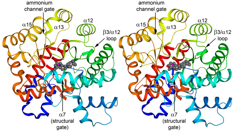
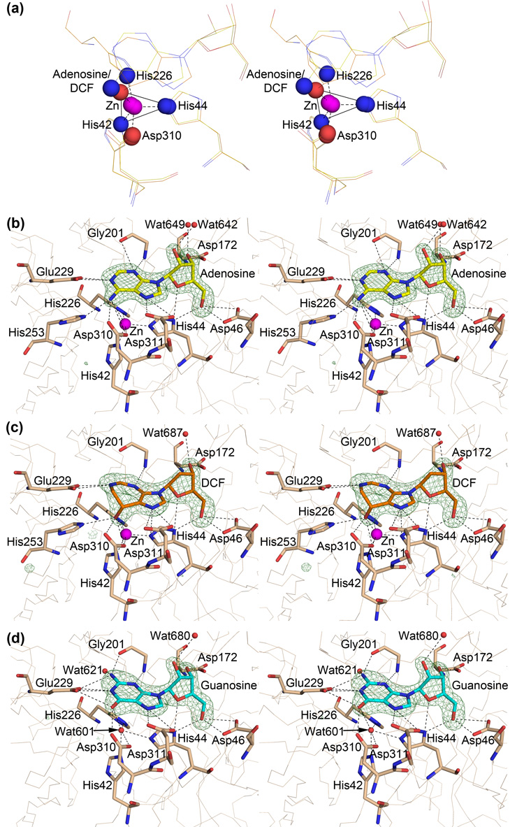
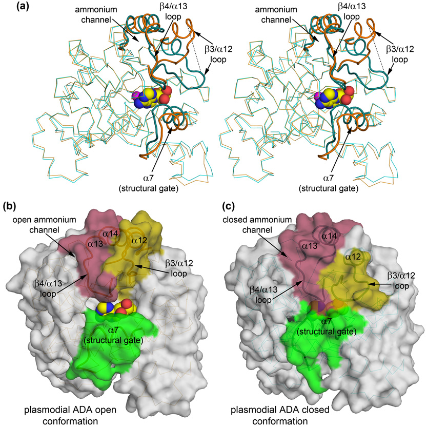
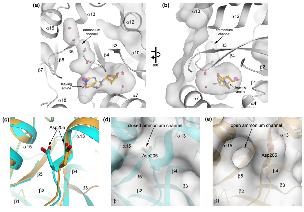
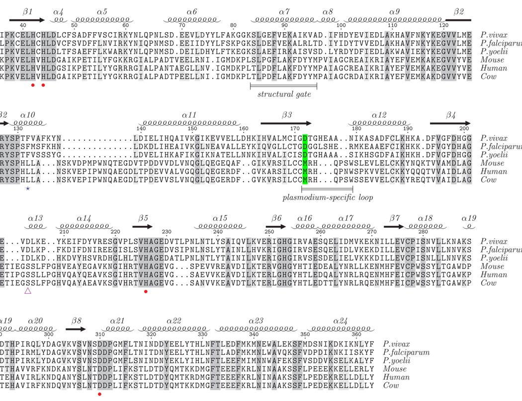
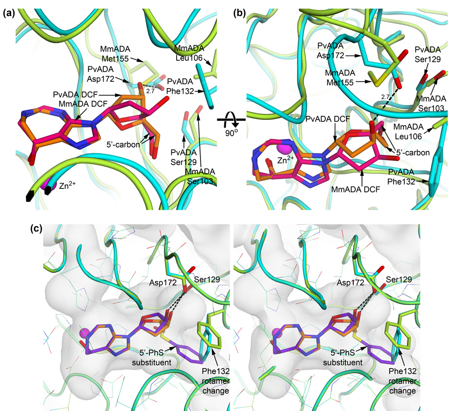
Plasmodium and other apicomplexan parasites are deficient in purine biosynthesis, relying instead on the salvage of purines from their host environment. Therefore, interference with the purine salvage pathway is an attractive therapeutic target. The plasmodial enzyme adenosine deaminase (ADA) plays a central role in purine salvage and, unlike mammalian ADA homologs, has a further secondary role in methylthiopurine recycling. For this reason, plasmodial ADA accepts a wider range of substrates, as it is responsible for deamination of both adenosine and 5'-methylthioadenosine. The latter substrate is not accepted by mammalian ADA homologs. The structural basis for this natural difference in specificity between plasmodial and mammalian ADA has not been well understood. We now report crystal structures of Plasmodium vivax ADA in complex with adenosine, guanosine, and the picomolar inhibitor 2'-deoxycoformycin. These structures highlight a drastic conformational change in plasmodial ADA upon substrate binding that has not been observed for mammalian ADA enzymes. Further, these complexes illuminate the structural basis for the differential substrate specificity and potential drug selectivity between mammalian and parasite enzymes.


Figures

Figure 1
Figure 1
Stereo view of Plasmodium vivax adenosine deaminase in complex with adenosine. The color of the protein ramps from blue at the N-terminus to red at the C-terminus. Since the structure of all the complexes is essentially identical, only the adenosine complex (2PGF) is shown. The bound adenosine is shown as a gray ball and stick model and the catalytic zinc ion is shown as a magenta sphere. Features discussed in the text are labeled. The view is into the active site. Figures were prepared with PyMol.
Figure 2
Figure 2
(a) Stereo view of the coordination environment of the catalytic Zn2+ ion (magenta sphere). Nitrogen atoms involved in coordination are shown as blue spheres and oxygen atoms as red spheres. The view is rotated −60° along the x-axis with respect to figure 1. (b–d) Stereo views of the ligand environments. Residues that make polar contacts within 3.5 Å of the ligand or that coordinate the catalytic zinc ion (magenta sphere; water 601 in the case of the guanosine complex) are shown as sticks. Waters that are in contact with the ligand are shown as red spheres. 2Fo-dFc ligand-omit difference density is shown as green mesh around the ligands at a contour level of 5σ. In all three cases the ribose 2′-endo sugar pucker is clearly defined by the electron density. The view is rotated −90° around the X-axis with respect to figure 1. (b) Adenosine environment (2PGF). (c) Guanosine environment (2QVN). (d) 2'-deoxycoformycin environment (2PGR).
Figure 3
Figure 3
Ligand-binding induces a large conformational change around the active site of the enzyme. (a) Stereo view highlighting the conformational changes associated with ligand-binding. The P. yoelii structure (2AMX; orange), corresponding to the open form, is superimposed onto the P. vivax structure (2PGF, 2PGR, 2QVN; cyan), corresponding to the closed form. In addition to the relatively small rigid body movements of α7 and β4/α13 loop-α13- α14 toward the substrate associated with the closed state of the mammalian enzymes, the β3/α12 loop of plasmodial ADA undergoes a reordering and is stabilized over the substrate, blocking access to the active site. The dashed line runs between PvADA Ala177 (cyan loop) and PyADA Ala190 (orange loop), which are the equivalent alpha carbons at the maximal displacement of this conformational change, approximately 15.5 Å. The features of greatest change between the apo, open, form and the substrate-bound, closed form are highlighted as tubes of slightly darker color. The view is rotated 45° around the Y-axis in relation to that of figure 1 to emphasize the changes upon substrate-binding. (b) Surface representation of PyADA (2AMX; the plasmodial open form) with adenosine (spheres) modeled in the active site pocket by superposition. (c) Surface representation of the PvADA: adenosine complex (2PGF; the plasmodial closed form). In panels b and c, the surface corresponding to the regions of greatest conformational change between the apo and ligand-bound forms are highlighted. α7, the structural gate, is highlighted green; the β3/α12 loop and the N-terminus of α12 are highlighted yellow; and the β4/α13 loop, α13, and the N-terminus of α14 is highlighted pink. The surface is transparent to allow visualization of the backbone and, in the case of the closed form, the bound substrate. The view is the same as depicted in figure 1.
Figure 4
Figure 4
The boot-shaped active site cavity and putative ammonium channel gate of the active conformation of plasmodial ADA. (a) Side view of the cavity, looking into the side opposite the catalytic zinc. The enclosed adenosine and DCF (yellow and orange sticks, respectively) and waters (red spheres) that occupy the cavity are displayed. The catalytic zinc (magenta spheres) makes up one wall of the "heel" of the boot. (b) The view has been rotated −100° along the Y-axis and is now into the "toe" of the boot. Note that the hydroxyl group of DCF that is equivalent to the leaving amine group of adenosine is oriented toward the putative ammonium channel. (c) The ammonia channel gate. Conformational changes in α13 and in the side chain of Asp205 exist between the closed, substrate-bound (d) and open, apo (e) forms of ADA. In the closed form, the solvent-filled channel leading to the surface from the active site is blocked by the side chain of Asp205. When the enzyme is not bound to ligand, the Asp205 side chain adopts an alternate conformation that allows the channel access to the surrounding solvent, presumably facilitating the release of the ammonia product.
Figure 5
Figure 5
Structure-based sequence alignment of the "closed" forms of plasmodial and mammalian ADAs. The structure-based sequence alignment was created using CE-MC with the structures of ADA enzymes reported to be in the closed, inhibitor-bound form; P. vivax (2PGR), cow (1KRM), and mouse (1A4L). The sequences of P. falciparum, P. yoelii, and human ADA, which lack structures in the closed form, were then manually aligned to the structure-based sequence alignment. For conciseness, residues at the termini that do not structurally align are not shown. Absolutely conserved residues are shaded dark gray and residues conserved in at least four of the sequences are shaded light gray. Secondary structural elements corresponding to the P. vivax structures are presented at the top of the alignment. Zn2+-coordinating residues and the structural gate, which are common to mammalian and plasmodial ADAs, are indicated by red dots and a labeled bar. The loop that makes a large, Plasmodium-specific conformational change upon substrate/inhibitor binding is indicated by a labeled bar. The critical amino acid difference between plasmodial and mammalian ADA that facilitates the broader substrate range for plasmodial ADA and its selective inhibition (D172M) is highlighted green. The putative ammonium channel gate is indicated by the purple triangle and the phenylalanine that adopts an alternate rotamer in plasmodial ADA to facilitate binding to 5′-functionalized substrates/inhibitors is indicated by a blue star.
Figure 6
Figure 6
(a) and (b) Alternate sugar pucker of substrate/inhibitor induced by the plasmodial ADA Asp172 : mammalian ADA Met155 sequence difference. Plasmodial ADA is cyan and its bound DCF in orange while mammalian ADA is green and its bound DCF in pink. Plasmodial ADA Asp172 hydrogen bonds with the ribose 3′-hydroxyl group, an interaction that mammalian Met155 is incapable of making. This causes the plasmodial ADA-bound inhibitor to adopt a C2′-endo sugar pucker while the mammalian ADA-bound inhibitor adopts a C4′-exo pucker. The result is that the 5′-carbon of the two riboses are oriented significantly differently with respect to the ribose ring although the 5′-hydroxyl groups occupy nearly the same location and are less than 0.4 Å apart. The different orientations of the 5′-carbon, however, has a great affect on the space that additions at this position may occupy while maintaining a biologically relevant glycosidic linkage with the purine ring. (c) Stereo view of 5′-PhS-DCF (purple sticks) docked into the active site cavity of plasmodial ADA and superimposed on the crystallographically observed DCF (orange sticks). The plasmodial ADA crystal structure is cyan, while the protein following docking is green. The most significant change in the structure of plasmodial ADA in order to accommodate the 5′-thiophenyl addition is an alternate rotamer adopted by Phe132, which both enlarges the cavity and stabilizes the 5′-addition.

References

    1. Fan E, Baker D, Gelb MH, Buckner FS, Van Voorhis WC, Phizicky E, Dumont M, Mehlin C, Grayhack EJ, Sullivan M, Verlinde CL, DeTitta G, Meldrum D, Merritt EA, Earnest TN, Soltis M, Zucker F, Myler P, Schoenfeld L, Kim D, Worthey EA, LaCount D, Vignali M, Li J, Mondal S, Massey A, Carroll B, Gulde S, Luft JR, DeSoto L, Holl M, Caruthers JM, Bosch J, Robien MA, Arakaki T, Holmes MA, Le Trong I, Hol WG. Structural Genomics of Pathogenic Protozoa: An Overview. In: Bostjan K, Mitchell G, Huber T, editors. Methods in Molecular Biology. Vol. 426. 2008. pp. 497–513. - PubMed
    1. Kicska GA, Tyler PC, Evans GB, Furneaux RH, Kim K, Schramm VL. Transition State Analogue Inhibitors of Purine Nucleoside Phosphorylase from Plasmodium falciparum. J. Biol. Chem. 2002;277:3219–3225. - PubMed
    1. Kicska GA, Tyler PC, Evans GB, Furneaux RH, Schramm VL, Kim K. Purine-less Death in Plasmodium falciparum Induced by Immucillin-H, a Transition State Analogue of Purine Nucleoside Phosphorylase. J. Biol. Chem. 2002;277:3226–3231. - PubMed
    1. Ting L-M, Shi W, Lewandowicz A, Singh V, Mwakingwe A, Birck MR, Ringia EAT, Bench G, Madrid DC, Tyler PC, Evans GB, Furneaux RH, Schramm VL, Kim K. Targeting a Novel Plasmodium falciparum Purine Recycling Pathway with Specific Immucillins. J. Biol. Chem. 2005;280:9547–9554. - PubMed
    1. Webster HK, Wiesmann WP, Pavia CS. Adenosine deaminase in malaria infection: effect of 2'-deoxycoformycin in vivo. Advances in Experimental Medicine and Biology. 1984;165(Pt A):225–229. - PubMed

Publication types

MeSH terms