. 2008 Sep 26;382(1):74-87.
doi: 10.1016/j.jmb.2008.06.049. Epub 2008 Jun 24.

SecA, the motor of the secretion machine, binds diverse partners on one interactive surface

Affiliations

SecA, the motor of the secretion machine, binds diverse partners on one interactive surface

Dylan B Cooper et al. J Mol Biol. .

Abstract

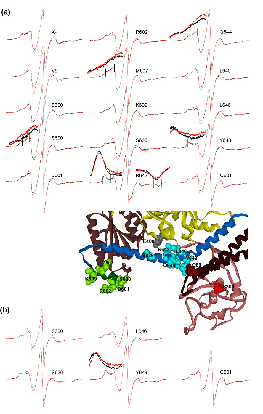
In all living cells, regulated passage across membranes of specific proteins occurs through a universally conserved secretory channel. In bacteria and chloroplasts, the energy for the mechanical work of moving polypeptides through that channel is provided by SecA, a regulated ATPase. Here, we use site-directed spin labeling and electron paramagnetic resonance spectroscopy to identify the interactive surface used by SecA for each of the diverse binding partners encountered during the dynamic cycle of export. Although the binding sites overlap, resolution at the level of aminoacyl side chains allows us to identify contacts that are unique to each partner. Patterns of constraint and mobilization of residues on that interactive surface suggest a conformational change that may underlie the coupling of ATP hydrolysis to precursor translocation.


Figures

Fig. 1
Fig. 1
Constraints and mobilizations. For key to the color scheme see text. (a) Venn Diagrams. Script font indicates residues constrained or mobilized depending on ligand. (b) Constrained sites, displayed as CPK models. (c) View in (b) rotated 180° around the x-axis. (d) Sites that are mobilized by lipids (orange) or by both lipids and SecB (blue-gray) displayed as CPK models. Note, residue K4 is not resolved in this structure. These structures and the subsequent structures, except those in Fig. 3, are PDB code 2FSF with the PBD modeled in, based on the B. subtilis SecA PBD (PDB code 1TF5).
Fig. 2
Fig. 2
Positions on SecA that show no significant change when in complex with any of the binding partners. Black traces, spin-labeled SecA alone; red traces, SecA with binding partner indicated. A subset of residues with the binding partners as indicated was chosen to illustrate the precise overlay of spectra. All spectra were acquired at 6 °C.
Fig. 3
Fig. 3
Comparison of the two conformations of SecA: closed, green, PDB code 1M6N; open, colored by domain, PDB code 1TF5. The PBD is shown in CPK to illustrate the change in accessibility of the nitroxide at position S350 (E. coli residue number). The nitroxide is colored by atom: carbon, gray; oxygen, red; nitrogen, blue; sulfur, yellow.
Fig. 4
Fig. 4
Spectra of constrained residues. Black traces, spin-labeled SecA alone; red traces, spin-labeled SecA in complex with ligand. (a) Binding of polypeptide ligands as indicated. The arrows indicate position of low field hyperfine extrema for residues I642 and G11. Spectra acquired at 6°C. (b) Binding of SecB. Temperature of acquisition indicated. The insets show the region of the spectra, indicated by vertical lines, magnified by a factor of four for both the intensity and the field.
Fig. 5
Fig. 5
Constraints require unfolded ligands. (a) Precursor galactose-binding protein. (b) Mature galactose-binding protein. Left panels: Black traces are SecAI641 alone; red traces are SecAI641 with ligand in 1 mM EGTA which maintains the ligands in an unfolded state. Right panels: black traces are SecA alone and green traces are SecAI641 with the ligands after addition of 3 mM CaCl2 which causes the ligands to partition to a folded state.
Fig. 6
Fig. 6
Spectra of constrained residues. (a) Binding of SecY. (b) Binding of lipid. Traces and insets are as described in Fig. 4.
Fig. 7
Fig. 7
Binding surface for polypeptide ligand. Contact sites identified in this study, CPK models; sites identified by others, ball and stick representations. The view in (b) is related to that in (a) by a 90° rotation about the x-axis away from the viewer. The arrow indicates the position of the groove originally suggested by Osborne and Rapoport as a potential binding site. The asterisk indicates the opening to the large cleft that we show interacts with ligands.
Fig. 8
Fig. 8
Comparison of spin-labeled species of SecA with and without zinc in complex with SecB. Samples containing 6 µM SecA dimer and 6 µM SecB tetramer were subjected to size-exclusion chromatography and the eluent was monitored to determine protein concentration by change in refractive index. Molar mass was determined by total intensity light scatter. The traces represent concentration and the symbols represent molar mass. (a) Complexes with zincless SecA. Samples applied were SecB (gray-green); zincless SecA (red); zincless SecA spin-labeled at S350 (green); SecB and zincless SecA (blue); SecB and zincless SecA spin-labeled at S350 (brown). (b) Complexes with SecA species containing zinc. Samples applied were SecB (gray-green); SecA containing zinc (red); SecA containing zinc labeled at S350 (green); SecB and SecA containing zinc (blue); SecB and SecA containing zinc spin-labeled at S350 (brown).
Fig. 9
Fig. 9
Binding surfaces for SecB on SecA. Views in right panels are related to those in the left by a 90° rotation away from the viewer about the x-axis. In the left panel sidechains of residues 337 through 339, which masked S350 visually, were removed. The central structure illustrates one possible way SecB could bind to SecA and make contacts at the residues which are constrained. The arrow indicates position 146 at which the resolution of the X-ray structure of SecB ends. Nine residues are missing.
Fig. 10
Fig. 10
Stereo view of contact sites on SecA for SecYEG. The CPK models are colored by domain as described in the main text. Spin-labeled residues on SecA showing constraints by interaction with SecYEG are colored light green in the linker helix, bright blue in the helical scaffold domain, red in the precursor binding domain and light brown in the nucleotide binding domains. The views in (a) through (d) are rotated sequentially 90° around the x-axis away from the viewer to display all surfaces. The residues shown in gray are those that showed no change with any binding partner.
Fig. 11
Fig. 11
Spectra of mobilized residues. (a) Lipids. (b) SecB. Mobilized residues are shown as CPK models and colored by domain, except for E400, gray, which is discussed in the text. All spectra were gathered at 27 °C. The insets are as described in Fig. 4.
Fig. 12
Fig. 12
Helical wheel showing disposition of constrained residues. The color scheme is: white, residues not tested; gray, residues that showed no constraint; red, residues constrained in complexes with SecYEG; green, residues constrained in complexes with precursor polypeptides; light blue, residues constrained in complexes with SecB. (a) Residues 636 through 648. The helix is oriented with the long axis from amino to carboxyl terminus pointing into the page (indicated by the X). NBD represents the nucleotide binding folds that are in contact with the HSD. The curved arrow within the wheel indicates the proposed direction of the rolling of the helix. (b) Residues 600 through 609. The helix is oriented with the long axis from amino to carboxyl terminus pointing out toward the reader (indicated by the dot).

References

    1. Papanikou E, Karamanou S, Economou A. Bacterial protein secretion through the translocase nanomachine. Nat. Rev. Microbiol. 2007;5:839–851. - PubMed
    1. Crane JM, Suo Y, Lilly AA, Mao C, Hubbell WL, Randall LL. Sites of interaction of a precursor polypeptide on the export chaperone SecB mapped by site-directed spin labeling. J. Mol. Biol. 2006;363:63–74. - PMC - PubMed
    1. Crane JM, Mao C, Lilly AA, Smith VF, Suo Y, Hubbell WL, Randall LL. Mapping of the docking of SecA onto the chaperone SecB by site-directed spin labeling: insight into the mechanism of ligand transfer during protein export. J. Mol. Biol. 2005;353:295–307. - PubMed
    1. Zimmer J, Li W, Rapoport TA. A novel dimer interface and conformational changes revealed by an X-ray structure of B. subtilis SecA. J. Mol. Biol. 2006;364:259–265. - PubMed
    1. Hunt JF, Weinkauf S, Henry L, Fak JJ, McNicholas P, Oliver DB, Deisenhofer J. Nucleotide control of interdomain interactions in the conformational reaction cycle of SecA. Science. 2002;297:2018–2026. - PubMed

Publication types

MeSH terms

LinkOut - more resources