. 2008 Sep;31(3):442-50.
doi: 10.1016/j.nbd.2008.06.001. Epub 2008 Jun 17.

Aggregation and catabolism of disease-associated intra-Abeta mutations: reduced proteolysis of AbetaA21G by neprilysin

Affiliations

Aggregation and catabolism of disease-associated intra-Abeta mutations: reduced proteolysis of AbetaA21G by neprilysin

Vicki Betts et al. Neurobiol Dis. 2008 Sep.

Abstract

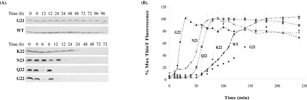
Five point mutations within the amyloid beta-protein (Abeta) sequence of the APP gene are associated with hereditary diseases which are similar or identical to Alzheimer's disease and encode: the A21G (Flemish), E22G (Arctic), E22K (Italian), E22Q (Dutch) and the D23N (Iowa) amino acid substitutions. Although a substantial body of data exists on the effects of these mutations on Abeta production, whether or not intra-Abeta mutations alter degradation and how this relates to their aggregation state remain unclear. Here we report that the E22G, E22Q and the D23N substitutions significantly increase fibril nucleation and extension, whereas the E22K substitution exhibits only an increased rate of extension and the A21G substitution actually causes a decrease in the extension rate. These substantial differences in aggregation together with our observation that aggregated wild type Abeta(1-40) was much less well degraded than monomeric wild type Abeta(1-40), prompted us to assess whether or not disease-associated intra-Abeta mutations alter proteolysis independent of their effects on aggregation. Neprilysin (NEP), insulin degrading enzyme (IDE) and plasmin play a major role in Abeta catabolism, therefore we compared the ability of these enzymes to degrade wild type and mutant monomeric Abeta peptides. Experiments investigating proteolysis revealed that all monomeric peptides are degraded similarly by IDE and plasmin, but that the Flemish peptide was degraded significantly more slowly by NEP than wild type Abeta or any of the other mutant peptides. This finding suggests that resistance to NEP-mediated proteolysis may underlie the pathogenicity associated with the A21G mutation.


Figures

Fig. 1
Fig. 1. Primary sequences of wt and disease-associated mutant Aβ peptides
The sequence of wt Aβ(1–40) is shown in black letters, with residues 21, 22 and 23 denoted in bold and larger type. Single amino acid substitutions corresponding to intra-Aβ mutations found in familial AD-related disorders are shown in grey: the A21G, Flemish; E22Q, Dutch; E22K, Italian; E22G, Arctic; D23N, Iowa. The α-cleavage site which facilitates APP ectodomain shedding and which precludes Aβ formation is indicated with an arrow.
Fig. 2
Fig. 2. Most disease-associated intra-Aβ mutations increase the aggregation kinetics of Aβ peptides
(A) Duplicate samples of Aβ peptides (10 µM) were incubated at 37°C and aggregation measured by loss of peptides from solution following centrifugation at 100,000 g for 1 h. Results shown are typical of three separate experiments. Gels have been cropped to show the region between 3 and 7 kDa, because no higher migrating species were detected. (B) For continuous assessment of aggregation, Aβ peptides were incubated with Thioflavin T in the wells of a 96 well micro-titre plate, shaken at 650 rpm, and fluorescence measurements made at regular intervals. The results shown are representative of two such experiments, in which 6 replicates were used per peptide and are presented as the mean +/− standard error.
Fig. 3
Fig. 3. Aggregated Aβ is degraded less well than monomeric Aβ
Incubation of monomeric wt Aβ(1–40) (20 µM) with IDE, NEP or Plasmin caused a time-dependent loss of peptide, whereas wt Aβ(1–40) levels remained constant when the protease had first been specifically inhibited (I). Aggregated wt Aβ(1–40) was not significantly degraded by IDE, NEP or plasmin. In experiments using active proteases, proteolysis was halted by addition of inhibitors (2 mM 1,10 Phenanthroline, 1 mM PEFA, 5 mM EDTA, 200 µM leupeptin, 1.5 µM aprotinin, 15 µM peptstatin, final concentrations); samples were resolved on 10–20% tris-tricine gels, and the proteins detected by silver stain. As in Fig. 2b gels have been cropped to show the region between 3 and 7 kDa, because no higher migrating species were detected.
Fig. 4
Fig. 4. IDE, NEP and Plasmin readily degrade Aβ peptides bearing disease-associated amino acid substitutions
Monomeric peptide solutions were incubated +/− IDE (A), NEP (B) or Plasmin (C) for 45, 60 and 30 min respectively, protease inhibitors were added and samples electrophoresed on 10–20% tricine gels and peptides detected by silver stain. Upper panels show results from a representative experiment and lower panels show average results +/− SD of three independent experiments (A–C). In a separate set of experiments monomeric peptide solutions were incubated +/− IDE (D), NEP (E) or Plasmin (F) for 45, 60 and 30 min respectively, protease inhibitors added and then chromatographed on a Discovery® C18 HPLC column (Supelco, Bellefonte, PA) column and the change in the parent Aβ peak area measured. The results shown are averages +/− SD of triplicate samples run in a single experiment. The difference in the extent of degradation between the experiments described in panels A–C and D–F is due to variances in enzyme activity, as experiments were carried out on different days, and the amount of enzyme used was determined by protein content and not activity.
Fig. 5
Fig. 5. NEP degrades Aβ(1–40)A21G less well than wt Aβ(1–40)
wt Aβ(1–40) and Flemish A21G peptides (20 µM) were incubated with NEP and degradation monitored over a 105 min interval by either densitometric quantitation of silver stained Aβ bands (A and B) or by HPLC (C). The results shown in (A) are representative of three experiments. The graph in (B) represents the mean ± SD of densitometric analysis of one such experiment in which triplicate samples were used. The data shown in (C) represents the mean ± SD of a typical experiment analysed in duplicate. In (D and E) HPLC chromatograms are shown, and the product peaks labelled a–d have the following elution times a: 11.30; b: 12.25; c: 12.52; d: 13.10 (min). Note the absence of two peaks (a and d) from the NEP digestion of the A21G peptide. Data shown in (D and E) are representative of 3 such experiments. The peaks that elute ~20 and 22 min were also present in the buffer controls. (F and G) Mass spectrometry analyses. Peptides were prepared as described above and adjusted to a concentration of 20 µM. NEP (1/10th volume) was added to the peptide solution and digestion allowed to proceed at 37°C until approximately 50% degradation was achieved (as assessed by SDS-PAGE/silver staining). Reactions were stopped by boiling and samples stored at −80°C until analysed by MALDI-TOF mass spectrometry.

References

    1. Beavis RC, Chait BT. Matrix-assisted laser desorption ionization mass-spectrometry of proteins. Methods Enzymol. 1996;270:519–551. - PubMed
    1. Brooks WS, Kwok JB, Halliday GM, Godbolt AK, Rossor MN, Creasey H, Jones AO, Schofield PR. Hemorrhage is uncommon in new Alzheimer family with Flemish amyloid precursor protein mutation. Neurology. 2004;63:1613–1617. - PubMed
    1. Cacquevel M, Launay S, Castel H, Benchenane K, Cheenne S, Buee L, Moons L, Delacourte A, Carmeliet P, Vivien D. Ageing and amyloid-beta peptide deposition contribute to an impaired brain tissue plasminogen activator activity by different mechanisms. Neurobiol Dis. 2007;27:164–173. - PubMed
    1. Carson JA, Turner AJ. beta-amyloid catabolism: roles for neprilysin (NEP) and other metallopeptidases? Journal of Neurochemistry. 2002;81:1–8. - PubMed
    1. Cheng IH, Palop JJ, Esposito LA, Bien-Ly N, Yan F, Mucke L. Aggressive amyloidosis in mice expressing human amyloid peptides with the Arctic mutation. Nat Med. 2004;10:1190–1192. - PubMed

Publication types

MeSH terms