

Hippocampal hyperperfusion in Alzheimer's disease
- PMID: 18602481
- PMCID: PMC2675915
- DOI: 10.1016/j.neuroimage.2008.06.006
Hippocampal hyperperfusion in Alzheimer's disease
Abstract
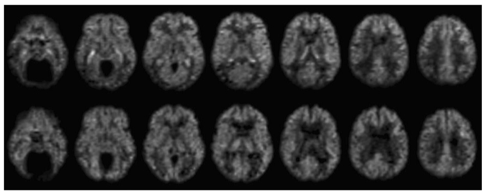
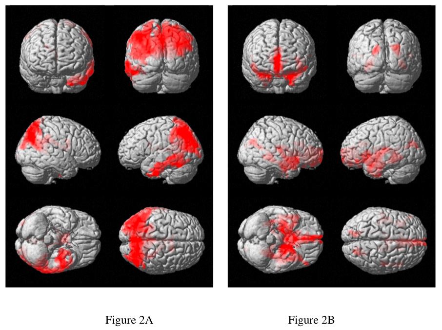
Many of the regions with the earliest atrophy in Alzheimer's Disease (AD) do not show prominent deficits on functional imaging studies of flow or metabolism. This paradox may provide unique insights into the pathophysiology of AD. We sought to examine the relationship between function and atrophy in AD using MRI blood flow and anatomic imaging. 22 subjects diagnosed with AD, mean Mini Mental State Exam (MMSE) score 22.2, and 16 healthy elderly controls were imaged with a volumetric arterial spin labeling blood flow MRI technique and an anatomical imaging method using the identical spatial resolution, image orientation, and spatial encoding strategy. Cerebral blood flow (CBF) and gray matter (GM) maps derived from the imaging were transformed to a standard anatomical space. GM and CBF maps were tested for significant differences between groups. Additionally, images were tested for regions with significant mismatch of the CBF and GM differences between groups. CBF was significantly lower in the bilateral precuneus, parietal association cortex and the left inferior temporal lobe but was non-significantly increased in the hippocampus and other medial temporal structures. After correction for GM loss, CBF was significantly elevated in the hippocampus and other medial temporal structures. The hippocampus and other regions affected early in AD are characterized by elevated atrophy-corrected perfusion per cm(3) of tissue. This suggests compensatory or pathological elevation of neural activity, inflammation, or elevated production of vasodilators.
Figures

References
-
- Alsop DC, Detre JA. Reduced Transit-Time Sensitivity in Non-invasive Magnetic Resonance Imaging of Human Cerebral Blood Flow. J Cereb Blood Flow Metab. 1996;16:1236–1249. - PubMed
-
- Alsop DC, Detre JA. Multisection Cerebral Blood Flow MR Imaging with Continuous Arterial Spin Labeling. Radiology. 1998;208:410–416. - PubMed
-
- Alsop DC, Detre JA, Grossman M. Assessment of cerebral blood flow in Alzheimer's disease by spin-labeled magnetic resonance imaging. Ann Neurol. 2000;47:93–100. - PubMed
-
- Atwood CS, Bowen RL, Smith MA, Perry G. Cerebrovascular requirement for sealant, anti-coagulant and remodeling molecules that allow for the maintenance of vascular integrity and blood supply. Brain Res Brain Res Rev. 2003;43:164–178. - PubMed
-
- Baron JC, Chetelat G, Desgranges B, Perchey G, Landeau B, de la Sayette V, Eustache F. In vivo mapping of gray matter loss with voxel-based morphometry in mild Alzheimer's disease. Neuroimage. 2001;14:298–309. - PubMed
Publication types
MeSH terms
Grants and funding
LinkOut - more resources
Full Text Sources
Medical
