. 2008 Sep;32(9):1044-56.
doi: 10.1016/j.cellbi.2008.06.003. Epub 2008 Jun 17.

Endopolyploidy in irradiated p53-deficient tumour cell lines: persistence of cell division activity in giant cells expressing Aurora-B kinase

Affiliations

Endopolyploidy in irradiated p53-deficient tumour cell lines: persistence of cell division activity in giant cells expressing Aurora-B kinase

Jekaterina Erenpreisa et al. Cell Biol Int. 2008 Sep.

Abstract

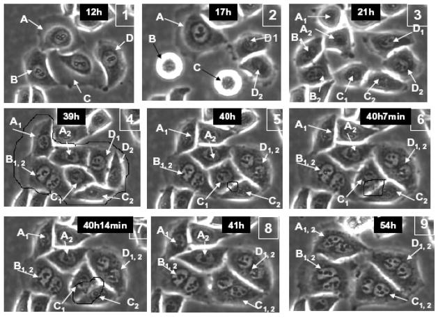
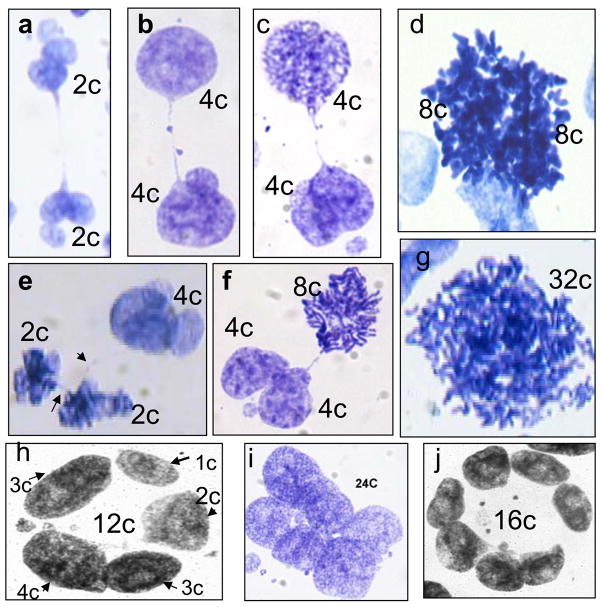
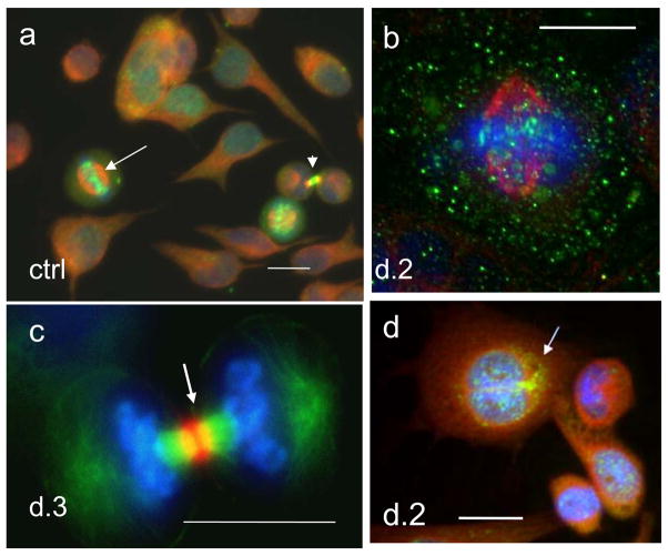
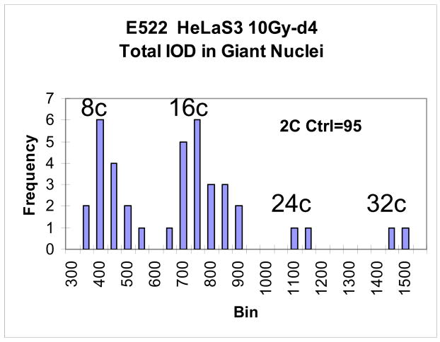
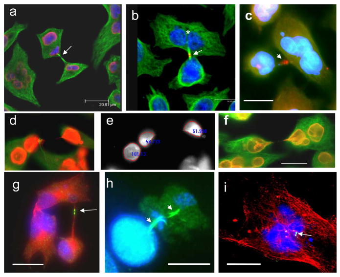
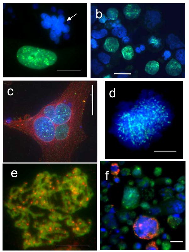
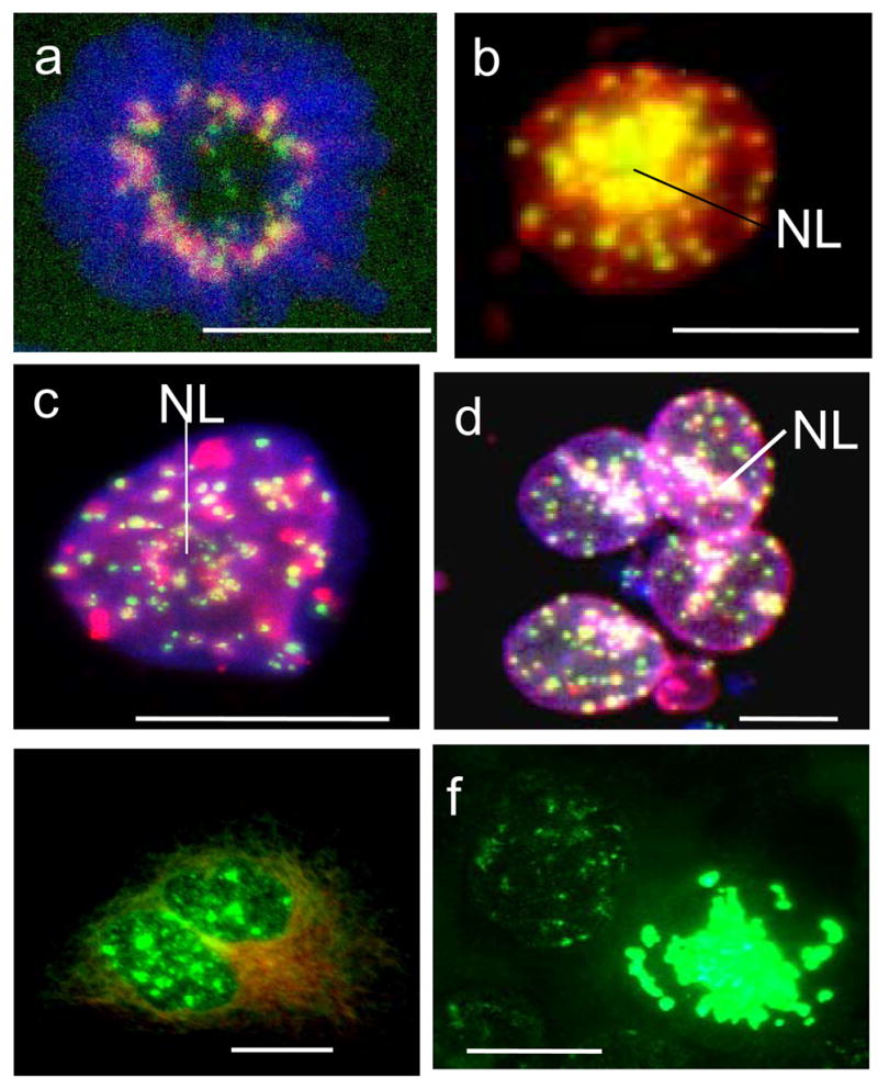
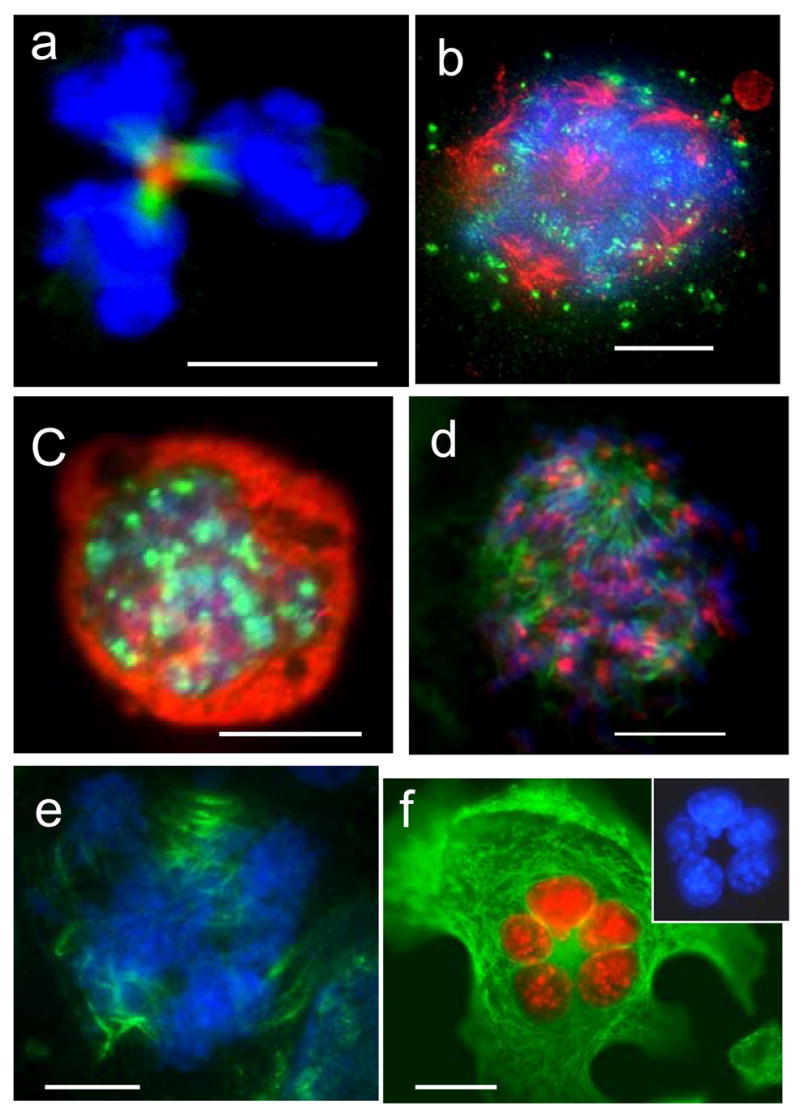
Recent findings including computerised live imaging suggest that polyploidy cells transiently emerging after severe genotoxic stress (and named 'endopolyploid cells') may have a role in tumour regrowth after anti-cancer treatment. Until now, mostly the factors enabling metaphase were studied in them. Here we investigate the mitotic activities and the role of Aurora-B, in view of potential depolyploidisation of these cells, because Aurora-B kinase is responsible for coordination and completion of mitosis. We observed that endopolyploid giant cells are formed via different means in irradiated p53 tumours, by: (1) division/fusion of daughter cells creating early multi-nucleated cells; (2) asynchronous division/fusion of sub-nuclei of these multi-nucleated cells; (3) a series of polyploidising mitoses reverting replicative interphase from aborted metaphase and forming giant cells with a single nucleus; (4) micronucleation of arrested metaphases enclosing genome fragments; or (5) incomplete division in the multi-polar mitoses forming late multi-nucleated giant cells. We also observed that these activities can release para-diploid cells, although infrequently. While apoptosis typically occurs after a substantial delay in these cells, we also found that approximately 2% of the endopolyploid cells evade apoptosis and senescence arrest and continue some form of mitotic activity. We describe here that catalytically active Aurora-B kinase is expressed in the nuclei of many endopolyploid cells in interphase, as well as being present at the centromeres, mitotic spindle and cleavage furrow during their attempted mitotes. The totally micronucleated giant cells (containing sub-genomic fragments in multiple micronuclei) represented only the minor fraction which failed to undergo mitosis, and Aurora-B was absent from it. These observations suggest that most endopolyploid tumour cells are not reproductively inert and that Aurora-B may contribute to the establishment of resistant tumours post-irradiation.


Figures

Fig. 1
Fig. 1
A series of images of irradiated HeLa cells in LINDCAS system starting live-imaging 12 h post-irradiation – four cells (A,B,C, D) divide remaining connected by nuclear bridges and then each daughter pair regresses cleavage furrow forming a bi-nucleated cell.
Fig. 2
Fig. 2
Characteristic patterns of the DNA cytometry images of HeLa cells: e–c,e,f – DNA- bridged karyoplasts arising by bi-polar mitoses in the first three days post-irradiation; h–j – late multinuclear cells on days 6–9 with circularly arranged subnuclei. ‘2c’ DNA content is an arbitrary value which correspond average of 50 anaphase halfs in the control not irradiated cells.
Fig. 3
Fig. 3
Aur B/a-tubulin/DNA cellular localisation in the first mitotic activities of HeLa cells: (a) non-treated control: in metaphase Aur B is equatorially attached to centrosomes (arrow), a mid-body connecting post-telophasic sister cells is arrowheaded (; (b–d) after irradiation: (b) in metaphase; (c) a proper mid-body in late anaphase; d) formation of a bi-nuclear cell enclosing mid-body in its cytoplasm (arrow) – a witness of post-anaphasic or post-mitotic re-union of sister nuclei/cells. For. 3a,b,d - Aurora B green, alfa-tubulin red, DAPI – blue; for 3c – Aurora B is red, alfa-tubulin green, DAPI – blue. Bars=20 μm
Fig. 4
Fig. 4
A histogram of the DNA content in giant HeLa cells on d.4 after irradiation (n=110). It is seen that 8C and 16C cells are in the cell cycle, while 24C and 32C represent apparently the end-point of polyploidisation.
Fig. 5
Fig. 5
Asymmetric bi-polar divisions of sub-nuclei in multinucleated HeLa and Namalwa cells after irradiation: (a–f) day 2; (g–i) days 4–7. (a,d,f) lamin B(red)/a-tubilin (green)/DAPI; (b,c,) Aur B (red)/a-tubulin (green)/DAPI; (g–i) Aur B (green)/a-tubulin (red)/DAPI; (e) DAPI only, fluorescence intensity measured. The presence of midbodies (arrowed) indicates to the mitotic character of these divisions. On (a) telophase chromatin figures are outlined by lamin B; (b)- after mitotic division of a presumably tetraploid HeLa cell, one of the daughter cell nuclei (asterics) has further divided a-mitotically; (c) - a multilobulated HeLa giant cell is joined with a smaller one by a tubulin bridge with remnant midbody; (d–e) is illustrating an equal division of one of the nuclei in a bi-nuclear cell; (g) - a low ploidy HeLa cell is still connected to the high ploidy multinuclear cell by a tubulin bridge with a mid-body. This well shaped mid-body displays a clear central split – the place of the centriolin ring, necessary for the completion of cytokinesis; (h) a sample was collected after 2h-treatment with lactocystin, proteasome inhibitor. On ‘h’ two subsequent asymmetric reduction bi-polar divisions of a giant Namalwa cell are seen displaying two persisting midbodies. These divisions result in a small ana-telophase. Persistence of the two subsequent midbodies and small final mitotic figures suggest that the second division rapidly followed the first, most probably without intermitting S-phase. (i) shows a midbody within a multinucleated cell – a probable result of the failed cytotomy and reverse of asymmetric mitotic division in a HeLa cell. Bars=20 μm.
Fig. 6
Fig. 6
Distribution of Aurora-B kinase in and among irradiated cells: (a) strongly positive MONGC, however always negative micronucleated HeLa cells (arrow); (b) a proportion of cells with Aurora-B-positive nuclei in Namalwa; (c) A positive MNGC of HeLa (green for Aurora, counterstained with red for a-tubulin); (d) thin dotted arrays of aur-B (green) in the chromatin of the nucleus restituting from the arrested polyploid metaphase: fixed 2 hs after application of Mg-132; (e) a rare finding: clear aurora-B dots over minimally condensed preprophasic chromosomes red – for Aur-B, green – a false colour for overexposed DAPI; (f) annexin-positive dying cells (red ) are negative for aurora-B kinase (Namalwa cells). Bars=20 μm.
Fig. 7
Fig. 7
Specific activities of aurora-B kinase in polyploid tumour cells after irradiation: (a) in control, foci of aurora-B (Alexa, red) colocalise with kinetochore proteins (FITC, green) in the metaphase plate (DAPI); (b–f) after irradiation: (b) nearly full colocalisation of aurora-B and kinetochores, especially heavy around central nucleolus (NL) in the interphase cell, presumably G2; DAPI channel is not overlaid; (c) large patches of aurora-B at centromere clusters and chains in a late giant cell; (d) heavy precipitation of aurora-B onto the centromeres around nucleoli (NL), tendency of colocalisation at the nuclear envelope, however absence of colocalisation in most centromeres inside the nuclei; (e,f) IF for substrate of aurora-B phosphorylation phospho-H3ser10(green) in interphase and mitotic giant cells shows catalytical activity of kinase. This activity is very high in the numerous polyploid aberrant mitoses arrested at this stage (on d.4 post-irradiation) in metaphase, one of them is seen on ‘f’. (a–d) sequential scanning by confocal microscope in three colour channels. Bars=20 μm.
Fig. 8
Fig. 8
Multipolar mitoses in irradiated samples: (a) clear central mid-body, aurora B (red) and a-tubulin (green) in a tri-twigged spindle, however telophase nuclei (DAPI) possess very irregular contours; (b) arrested multipolar metaphase: alfa-tubulin revealing poles is red, aurora B (green) is poorly attached to chromosomes (blue), which are not sufficiently condensed and seem polytenised; (c) true endomitosis with condensed chromosomes (DAPI) and richly bound to them aurora-B (green), a-tubulin is red; (d,e) the cells were fixed 2hs after application of the calpain inhibitor: (d) a tri-polar metaphase, chromosomes are well condensed and aurora B (red) is richly attached to them; a-tubulin of the poles is green; (e) three spindles are seen decorated in their central part with the tandemly arranged aurora-B-positive grains (green; tubulins were not stained), while DAPI reveals several anaphase figures; (f) radial arrangement of the equal-sized aurora-B-imunopositive (red) nuclei in the large adherent HeLa cell with active microtubular skeleton (green). On insert – the nuclei in DAPI channel. (a) 2days; (b) 5 days; (c–e) 6 days; (f) 8 days post-irradiation. Bars=20 μm.

Comment in

References

    1. Adams RR, Eckley DM, Vagnarelli P, Wheatley SP, Gerloff DL, Mackay AM, Svingen PA, Kaufmann SH, Earnshaw WC. Human INCENP colocalizes with the Aurora-B/AIRK2 kinase on chromosomes and is overexpressed in tumour cells. Chromosoma. 2001;110:65–74. - PubMed
    1. Andreassen PR, Lacroix FB, Lohez OD, Margolis RL. Neither p21WAF1 nor 14–3–3 sigma prevents G2 progression to mitotic catastrophe in human colon carcinoma cells after DNA damage, but p21WAF1 induces stable G1 arrest in resulting tetraploid cells. Cancer Res. 2001;61:7660–68. - PubMed
    1. Baroja A, de la Hoz C, Alvarez A, Ispizua A, Bilbao J, de Gandarias JM. Genesis and evolution of high-ploidy tumour cells evaluated by means of the proliferation markers p34(cdc2), cyclin B1, PCNA and 3[H]-thymidine. Cell Prolif. 1996;29:89–100. - PubMed
    1. Baroja A, de la Hoz C, Alvarez A, Vielba R, Sarrat R, Aréchaga J, de Gandarias JM. Polyploidization and exit from cell cycle as mechanisms of cultured melanoma cell resistance to methotrexate. Life Sci. 1998;62:2275–82. - PubMed
    1. Carmena M, Earnshaw WC. The cellular geography of aurora kinases. Nature Rev Mol Cell Biol. 2003;4:842–54. - PubMed

Publication types

MeSH terms