

Mechanisms for suppression of interleukin-6 expression in peritoneal macrophages from docosahexaenoic acid-fed mice
- PMID: 18602807
- PMCID: PMC2727136
- DOI: 10.1016/j.jnutbio.2008.04.006
Mechanisms for suppression of interleukin-6 expression in peritoneal macrophages from docosahexaenoic acid-fed mice
Abstract
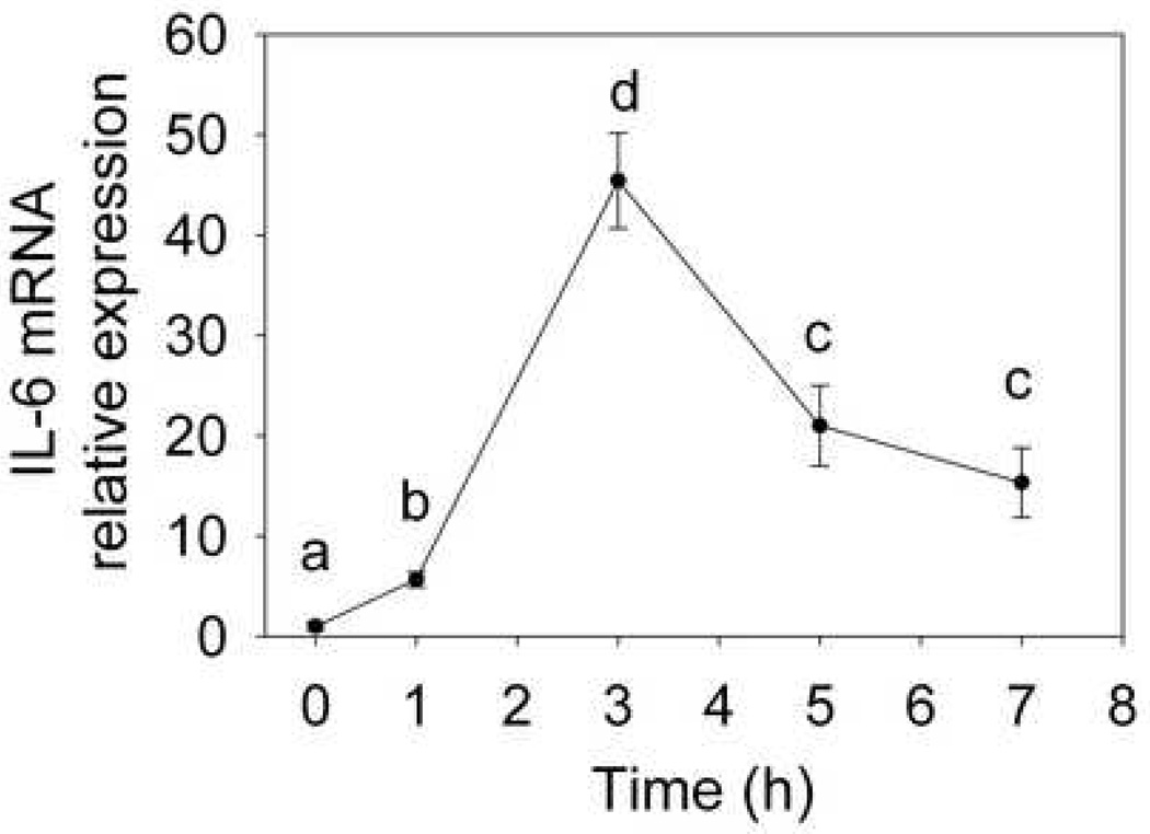
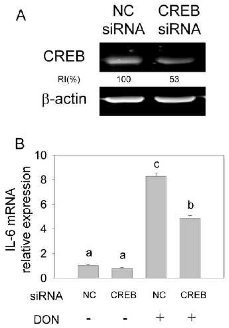
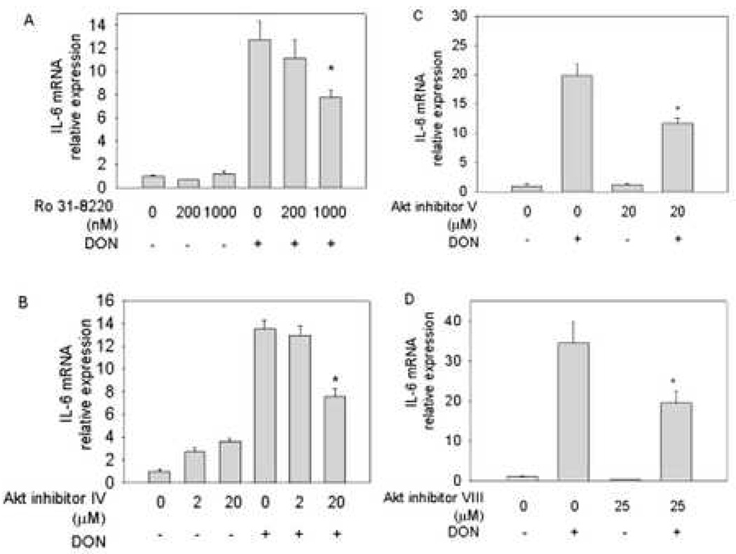
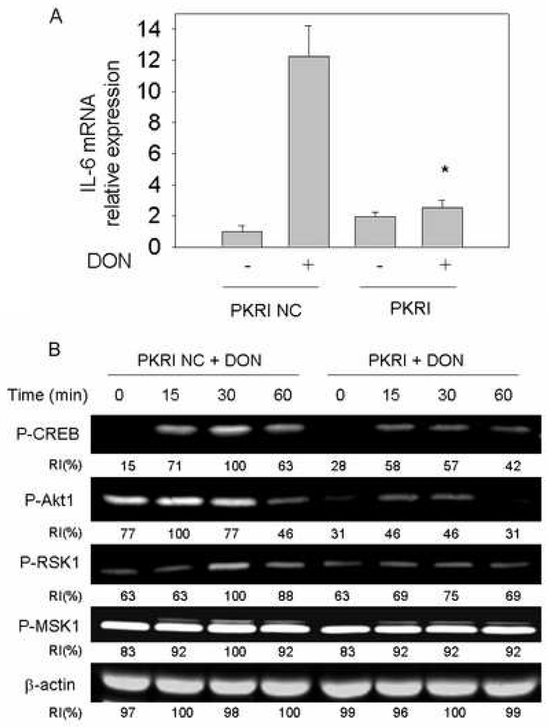
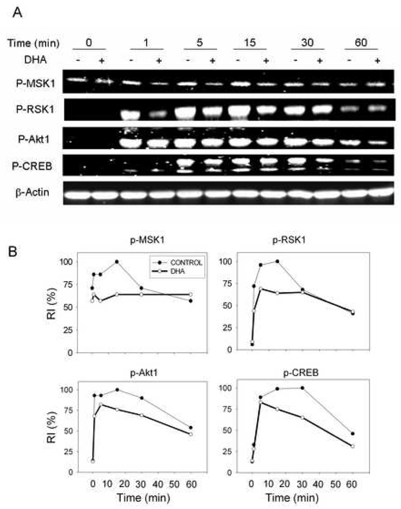
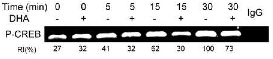
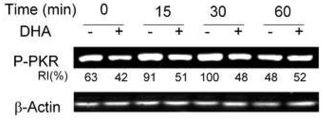
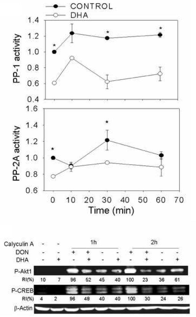
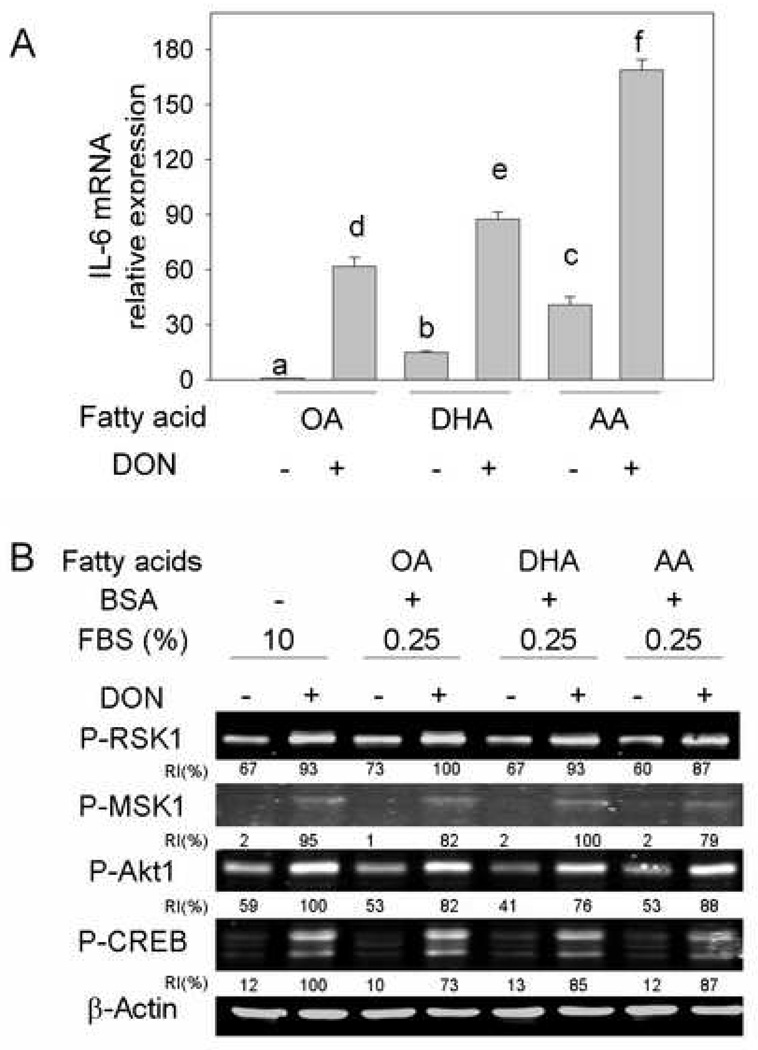
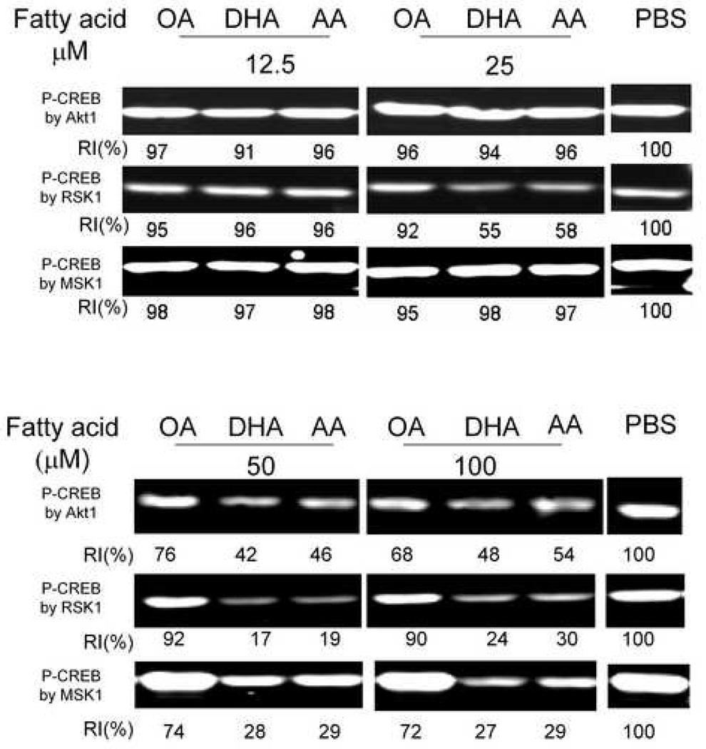
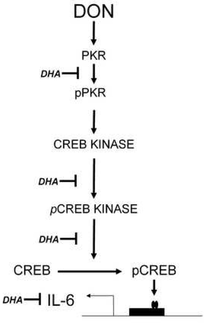
Consumption of the trichothecene mycotoxin deoxynivalenol (DON) induces interleukin-6 (IL-6)-dependent IgA nephropathy (IgAN) in mice. This effect can be prevented by feeding long-chain n-3 polyunsaturated fatty acids (PUFAs) found in fish oil. The purpose of this study was to identify the signal transduction pathways by which DON up-regulates IL-6 in the peritoneal macrophage and how consumption of fish oil enriched with the n-3 PUFA docosahexaenoic acid (DHA) suppresses these processes. Incubation with DON induced IL-6 expression in naïve macrophages maximally at 3 h. Knockdown of the transcription factor cAMP response element-binding protein (CREB) or pharmacologic inhibition of the CREB kinases Akt1/2, MSK1 and RSK1 down-regulated this expression. Inhibition of double-stranded RNA-activated protein kinase (PKR) suppressed not only IL-6 expression but also phosphorylation of CREB and its upstream kinases, Akt1, MSK1 and RSK1. Phosphorylations of PKR, CREB kinases and CREB were markedly impaired in peritoneal macrophages isolated from mice that consumed DHA-enriched fish oil for 6 to 8 weeks. DHA's effects were not explainable by increased activity of protein phosphatase 1 and 2A since both were suppressed in mice consuming the DHA diet. Although cells cultured directly with DHA expressed less IL-6 compared to cells cultured with arachidonic acid (AA), neither fatty acid treatment affected DON-induced protein phosphorylation. Furthermore, DHA and AA similarly inhibited cell-free protein kinase activity. These data suggest that DON-induced IL-6 expression is CREB mediated and PKR dependent, and that requisite kinase activities for these pathways were suppressed in macrophages from mice fed DHA for an extended period.
Figures
Similar articles
-
Docosahexaenoic acid consumption inhibits deoxynivalenol-induced CREB/ATF1 activation and IL-6 gene transcription in mouse macrophages.J Nutr. 2006 Feb;136(2):366-72. doi: 10.1093/jn/136.2.366. J Nutr. 2006. PMID: 16424113
-
Deoxynivalenol-induced mitogen-activated protein kinase phosphorylation and IL-6 expression in mice suppressed by fish oil.J Nutr Biochem. 2003 Dec;14(12):717-26. doi: 10.1016/j.jnutbio.2003.08.009. J Nutr Biochem. 2003. PMID: 14690764
-
Docosahexaenoic acid and eicosapentaenoic acid, but not alpha-linolenic acid, suppress deoxynivalenol-induced experimental IgA nephropathy in mice.J Nutr. 2004 Jun;134(6):1353-61. doi: 10.1093/jn/134.6.1353. J Nutr. 2004. PMID: 15173396
-
Docosahexaenoic acid attenuates mycotoxin-induced immunoglobulin a nephropathy, interleukin-6 transcription, and mitogen-activated protein kinase phosphorylation in mice.J Nutr. 2004 Dec;134(12):3343-9. doi: 10.1093/jn/134.12.3343. J Nutr. 2004. PMID: 15570035
-
Attenuation of mycotoxin-induced IgA nephropathy by eicosapentaenoic acid in the mouse: dose response and relation to IL-6 expression.J Nutr Biochem. 2006 Oct;17(10):697-706. doi: 10.1016/j.jnutbio.2005.12.002. Epub 2006 Jan 9. J Nutr Biochem. 2006. PMID: 16524712
Cited by
-
Body composition and hormonal effects following exposure to mycotoxin deoxynivalenol in the high-fat diet-induced obese mouse.Mol Nutr Food Res. 2011 Jul;55(7):1070-8. doi: 10.1002/mnfr.201000640. Epub 2011 May 2. Mol Nutr Food Res. 2011. PMID: 21538849 Free PMC article.
-
Dynamic changes in ribosome-associated proteome and phosphoproteome during deoxynivalenol-induced translation inhibition and ribotoxic stress.Toxicol Sci. 2014 Mar;138(1):217-33. doi: 10.1093/toxsci/kft270. Epub 2013 Nov 27. Toxicol Sci. 2014. PMID: 24284785 Free PMC article.
-
Oral administration of oleic or linoleic acids modulates the production of inflammatory mediators by rat macrophages.Lipids. 2012 Aug;47(8):803-12. doi: 10.1007/s11745-012-3687-9. Epub 2012 Jun 14. Lipids. 2012. PMID: 22695743
-
Role of GRP78/BiP degradation and ER stress in deoxynivalenol-induced interleukin-6 upregulation in the macrophage.Toxicol Sci. 2009 Jun;109(2):247-55. doi: 10.1093/toxsci/kfp060. Epub 2009 Mar 31. Toxicol Sci. 2009. PMID: 19336499 Free PMC article.
-
The antiatherogenic, renal protective and immunomodulatory effects of purslane, pumpkin and flax seeds on hypercholesterolemic rats.N Am J Med Sci. 2011 Sep;3(9):411-7. doi: 10.4297/najms.2011.3351. N Am J Med Sci. 2011. PMID: 22362450 Free PMC article.
References
-
- Pestka JJ, Zhou HR, Moon Y, Chung YJ. Cellular and molecular mechanisms for immune modulation by deoxynivalenol and other trichothecenes: unraveling a paradox. Toxicol Lett. 2004;153:61–73. - PubMed
-
- Pestka JJ. Deoxynivalenol-induced IgA production and IgA nephropathy-aberrant mucosal immune response with systemic repercussions. Toxicol Lett. 2003;140–141:287–295. - PubMed
-
- Pestka JJ, Moorman MA, Warner RL. Dysregulation of IgA production and IgA nephropathy induced by the trichothecene vomitoxin. Food Chem Toxicol. 1989;27:361–368. - PubMed
-
- Yan D, Zhou HR, Brooks KH, Pestka JJ. Potential role for IL-5 and IL-6 in enhanced IgA secretion by Peyer's patch cells isolated from mice acutely exposed to vomitoxin. Toxicol. 1997;122:145–158. - PubMed
-
- Pestka JJ, Zhou HR. Interleukin-6-deficient mice refractory to IgA dysregulation but not anorexia induction by vomitoxin (deoxynivalenol) ingestion. Food Chem Toxicol. 2000;38:565–575. - PubMed
Publication types
MeSH terms
Substances
Grants and funding
LinkOut - more resources
Full Text Sources
Miscellaneous
