. 2008 Sep;87(3):226-41.
doi: 10.1016/j.exer.2008.06.003. Epub 2008 Jun 17.

PEDF-deficient mice exhibit an enhanced rate of retinal vascular expansion and are more sensitive to hyperoxia-mediated vessel obliteration

Affiliations

PEDF-deficient mice exhibit an enhanced rate of retinal vascular expansion and are more sensitive to hyperoxia-mediated vessel obliteration

Qiong Huang et al. Exp Eye Res. 2008 Sep.

Abstract

Pigment epithelium derived factor (PEDF) is an endogenous inhibitor of angiogenesis. However, its physiological role during vascular development and neovascularization remains elusive. Here we investigated the role of PEDF in normal postnatal vascularization of retina and retinal neovascularization during oxygen-induced ischemic retinopathy (OIR) using PEDF-deficient (PEDF-/-) mice. The beta-galactosidase staining of eye sections from PEDF-/- mice confirmed the expression pattern of endogenous PEDF previously reported in mouse retina. However, strongest staining was observed in the retinal outer plexiform layer. Retinal trypsin digests indicated that the ratio of endothelial cells (EC) to pericytes (PC) was significantly higher in PEDF-/- mice compared to wild type (PEDF+/+) mice at postnatal day 21 (P21). This was mainly attributed to increased numbers of EC in the absence of PEDF. There was no significant difference in the number of PC. We observed an increased rate of proliferation in retinal vasculature of PEDF-/- mice, which was somewhat compensated for by an increase in the rate of apoptosis. Staining of the retinal wholemounts and eye frozen sections indicated postnatal retinal vascularization expansion occurred at a faster rate in the absence of PEDF, and was more prominent at early time points (prior to P21). The retinal vascularization in PEDF+/+ mice reaches that of PEDF-/- mice such that no significant difference in vascular densities was observed by P42. Lack of PEDF had a minimal effect on the regression of hyaloid vasculature and VEGF levels. PEDF-/- mice also exhibited enhanced sensitivity to hyperoxia-mediated vessel obliteration during OIR compared to PEDF+/+ mice despite higher levels of VEGF. However, there was no significant difference in the degree of retinal neovascularization. Our studies indicate that PEDF is an important modulator of early postnatal retinal vascularization and in its absence retinal vascularization proceeds at a faster rate and is more susceptible to hyperoxia-mediated vessel obliteration.


Figures
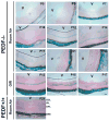
Fig. 1
Fig. 1
The spatial and temporal pattern of PEDF expression in the eye. Frozen eye sections prepared from PEDFminus;/minus; mice, at indicated time points, were stained for β-galactosidase activity. Eyes from PEDF+/+ mice were used as negative control. Please note strong staining in the outer plexiform layer of eyes from P14 mice and, increased staining upon exposure to hyperoxia in P12 mice. These experiments were repeated with eyes from 3 different mice with similar results. A representative image is shown. V: vitreous; Bar = 100 μm. GCL: ganglion cell layer; IPL: inner plexiform layer; OPL: outer plexiform layer; and RPE: retinal pigment epithelium.
Fig. 2
Fig. 2
PEDFminus;/minus; mice exhibit increased retinal vascular density during normal postnatal retinal vascularization. Mice retinal vasculature was prepared by trypsin digestion of retinal wholemounts. Retinas were obtained from P21 PEDF+/+ (A, C) and PEDFminus;/minus; (B, D) mice. Lower panels (C and D; x400) are higher magnification of upper panels (A and B; x100). The arrows indicate endothelial cells, while arrowheads show pericytes. Please note the increased densities of capillaries (see Tables 1–3) in retinas from PEDFminus;/minus; mice. Bars, A and B=100 μm; C and D=20 μm.
Fig. 3
Fig. 3
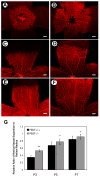
The primary retinal vasculature expands at a faster-rate in PEDFminus;/minus; mice. Collagen IV staining of the retinal wholemounts prepared from PEDF+/+ (A, C, E) or PEDFminus;/minus; (B, D, F) mice at postnatal day 3 (P3) (A, B), P5 (C, D) and P7 (E, F). The quantitative assessment of the data is shown in G. Please note that the spreading of retinal vessels is significantly faster in PEDFminus;/minus;mice compared to PEDF+/+ mice (** P < 0.01; n=10, * P < 0.05; n=10). Bar = 200 μm.
Fig. 4
Fig. 4
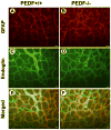
Similar astrocytic organization during retinal vascularization in PEDF+/+ and PEDFminus;/minus; mice. GFAP (astrocytes, red; A and B) and endoglin (EC, green; C and D) staining of the retinal wholemounts prepared from P7 PEDF+/+ (A, C, E) or PEDFminus;/minus; (B, D, F) mice. The merged images are shown in panels E and F. Please note similar astrocytic and EC organization in these mice. Similar astrocytic organizations were observed in areas not covered by EC (not shown). These experiments were repeated with eyes from 5 different mice with similar results. A representative image is shown. Bar=20 μm.
Fig. 5
Fig. 5
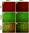
Similar organization of pericytes during retinal vascularization of PEDF+/+ and PEDFminus;/minus; mice. NG2 (PC, red; A and B) and endoglin (EC, green; C and D) staining of the retinal wholemounts prepared from P7 PEDF+/+ (A, C, E) or PEDFminus;/minus; (B, D, F) mice. The merged images are shown in panels E and F. Please note similar PC and EC organization in these mice. These experiments were repeated with eyes from 5 different mice with similar results. A representative image is shown. Bar = 20 μm.
Fig. 6
Fig. 6
Organization of the deep retinal vascular plexuses. Collagen IV staining of the frozen eye sections from P10 PEDF+/+ (A) and PEDF minus;/minus; (B) mice are shown. The quantitative assessment of the data is shown in C. Please note significantly more intermediate retinal vessels are formed in PEDFminus;/minus; mice (arrow) compared to PEDF+/+ mice (*P<0.01; n=10). Bar = 50 μm.
Fig. 7
Fig. 7
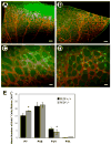
Enhanced retinal vascular cell proliferation in PEDFminus;/minus; mice. BrdU incorporation of retinal vascular cells was used to determine the rates of proliferating cells in the postnatal developing retinal vasculature from PEDF+/+ and PEDFminus;/minus; mice at indicated postnatal days. A and B show retinas from P7 PEDF+/+ and PEDF minus;/minus; mice, respectively. C and D are higher magnification of above images. The quantitative assessment of the data is shown in E (*P < 0.05; n=10). Bar = 100 μm (A and B) and 20 μm (C and D).
Fig. 8
Fig. 8
Enhanced retinal vascular cell apoptosis in PEDFminus;/minus; mice. TUNEL staining was used to assess the rate of apoptotic vascular cells in the trypsin digested retinal vasculature from PEDF+/+ and PEDFminus;/minus; mice at indicated postnatal days. A and B show retinas from PEDF+/+ and PEDF minus;/minus; mice at postnatal day 10 (P10), respectively. C and D are merged images following Hoechst 33258 staining. The quantitative assessment of the data is shown in E (** P < 0.01, n=8; * P < 0.05, n=8). Bar = 20 μm.
Fig. 9
Fig. 9
Assessment of hyaloid vasculature in PEDF+/+ and PEDF minus;/minus; mice. Collagen IV wholemount staining of hyaloid vasculature from PEDF+/+ (A, C, E) and PEDF minus;/minus; mice (B, D, F) at P10 (A, B), P21 (C, D) and P42 (E, F). Please note similar degrees of vessel regression in PEDF+/+ and PEDFminus;/minus; mice at all time points. These experiments were repeated with eyes from 5 different mice with similar results. A representative image is shown. Bar = 500 μm.
Fig. 10
Fig. 10
PEDFminus;/minus; mice are more sensitive to hyperoxia-mediated vessel obliteration during OIR. P7 PEDF+/+ and PEDFminus;/minus; mice were exposed to hyperoxia (75% oxygen) for 5 days. A and B show the wholemount collagen IV staining of retinal vasculature from P12 PEDF+/+ and PEDFminus;/minus; mice during OIR, respectively. The quantitative assessment of the non-perfused area is shown in C (*P < 0.05, n=12). Bar = 500 μm.
Fig. 11
Fig. 11
Preretinal neovascularization is not impacted by the lack of PEDF. A and B show the wholemount collagen IV staining of retinal vasculature from P17 PEDF+/+ and PEDFminus;/minus; mice during OIR, respectively. The number of neovascular cell nuclei projecting into the vitreous from the retina was determined from ocular serial sections as described in Methods and shown in C. The differences were not significant (P> 0.05, n=16). Bar = 500 μm.
Fig. 12
Fig. 12
Assessment of VEGF levels in PEDF+/+ and PEDFminus;/minus; mice. Eye extracts prepared from P15 PEDF+/+ and PEDF minus;/minus; mice (5 days of hyperoxia and 3 days of normoxia) were analyzed by SDS-PAGE and Western blotting as described in Methods. A representative blot is shown (A). Please note similar levels of VEGF in eye extracts from PEDF+/+ and PEDFminus;/minus; mice. β-actin was used for loading control. B shows VEGF levels in retina extracts prepared from PEDF+/+ and PEDFminus;/minus; mice at indicated postnatal days. The level of VEGF was determined using a mouse VEGF immunoassay as described in Methods. C shows VEGF levels in retinal extracts prepared from PEDF+/+ and PEDFminus;/minus; mice at indicated postnatal days during OIR; (P12, 5 days of hyperoxia; P15, 5 days of hyperoxia and 3 days of normoxia; P21, 5 days of hyperoxia and 9 days of normoxia). The differences were not significant in room air and P15 OIR (P> 0.05; n=5), but significant differences were observed at P12 and P21 during OIR (*P<0.05; n=5).
Fig. 13
Fig. 13
Histological and functional analysis of retinas from PEDF+/+ and PEDFminus;/minus; mice. A and B show the PAS/HE staining of retinas from P21 PEDF+/+ and PEDFminus;/minus; mice, respectively. The ganglion cells, inner nuclear cell layer and photoreceptors of the PEDF+/+ eyes are histologically similar to that of PEDFminus;/minus; retinas. C shows dark-adapted ERGs from P42 PEDF+/+ and PEDFminus;/minus; mice. Traces show representative ERGs to a series of flash intensities from 0.5 cd-s m−2 to 5.0 cd-s m−2. Both PEDF+/+ and PEDFminus;/minus; mice exhibited normal growth of the a-wave amplitude and decrease in implicit time with flash intensity. Normal amplitude oscillatory potentials on the ascending limb of the b-wave are evident in PEDF+/+ and PEDFminus;/minus; mice. These experiments were repeated with eyes from 5 different mice with similar results. A representative image is shown. Bar = 50 μm.

References

    1. Amano S, Yamagishi S, Inagaki Y, Nakamura K, Takeuchi M, Inoue H, Imaizumi T. Pigment epithelium-derived factor inhibits oxidative stress-induced apoptosis and dysfunction of cultured retinal pericytes. Microvasc Res. 2005;69(1–2):45–55. - PubMed
    1. Becerra SP. Structure-function studies on PEDF. A noninhibitory serpin with neurotrophic activity. Adv Exp Med Biol. 1997;425:223–237. - PubMed
    1. Becerra SP, Sagasti A, Spinella P, Notario V. Pigment epithelium-derived factor behaves like a noninhibitory serpin. Neurotrophic activity does not require the serpin reactive loop. J Biol Chem. 1995;270(43):25992–25999. - PubMed
    1. Behling KC, Surace EM, Bennett J. Pigment epithelium-derived factor expression in the developing mouse eye. Mol Vis. 2002;8:449–454. - PubMed
    1. Bhutto IA, Kim SY, McLeod DS, Merges C, Fukai N, Olsen BR, Lutty GA. Localization of collagen XVIII and the endostatin portion of collagen XVIII in aged human control eyes and eyes with age-related macular degeneration. Invest Ophthalmol Vis Sci. 2004;45(5):1544–1552. - PubMed

Publication types

MeSH terms

Substances