

Angiogenic laminin-derived peptides stimulate wound healing
- PMID: 18603014
- PMCID: PMC2707597
- DOI: 10.1016/j.biocel.2008.05.025
Angiogenic laminin-derived peptides stimulate wound healing
Abstract
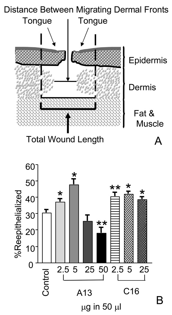
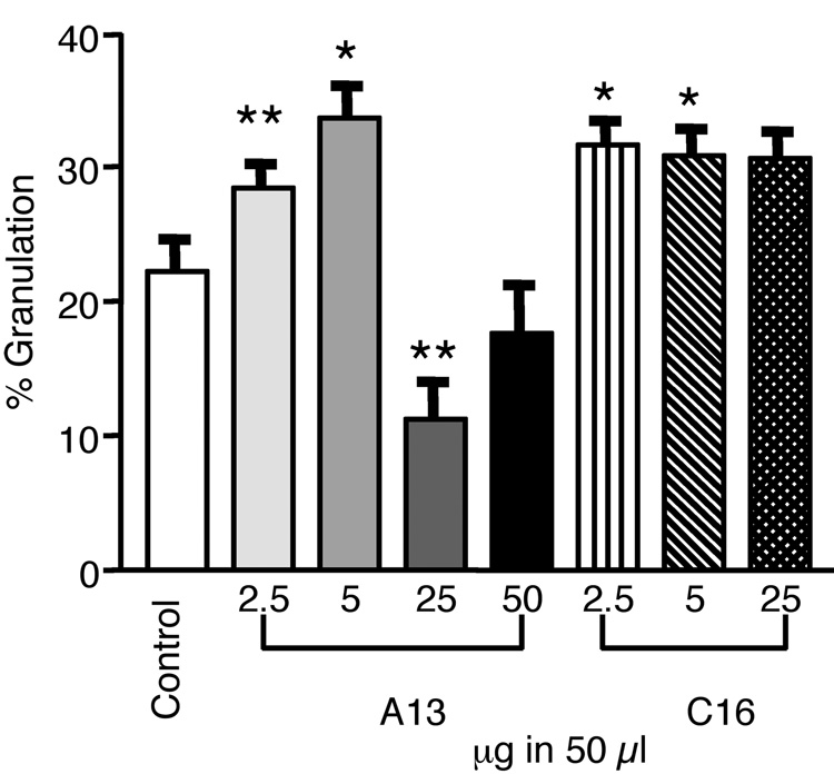
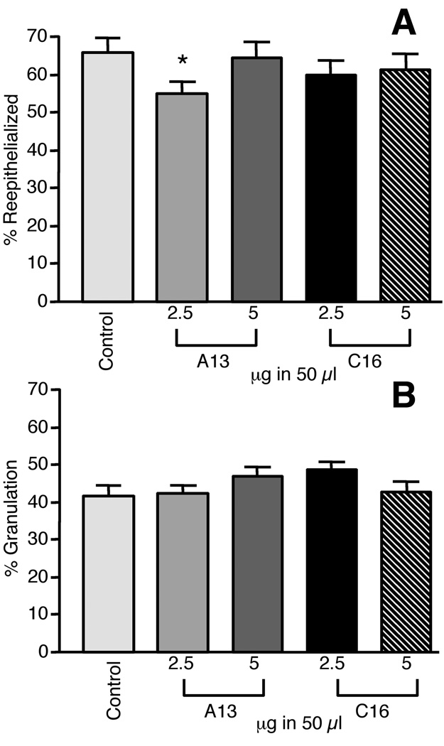
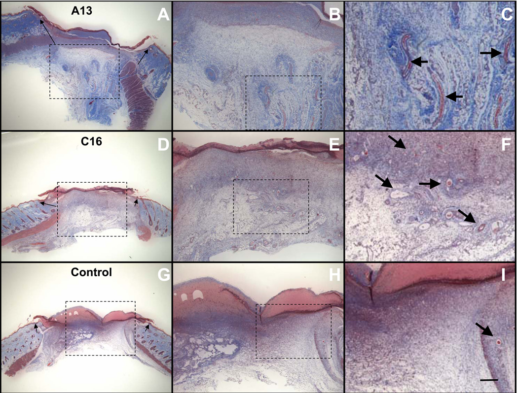
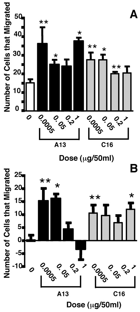
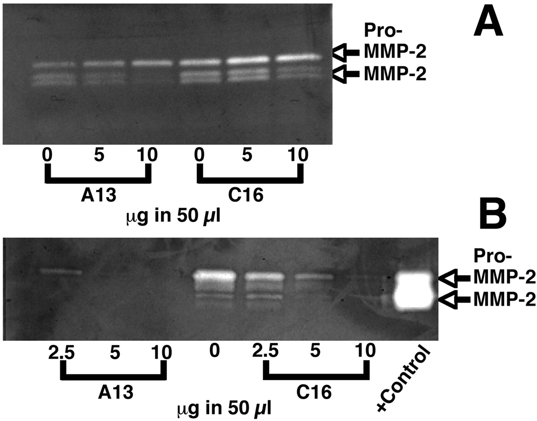
Acceleration of the wound healing process by using angiogenic peptides has been demonstrated previously. Here we used select laminin-111 peptides, A13 and C16, from the laminin alpha1 and gamma1 chain, respectively, to test whether they are able to stimulate wound healing in a rat full thickness wound model. The 12-mer peptides C16 and A13 are highly angiogenic and bind to integrins alphavbeta3 and alpha5beta1. We show that A13 increases wound re-epithelialization as much as 17% over controls by day 4 and C16 increases coverage by 11%. Contraction of the treated wounds was increased as much as 11% for A13 and 8% for C16 at day 4. No differences were observed at day 7 with either peptide. The peptides also stimulated fibroblast migration in Boyden chamber assays. A13 increased cell migration as much as 2.4-fold on uncoated filters and as much as 16-fold on collagen type IV-coated filters over negative controls. Similarly, C16 also stimulated migration 1.8-fold on uncoated filters and as much as 12-fold on collagen-coated filters. A13 and C16 significantly decreased expression of the pro and active forms of matrix metalloproteinase 2 in foreskin fibroblasts indicating their role in collagen accumulation. We conclude that small bioactive angiogenic peptides can promote dermal wound healing and may offer a new class of stable and chemically manipulable therapeutics for wound healing.
Figures
Similar articles
-
Identification of redundant angiogenic sites in laminin alpha1 and gamma1 chains.Exp Cell Res. 2003 May 1;285(2):189-95. doi: 10.1016/s0014-4827(03)00056-9. Exp Cell Res. 2003. PMID: 12706114
-
Laminin mimetic angiogenic and collagen peptide hydrogel for enhance dermal wound healing.Biomater Adv. 2024 Apr;158:213761. doi: 10.1016/j.bioadv.2024.213761. Epub 2024 Jan 17. Biomater Adv. 2024. PMID: 38281321
-
An angiogenic laminin site and its antagonist bind through the alpha(v)beta3 and alpha5beta1 integrins.FASEB J. 2001 Jun;15(8):1389-97. doi: 10.1096/fj.00-0736com. FASEB J. 2001. PMID: 11387236
-
Thrombin peptide, TP508, stimulates angiogenic responses in animal models of dermal wound healing, in chick chorioallantoic membranes, and in cultured human aortic and microvascular endothelial cells.Gen Pharmacol. 2000 Nov;35(5):249-54. doi: 10.1016/s0306-3623(01)00118-5. Gen Pharmacol. 2000. PMID: 11888680 Review.
-
Angiogenesis in wound repair: angiogenic growth factors and the extracellular matrix.Microsc Res Tech. 2003 Jan 1;60(1):107-14. doi: 10.1002/jemt.10249. Microsc Res Tech. 2003. PMID: 12500267 Review.
Cited by
-
Human platelet-rich plasma- and extracellular matrix-derived peptides promote impaired cutaneous wound healing in vivo.PLoS One. 2012;7(2):e32146. doi: 10.1371/journal.pone.0032146. Epub 2012 Feb 23. PLoS One. 2012. PMID: 22384158 Free PMC article.
-
Propolis modulates vitronectin, laminin, and heparan sulfate/heparin expression during experimental burn healing.J Zhejiang Univ Sci B. 2012 Nov;13(11):932-41. doi: 10.1631/jzus.B1100310. J Zhejiang Univ Sci B. 2012. PMID: 23125086 Free PMC article.
-
A Novel Collagen Matricryptin Reduces Left Ventricular Dilation Post-Myocardial Infarction by Promoting Scar Formation and Angiogenesis.J Am Coll Cardiol. 2015 Sep 22;66(12):1364-74. doi: 10.1016/j.jacc.2015.07.035. J Am Coll Cardiol. 2015. PMID: 26383724 Free PMC article.
-
Host matrix modulation by tumor exosomes promotes motility and invasiveness.Neoplasia. 2013 Aug;15(8):875-87. doi: 10.1593/neo.13786. Neoplasia. 2013. PMID: 23908589 Free PMC article.
-
Laminin-111: a potential therapeutic agent for Duchenne muscular dystrophy.Mol Ther. 2010 Dec;18(12):2155-63. doi: 10.1038/mt.2010.165. Epub 2010 Aug 3. Mol Ther. 2010. PMID: 20683444 Free PMC article.
References
-
- Amano S, Akutsu N, Ogura Y, Nishiyama T. Increase of laminin 5 synthesis in human keratinocytes by acute wound fluid, inflammatory cytokines and growth factors, and lysophospholipids. Br J Dermatol. 2004;151:961–970. - PubMed
-
- Aumailley M, Bruckner-Tuderman L, Carter WG, Deutzmann R, Edgar D, Ekblom P, et al. A simplified laminin nomenclature. Matrix Biol. 2005;24:326–332. - PubMed
-
- Bhartiya D, Sklarsh JW, Maheshwari RK. Enhanced wound healing in animal models by interferon and an interferon inducer. J Cell Physiol. 1992;150:312–319. - PubMed
-
- Blinova MI, Paramonov BA, Kukhareva LV, Gorelik Iu V, Nikitina Iu M, Voronkina IV. [Effect of fibroblasts, collagen, and laminin on the healing of wounds formed after dissection of split skin flaps in rats] Biull Eksp Biol Med. 1997;124:229–232. - PubMed
-
- Chan LS. Human skin basement membrane in health and in autoimmune diseases. Front Biosci. 1997;2:d343–d352. - PubMed
MeSH terms
Substances
Grants and funding
LinkOut - more resources
Full Text Sources
Other Literature Sources
Miscellaneous
