Review
. 2008 Aug;45(2):128-47.
doi: 10.1016/j.yjmcc.2008.05.014. Epub 2008 Jun 15.

Emerging roles of inositol 1,4,5-trisphosphate signaling in cardiac myocytes

Affiliations
Review

Emerging roles of inositol 1,4,5-trisphosphate signaling in cardiac myocytes

Jens Kockskämper et al. J Mol Cell Cardiol. 2008 Aug.

Abstract

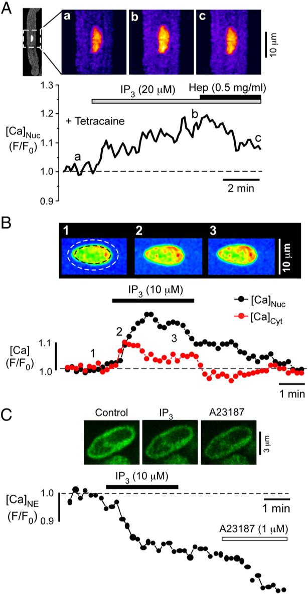
Inositol 1,4,5-trisphosphate (IP(3)) is a ubiquitous intracellular messenger regulating diverse functions in almost all mammalian cell types. It is generated by membrane receptors that couple to phospholipase C (PLC), an enzyme which liberates IP(3) from phosphatidylinositol 4,5-bisphosphate (PIP(2)). The major action of IP(3), which is hydrophilic and thus translocates from the membrane into the cytoplasm, is to induce Ca(2+) release from endogenous stores through IP(3) receptors (IP(3)Rs). Cardiac excitation-contraction coupling relies largely on ryanodine receptor (RyR)-induced Ca(2+) release from the sarcoplasmic reticulum. Myocytes express a significantly larger number of RyRs compared to IP(3)Rs (~100:1), and furthermore they experience substantial fluxes of Ca(2+) with each heartbeat. Therefore, the role of IP(3) and IP(3)-mediated Ca(2+) signaling in cardiac myocytes has long been enigmatic. Recent evidence, however, indicates that despite their paucity cardiac IP(3)Rs may play crucial roles in regulating diverse cardiac functions. Strategic localization of IP(3)Rs in cytoplasmic compartments and the nucleus enables them to participate in subsarcolemmal, bulk cytoplasmic and nuclear Ca(2+) signaling in embryonic stem cell-derived and neonatal cardiomyocytes, and in adult cardiac myocytes from the atria and ventricles. Intriguingly, expression of both IP(3)Rs and membrane receptors that couple to PLC/IP(3) signaling is altered in cardiac disease such as atrial fibrillation or heart failure, suggesting the involvement of IP(3) signaling in the pathology of these diseases. Thus, IP(3) exerts important physiological and pathological functions in the heart, ranging from the regulation of pacemaking, excitation-contraction and excitation-transcription coupling to the initiation and/or progression of arrhythmias, hypertrophy and heart failure.


Figures

Fig. 1
Fig. 1
Elementary IP3-induced Ca2+ release events in cardiac myocytes. (A) Confocal linescan image of Ca2+ sparks in a permeabilized cat atrial myocyte (control). Tetracaine (Tetrac) abolishes Ca2+ sparks. Addition of IP3 elevates baseline Ca2+ concentration and causes the appearance of smaller Ca2+ release events, i.e. Ca2+ puffs, which are suppressed by heparin. (B) Averaged Ca2+ spark and Ca2+ puff. (C) Normalized Ca2+ spark (black) and Ca2+ puff (red) (top) and first derivative thereof (bottom), a measure for the underlying Ca2+ release flux. The Ca2+ puff exhibits slower kinetics and lower Ca2+ release flux than the Ca2+ spark. (D) Surface plots of averaged linescan images of Ca2+ sparks and Ca2+ puffs. The Ca2+ puff displays lower amplitude, longer rise time and duration, and similar width as compared to the Ca2+ spark. Modified from [70]. Copyright of the Journal of Physiology; used by kind permission.
Fig. 2
Fig. 2
Colocalization of subsarcolemmal IP3Rs and RyRs in atrial myocytes. (A) Confocal image of a rat atrial myocyte immunostained for the expression of IP3Rs (red) and RyRs (green). N denotes the nucleus. The area marked by the dashed white rectangular is shown enlarged in (B). Colocalization of IP3Rs and RyRs (yellow) occurs in some areas underneath the sarcolemma. (C) Schematic illustration of colocalization of subsarcolemmal IP3Rs and RyRs. Modified from [63]. Copyright of the Journal of Physiology; used by kind permission.
Fig. 3
Fig. 3
IP3-dependent nuclear Ca2+ signaling in cardiac myocytes. (A) Confocal images of a permeabilized cat atrial myocyte. In the presence of 0.7 mM tetracaine, IP3 increases nuclear Ca2+ concentration. The IP3-induced nuclear Ca2+ increase is partly reversed by the addition of heparin (Hep). (B) Confocal images of an isolated rat cardiac nucleus. The nucleus and the solution surrounding the nucleus contained fluo-4 dextran. Fluorescence changes from the regions marked by the dashed black line (nucleus) and the area delimited by the nuclear border and the dashed white line (‘cytoplasm’) are shown below. The IP3-induced Ca2+ increase is larger in the nucleus (black) than in the adjacent cytoplasm (red). (C) Confocal images of an isolated rat cardiac nucleus. The nuclear envelope (NE) has been loaded with the low affinity Ca2+ dye fluo-5N. Addition of IP3 causes a reduction of Ca2+ stored in the NE. The Ca2+ ionophore A23187 further depletes NE Ca2+. Modified from [137]. Copyright of the Journal of Physiology; used by kind permission.
Fig. 4
Fig. 4
Overview on IP3 signaling and its involvement in excitation–contraction coupling, excitation–transcription coupling and arrhythmias in adult cardiac myocytes. Schematic drawing of an adult cardiac myocyte showing parts of the sarcolemma, the nucleus, the SR and the myofilaments. G protein-coupled receptors (GPCR), e.g. for angiotensin II, endothelin-1 or phenylephrine, as well as growth factor receptors (GFR) increase cytoplasmic IP3 concentration via activation of phospholipase C (PLC). IP3Rs are found in the SR (in atrial myocytes both in the subsarcolemmal and central SR) and in the nuclear envelope, where they face both the cytoplasm and the nucleoplasm. By contrast, RyR2s are found predominantly in the SR. IP3 signaling is involved in excitation–contraction coupling, in excitation–transcription coupling and in the generation of arrhythmias. Excitation–contraction coupling is mediated by Ca2+-induced Ca2+ release (CICR) from the SR through RyR2. Cytoplasmic Ca2+ binds to the myofilaments and initiates contraction. IP3-induced Ca2+ release from the SR through IP3Rs may contribute to excitation–contraction coupling by increasing the Ca2+ transient and thus contraction. On the other hand, IP3-induced Ca2+ release from (subsarcolemmal) SR may trigger further Ca2+ release through neighboring RyR2 with subsequent activation of sarcolemmal NCX. This induces arrhythmias via generation of delayed afterdepolarizations and triggered activity. IP3-induced SR Ca2+ release also generates pro-arrhythmic Ca2+ alternans. Finally, IP3 is involved in excitation–transcription coupling via modulation of nuclear Ca2+ concentration. Nuclear Ca2+ may be increased by IP3 in three ways: (1) IP3 may increase the cytoplasmic Ca2+ transient. Cytoplasmic Ca2+ then diffuses through the nuclear pores into the nucleoplasm to elicit a delayed nuclear Ca2+ transient. (2) Cytoplasmic IP3 may itself diffuse through the nuclear pores into the nucleus to activate nuclear IP3Rs facing the nucleoplasm. Ca2+ release from the nuclear envelope, a Ca2+ store continuous with the SR, into the nucleoplasm then increases the nuclear Ca2+ concentration directly. (3) Finally, IP3 may also activate IP3Rs on the nuclear envelope facing the cytoplasm. Ca2+ release from the nuclear envelope into the cytoplasm may then increase nuclear Ca2+ concentration indirectly via cytoplasmic Ca2+ diffusing into the nucleus through nuclear pores. A fourth alternative (not shown in the cartoon) is the generation of IP3 in the nucleus via nuclear GPCR coupling to the nuclear PLC-IP3 cascade. Abbreviations: CICR, Ca2+-induced Ca2+ release; GFR, growth factor receptor; GPCR, G protein-coupled receptor; LTCC, L-type Ca2+ channel; NPC, nuclear pore complex; NCX, Na+/Ca2+ exchanger; PLC, phospholipase C.
Fig. 5
Fig. 5
Endothelin-1 enhances nuclear Ca2+ transients in atrial myocytes. (A) Two-dimensional confocal images of a rabbit atrial myocyte during an electrically-stimulated Ca2+ transient before (Control) and after the addition of 10 nM endothelin-1. The nucleus is visible as the oval area in the cell center with a delayed Ca2+ increase during the rising phase and with a delayed Ca2+ decrease during the decaying phase of the Ca2+ transient. (B) Ca2+ transients (from the cell shown in A) obtained from the entire cytoplasm (black) and the nucleus (red). Addition of 10 nM endothelin-1 (ET-1) increases the cytoplasmic and, even more pronounced, the nuclear Ca2+ transient. When a threshold concentration of 0.1 nM endothelin-1 is applied (right; different cell), the cytoplasmic Ca2+ transient remains unaltered, whereas the nuclear Ca2+ transient is augmented selectively. Modified from [72]. Copyright of the Journal of Cell Science; used by kind permission.d

References

    1. Streb H, Irvine RF, Berridge MJ, Schulz I. Release of Ca2+ from a nonmitochondrial intracellular store in pancreatic acinar cells by inositol-1,4,5-trisphosphate. Nature. 1983;306:67–9. - PubMed
    1. Rizzuto R, Pozzan T. Microdomains of intracellular Ca2+: molecular determinants and functional consequences. Physiol Rev. 2006;86:369–408. - PubMed
    1. Vazquez G, Wedel BJ, Bird GS, Joseph SK, Putney JW. An inositol 1,4,5-trisphosphate receptor-dependent cation entry pathway in DT40 B lymphocytes. Embo J. 2002;21:4531–8. - PMC - PubMed
    1. Woodcock EA, Lambert KA, Phan T, Jacobsen AN. Inositol phosphate metabolism during myocardial ischemia. J Mol Cell Cardiol. 1997;29:449–60. - PubMed
    1. Nasuhoglu C, Feng S, Mao Y, Shammat I, Yamamato M, Earnest S, et al. Modulation of cardiac PIP2 by cardioactive hormones and other physiologically relevant interventions. Am J Physiol Cell Physiol. 2002;283:C223–34. - PubMed

Publication types

MeSH terms

Substances