. 2008 Sep 1;7(9):1455-70.
doi: 10.1016/j.dnarep.2008.05.009. Epub 2008 Jul 7.

Novel conserved motifs in Rev1 C-terminus are required for mutagenic DNA damage tolerance

Affiliations

Novel conserved motifs in Rev1 C-terminus are required for mutagenic DNA damage tolerance

Sanjay D'Souza et al. DNA Repair (Amst). .

Abstract

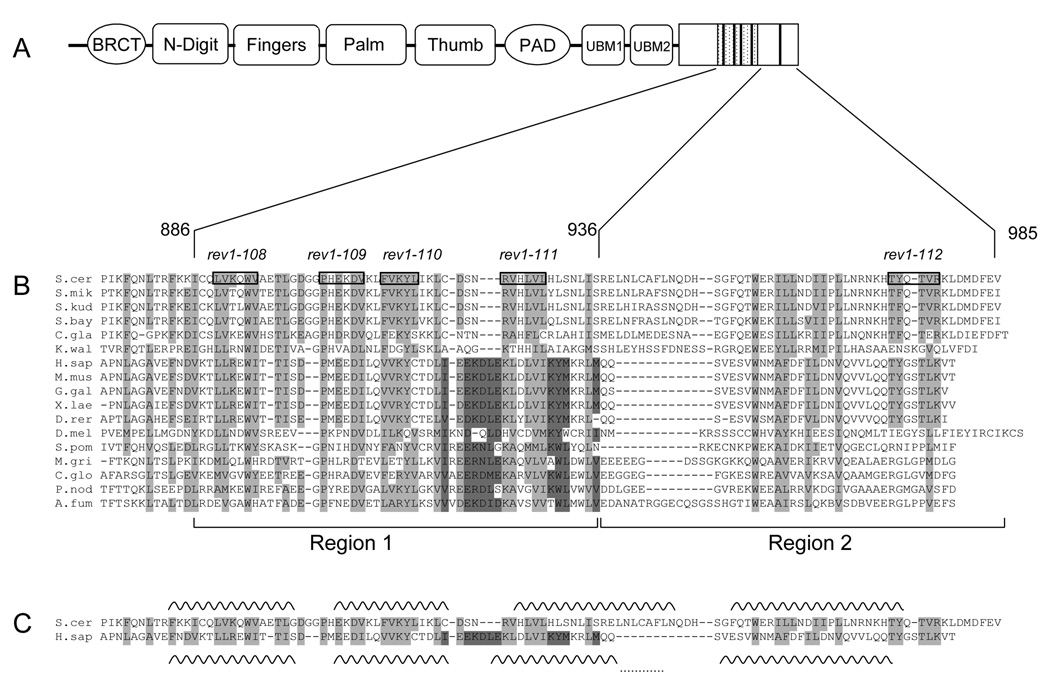
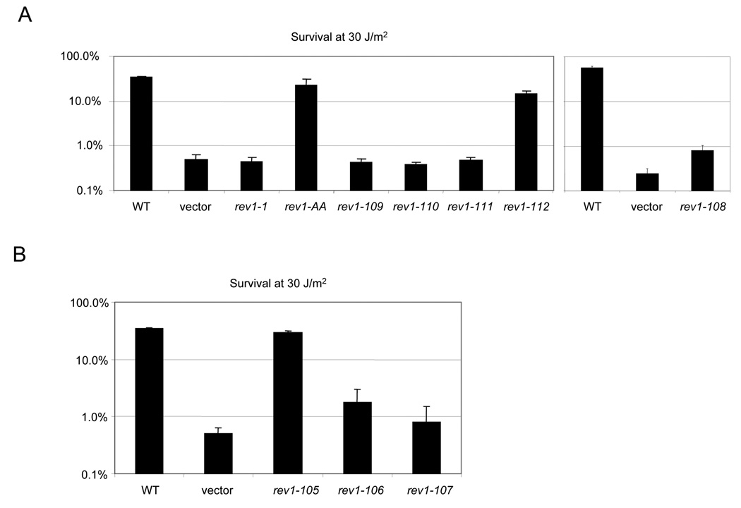
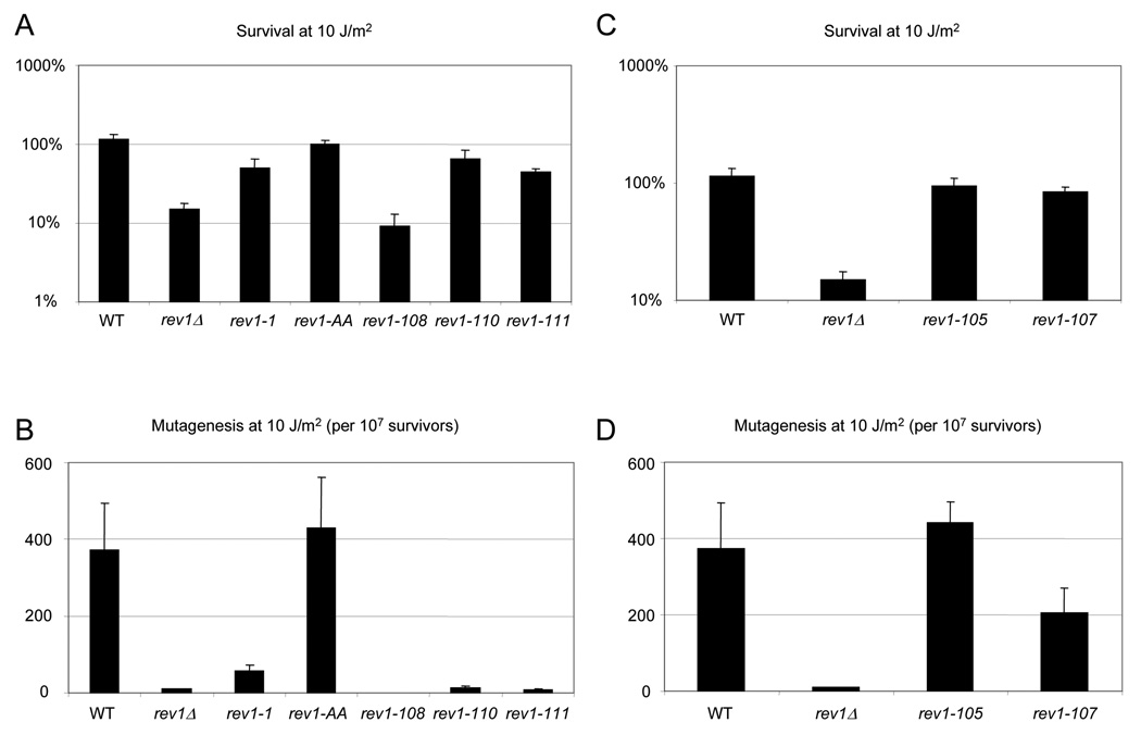
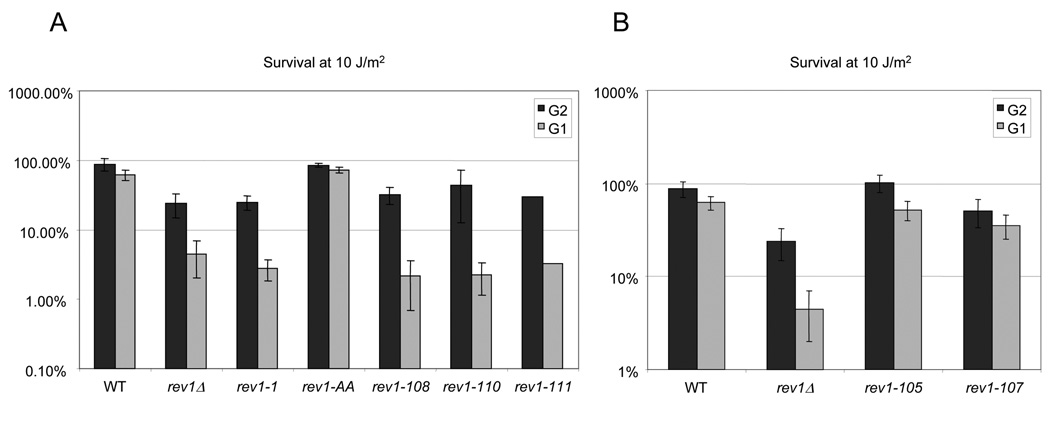
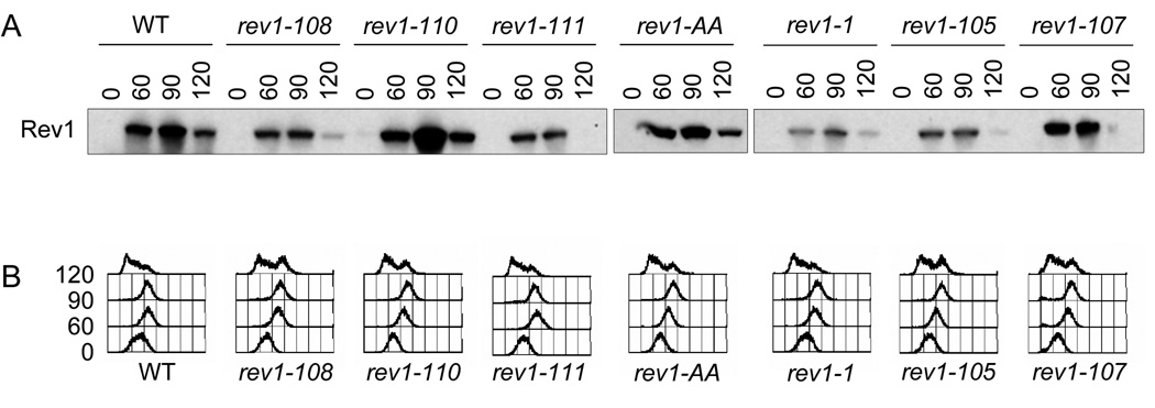
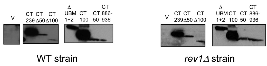
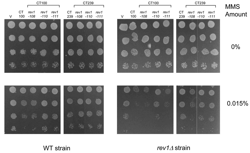
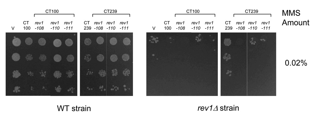
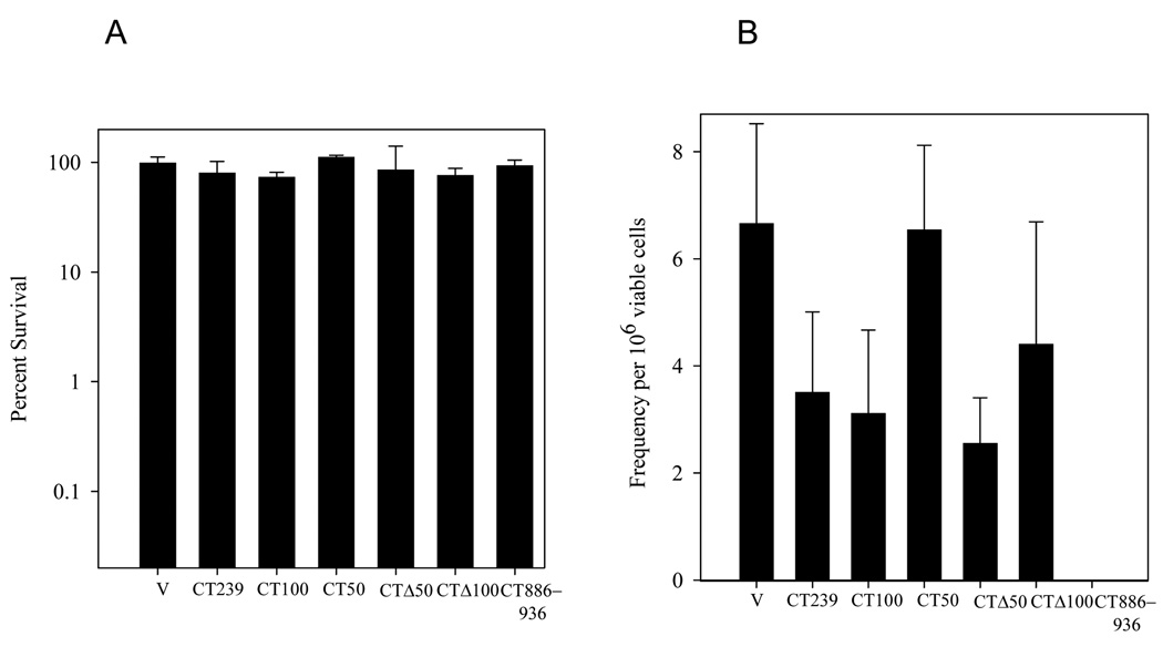
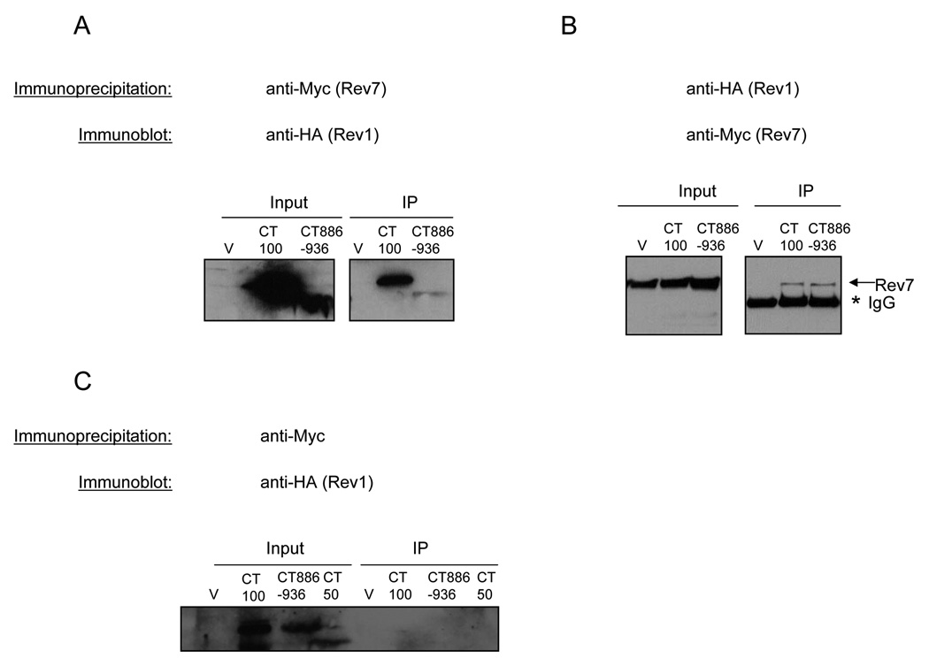
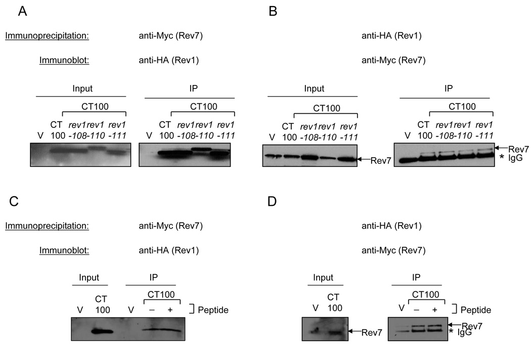
The genes encoding Rev1 and DNA polymerase zeta (Rev3/Rev7) are together required for the vast majority of DNA damage-induced mutations in eukaryotes from yeast to humans. Here, we provide insight into the critical role that the Saccharomyces cerevisiae Rev1 C-terminus plays in the process of mutagenic DNA damage tolerance. The Rev1 C-terminus was previously thought to be poorly conserved and therefore not likely to be important for mediating protein-protein interactions. However, through comprehensive alignments of the Rev1 C-terminus, we have identified novel and hitherto unrecognized conserved motifs that we show play an essential role in REV1-dependent survival and mutagenesis in S. cerevisiae, likely in its post-replicative gap-filling mode. We further show that the minimal C-terminal fragment of Rev1 containing these highly conserved motifs is sufficient to interact with Rev7.


Figures

Fig. 1
Fig. 1
The C-terminal ~100 amino acids of Rev1 are conserved across a wide range of organisms. (A) Schematic of the domain structure of S. cerevisiae Rev1 with the minimal Rev7-interacting 50 amino acid Region 1 indicated by the hatched box and the conserved motifs indicated by dark bars. (B) Multiple sequence alignment of Rev1 sequences from selected species. Boxed residues and the text above indicate the alanine-patch mutations. (See Table 3 for more details). Amino acids highlighted in light grey show conservation across all species, while amino acids highlighted in dark grey indicate a conserved region not found in S. cerevisiae and closely related yeasts. Species abbreviation: S. cer, S. cerevisiae; S. mik, S. mikatae; S. kud, S. kudriavzevii; S. bay, S. bayanus; C. gla, C. glabrata; K. wal, K. waltii; H. sap, H. sapiens; M. mus, M. musculus; G. gal, Gallus gallus ; X. lae, X. laevis; D. rer, D. rerio; D. mel, D. melanogaster; S. pom, S. pombe; M. gri, M. grisea; C. glo, C. globosum; P. nod, P. nodorum; A. fum, A. fumigatus. Rev1 sequences from over 50 organisms were used to generate the alignment, but only 17 are shown due to space considerations. (C) Predicted secondary structure of the C-terminus of Rev1. Alignment of yeast and human Rev1 excerpted from (B) for clarity with the predicted helices for each sequence indicated by wavy lines. The dots connecting the last two helices of human Rev1 indicate that this helix is predicted to be continuous. (D) Helical wheel projections of the first predicted helix. Dark grey indicates hydrophobic residues, red indicates positively charged residues, blue indicates negatively charged residues and yellow indicates polar residues.
Fig. 1
Fig. 1
The C-terminal ~100 amino acids of Rev1 are conserved across a wide range of organisms. (A) Schematic of the domain structure of S. cerevisiae Rev1 with the minimal Rev7-interacting 50 amino acid Region 1 indicated by the hatched box and the conserved motifs indicated by dark bars. (B) Multiple sequence alignment of Rev1 sequences from selected species. Boxed residues and the text above indicate the alanine-patch mutations. (See Table 3 for more details). Amino acids highlighted in light grey show conservation across all species, while amino acids highlighted in dark grey indicate a conserved region not found in S. cerevisiae and closely related yeasts. Species abbreviation: S. cer, S. cerevisiae; S. mik, S. mikatae; S. kud, S. kudriavzevii; S. bay, S. bayanus; C. gla, C. glabrata; K. wal, K. waltii; H. sap, H. sapiens; M. mus, M. musculus; G. gal, Gallus gallus ; X. lae, X. laevis; D. rer, D. rerio; D. mel, D. melanogaster; S. pom, S. pombe; M. gri, M. grisea; C. glo, C. globosum; P. nod, P. nodorum; A. fum, A. fumigatus. Rev1 sequences from over 50 organisms were used to generate the alignment, but only 17 are shown due to space considerations. (C) Predicted secondary structure of the C-terminus of Rev1. Alignment of yeast and human Rev1 excerpted from (B) for clarity with the predicted helices for each sequence indicated by wavy lines. The dots connecting the last two helices of human Rev1 indicate that this helix is predicted to be continuous. (D) Helical wheel projections of the first predicted helix. Dark grey indicates hydrophobic residues, red indicates positively charged residues, blue indicates negatively charged residues and yellow indicates polar residues.
Fig. 2
Fig. 2
Mutations in predicted polymerase interaction motifs and UBM2 disrupt REV1-mediated survival. (A) Survival after a dose of 30 J/m2 UV irradiation of the rev1Δ strain bearing a low-copy plasmid expressing WT REV1 or the rev1-1, rev1-AA, or rev1 C-terminal mutants under the native REV1 promoter (B). Survival after a dose of 30 J/m2 UV irradiation of the rev1Δ strain transformed with plasmids expressing the WT or rev1 UBM mutants.
Fig. 3
Fig. 3
Chromosomal mutations disrupting C-terminal motifs and UBM2 lead to impaired REV1-mediated survival and mutagenesis. (A) Survival of the rev1-1, rev1-AA, and indicated C-terminal mutants after UV irradiation with 10 J/m2. (B) Reversion frequency for the rev1-1, rev1-AA, and indicated C-terminal mutants, monitored by reversion of the trp1-1 allele, after 10 J/m2 UV irradiation. Error bars represent the standard deviation of the results derived from three independent colonies. Note that as no Trp+ colonies were recovered for the rev1-108 strain, the mutation frequency was calculated to be at or below the limit of detection for this strain of 22.2 revertants per 107 survivors. (C) Survival of the indicated UBM mutants after UV irradiation with 10 J/m2. (D) Reversion frequency for the indicated UBM mutants after UV irradiation with 10 J/m2.
Fig. 4
Fig. 4
Differential survival of the chromosomal rev1 mutants after UV irradiation throughout the cell cycle. (A) Survival after a UV dose of 10 J/m2 of the rev1-1, rev1-AA, or indicated C-terminal rev1 mutant strains, after release from G1 or G2 arrest. The values for rev1-111 are the average of two replicates. (B) Survival after a UV dose of 10 J/m2 of rev1 strains mutated in the UBMs, showing no hypersensitivity to UV irradiation after release from G1 or G2 arrest. Error bars represent the standard deviation of the results derived from three independent colonies.
Fig. 5
Fig. 5
Rev1 protein levels expressed by the chromosomal rev1 mutants during the cell-cycle. (A) Immunoblot analysis using the protein A epitope tag to visualize the WT and indicated mutant Rev1 protein levels throughout the cell cycle. The membrane was stained with Ponceau-S prior to immunoblotting to confirm equal loading. (B) FACS profiles of time points after α-factor release showing progression through the cell cycle.
Fig. 6
Fig. 6
Delineation of the minimal region of Rev1 required to manifest the dominant-negative phenotype of decreased survival upon overproduction after DNA damage. (A) Survival of WT and rev1Δ strains transformed with the indicated Rev1 truncation constructs after growth on plates containing the indicated amounts of MMS. Error bars represent the standard deviation of the results derived from three independent colonies. (B) Immunoblot analysis from WT and rev1Δ strains expressing the indicated Rev1 fragment using an anti-HA antibody.
Fig. 6
Fig. 6
Delineation of the minimal region of Rev1 required to manifest the dominant-negative phenotype of decreased survival upon overproduction after DNA damage. (A) Survival of WT and rev1Δ strains transformed with the indicated Rev1 truncation constructs after growth on plates containing the indicated amounts of MMS. Error bars represent the standard deviation of the results derived from three independent colonies. (B) Immunoblot analysis from WT and rev1Δ strains expressing the indicated Rev1 fragment using an anti-HA antibody.
Fig. 7
Fig. 7
Effect of mutations in conserved C-terminal motifs on survival of strains after DNA damage. (A) WT and rev1Δ strains transformed with the empty vector (V) or WT and mutant derivatives of the CT100 and CT239 fragments were grown on plates containing the indicated amounts of MMS. (B) Immunoblot analysis from the WT and rev1Δ strains transformed with the Rev1 constructs as in (A).
Fig. 7
Fig. 7
Effect of mutations in conserved C-terminal motifs on survival of strains after DNA damage. (A) WT and rev1Δ strains transformed with the empty vector (V) or WT and mutant derivatives of the CT100 and CT239 fragments were grown on plates containing the indicated amounts of MMS. (B) Immunoblot analysis from the WT and rev1Δ strains transformed with the Rev1 constructs as in (A).
Fig. 8
Fig. 8
Effect of overproduction of Rev1 C-terminal deletion constructs on UV-survival and UV-induced mutagenesis. (A) Survival of WT yeast strains transformed with either the empty vector (V) or expressing the indicated Rev1 C-terminal constructs after irradiation with 10 J/m2 UV. Error bars represent the standard deviation of the results derived from three independent colonies. (B) Mutation frequency of ade2-1 for the same strains as in (A). Note that due to the growth defect of WT cells overproducing the CT886-936 fragment, no colonies were obtained on the SC−Ade plates. Error bars represent the standard deviation of the results derived from three independent colonies.
Fig. 9
Fig. 9
Interaction between Rev1 C-terminal fragments and Rev7. (A, B) Co-immunoprecipitations of Rev7-13Myc and the indicated HA-tagged Rev1 C-terminal truncation constructs. Immunoprecipitated proteins were subjected to immunoblot analysis using an anti-HA antibody that detects Rev1 (A) or an anti-Myc antibody that detects Rev7 (B). (C) Co-immunoprecipitations from WT strain (untagged Rev7) overproducing the indicated HA-tagged Rev1 fragment. Immunoprecipitated proteins were immunoblotted using an anti-HA antibody.
Fig. 10
Fig. 10
Effect of mutations in conserved C-terminal motifs on the Rev1-Rev7 interaction. (A) Lysates from the Rev7-13Myc strain transformed with the empty vector (V) or expressing WT and the indicated mutant derivatives of CT100 were immunoprecipitated using an anti-Myc antibody to pull down Rev7 and immunoblotted with an anti-HA antibody to detect Rev1. A portion of the lysate was run as the input. (B) Lysates from the Rev7-13Myc strain transformed with the empty vector (V) or expressing WT and the indicated mutant derivatives of CT100 were immunoprecipitated using an anti-HA antibody to pull down the Rev1 C-terminal fragments and immunoblotted with an anti-Myc antibody to detect Rev7. A portion of the lysate was run as the input. (C) Immunoprecipitations performed as above with an anti-Myc antibody (C) or an anti-HA antibody (D) were carried out in the presence or absence of the Rev1 peptide described in the materials and methods. Immunoprecipitated proteins were immunoblotted using an anti-HA antibody (C) or an anti-Myc antibody (D).
Fig. 11
Fig. 11
Schematic representation of the various Rev1 C-terminal deletion fragments. The 985 amino acid Rev1 protein consists of the N-terminal BRCT region, the polymerase domain (amino acids 297–746) comprising the N-digit that interacts with the incoming dCTP and the fingers, palm, thumb and PAD domains conserved among Y-family polymerases. The Rev1 C-terminal region is composed of two copies of the Ubiquitin-Binding Motif (UBM1 and UBM2). The hatched region in the figure represents Region 1 of the Rev1 C-terminus and the bars within this region, the short peptide motifs. On the right is a summary of the regions of Rev1 that confer the dominant–negative phenotype on survival after DNA damage and that interact with Rev7. ND: Not determined.

References

    1. Friedberg EC, Walker GC, Siede W, Wood RD, Schultz RA, Ellenberger T. DNA Repair and Mutagenesis. Washington, D. C.: ASM Press; 2005.
    1. Goodman MF. Error-prone repair DNA polymerases in prokaryotes and eukaryotes. Annu Rev Biochem. 2002;71:17–50. - PubMed
    1. Prakash S, Johnson RE, Prakash L. Eukaryotic translesion synthesis DNA polymerases: specificity of structure and function. Annu Rev Biochem. 2005;74:317–353. - PubMed
    1. Kunkel TA. DNA replication fidelity. J Biol Chem. 2004;279:16895–16898. - PubMed
    1. Friedberg EC, Wagner R, Radman M. Specialized DNA polymerases, cellular survival, and the genesis of mutations. Science. 2002;296:1627–1630. - PubMed

Publication types

MeSH terms

Substances

LinkOut - more resources