Review
. 2008 Sep 1;24(17):1827-36.
doi: 10.1093/bioinformatics/btn346. Epub 2008 Jul 4.

Bioimage informatics: a new area of engineering biology

Affiliations
Review

Bioimage informatics: a new area of engineering biology

Hanchuan Peng. Bioinformatics. .

Abstract

In recent years, the deluge of complicated molecular and cellular microscopic images creates compelling challenges for the image computing community. There has been an increasing focus on developing novel image processing, data mining, database and visualization techniques to extract, compare, search and manage the biological knowledge in these data-intensive problems. This emerging new area of bioinformatics can be called 'bioimage informatics'. This article reviews the advances of this field from several aspects, including applications, key techniques, available tools and resources. Application examples such as high-throughput/high-content phenotyping and atlas building for model organisms demonstrate the importance of bioimage informatics. The essential techniques to the success of these applications, such as bioimage feature identification, segmentation and tracking, registration, annotation, mining, image data management and visualization, are further summarized, along with a brief overview of the available bioimage databases, analysis tools and other resources.


Figures

Fig. 1.
Fig. 1.
(A) Maximum projection of a 5-channel confocal 3D image of a 100 μm thick section of rat hippocampus. Red: GFAP-labeled astrocytes; green: EBA-labeled blood vessels; yellow: Iba1-labeled microglia; cyan: CyQuant-labeled cell nuclei; purple: NeuroTrace-labeled Nissl substance; scale bar=50 μm. (B) 3D rendering (with a similar color scheme) of the segmented and classified cells produced using the FARSIGHT techniques for (A). Image courtesy of Badrinath Roysam (Bjornsson et al., 2008)
Fig. 2.
Fig. 2.
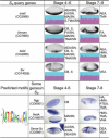
Clustering analysis of embryonic in situ mRNA gene expression patterns of fruit fly genes and its utility in assisting prediction of the regulatory sequence motifs. Based on clustering the eigen-embryo profiles (purple–cyan plot) of representative gene expression patterns, four genes in SQ are detected to be co-expressed genes. This prediction is consistent with their known gene regulation relationship for fly mesoderm patterning. Further, SQ can be used to predict sequence motifs. The motif example shown is detected using the entire upstream regions of the homologous genes in eight fly species D.melanogaster, D.simulans, D.yakuba, D.erecta, and D.ananassae, D.pseudoobscura, D.virilis, and D.mojavensis, along with three randomly selected example genes in the subsequent genome-wide motif scanning results. BDGP (fruitfly.org) ISH images (in blue) and annotations are also shown, without image cropping or orientation correction. Short terms of annotations: AAISN, amnioserosa anlage in statu nascendi; AISN, anlage in statu nascendi; AEA, anterior endoderm anlage; AEAISN, anterior endoderm anlage in statu nascendi; CB, cellular blastoderm; DEA, dorsal ectoderm anlage; DEAISN, dorsal ectoderm anlage in statu nascendi; EAISN, endoderm anlage in statu nascendi; FA, foregut anlage; FAISN, foregut anlage in statu nascendi; HMA, head mesoderm anlage; HA, hindgut anlage; MAISN, mesoderm anlage in statu nascendi; PTEA, posterior endoderm anlage; S, subset; TMA, trunk mesoderm anlage; TMAISN, trunk mesoderm anlage in statu nascendi; VEA, ventral ectoderm anlage; VNA, ventral neuroderm anlage. Original image source: (Peng et al., 2007)
Fig. 3.
Fig. 3.
Maximum projection of 3D registered and overlaid neuronal patterns of multiple fruit fly central complexes (top) and thoracic ganglia (bottom), each with a different GAL4 line (Peng et al., unpublished data). Red: a205; Green: EB1; Cyan: NP2320; Yellow: NP6510; gray: NC82-labeled neuropil. Raw confocal images were produced by Julie Simpson and Phuong Chung.

References

    1. Abramoff MD, et al. Image processing with ImageJ. Biophoto. Int. 2004;11:36–42.
    1. Ahammad P, et al. Joint nonparametric alignment for analyzing spatial gene expression patterns in Drosophila imaginal discs. IEEE CVPR 2005. 2005;2:20–25.
    1. Ai Z, et al. Reconstruction and exploration of three-dimensional confocal microscopy data in an immersive virtual environment. Comput. Med. Imaging Graph. 2005;29:313–318. - PubMed
    1. Al-Kofahi K, et al. Rapid automated three-dimensional tracing of neurons from confocal image stacks. IEEE Trans. Inf. Technol. Biomed. 2002;6:171–187. - PubMed
    1. Al-Kofahi K, et al. Median based robust algorithms for tracing neurons from noisy confocal microscope images. IEEE Trans. Inf. Technol. Biomed. 2003;7:302–317. - PubMed

MeSH terms