. 2008 Oct;22(10):3648-60.
doi: 10.1096/fj.08-110171. Epub 2008 Jul 4.

Targeted deletion of kcne2 impairs ventricular repolarization via disruption of I(K,slow1) and I(to,f)

Affiliations

Targeted deletion of kcne2 impairs ventricular repolarization via disruption of I(K,slow1) and I(to,f)

Torsten K Roepke et al. FASEB J. 2008 Oct.

Abstract

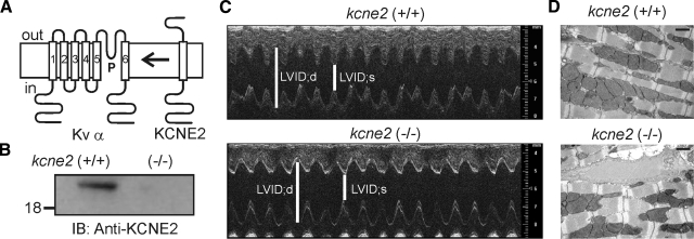
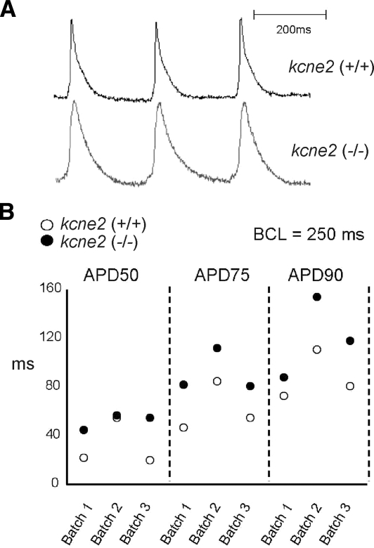
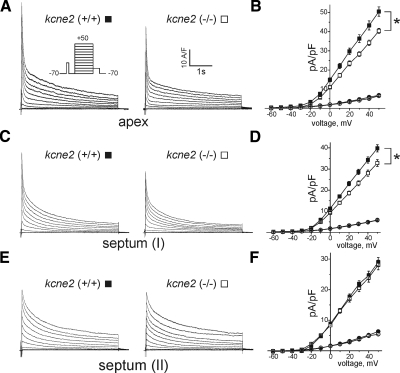
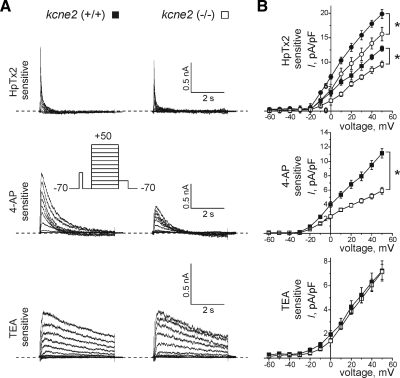
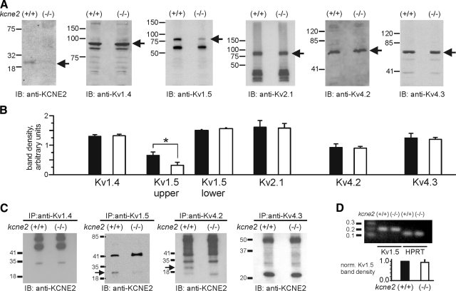
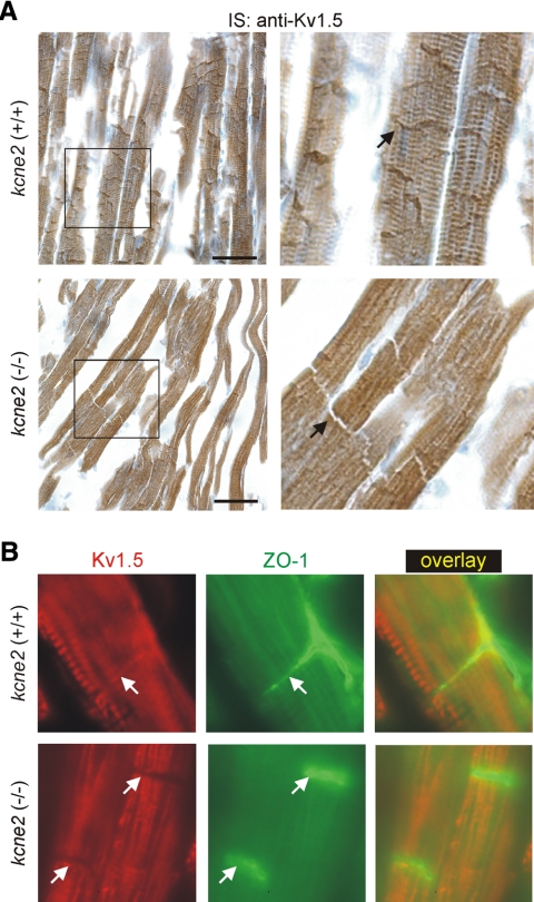
Mutations in human KCNE2, which encodes the MiRP1 potassium channel ancillary subunit, associate with long QT syndrome (LQTS), a defect in ventricular repolarization. The precise cardiac role of MiRP1 remains controversial, in part, because it has marked functional promiscuity in vitro. Here, we disrupted the murine kcne2 gene to define the role of MiRP1 in murine ventricles. kcne2 disruption prolonged ventricular action potential duration (APD), suggestive of reduced repolarization capacity. Accordingly, kcne2 (-/-) ventricles exhibited a 50% reduction in I(K,slow1), generated by Kv1.5--a previously unknown partner for MiRP1. I(to,f), generated by Kv4 alpha subunits, was also diminished, by approximately 25%. Ventricular MiRP1 protein coimmunoprecipitated with native Kv1.5 and Kv4.2 but not Kv1.4 or Kv4.3. Unexpectedly, kcne2 (-/-) ventricular membrane fractions exhibited 50% less mature Kv1.5 protein than wild type, and disruption of Kv1.5 trafficking to the intercalated discs. Consistent with the reduction in ventricular K(+) currents and prolonged ventricular APD, kcne2 deletion lengthened the QT(c) under sevoflurane anesthesia. Thus, targeted disruption of kcne2 has revealed a novel cardiac partner for MiRP1, a novel role for MiRPs in alpha subunit targeting in vivo, and a role for MiRP1 in murine ventricular repolarization with parallels to that proposed for the human heart.


Figures

Figure 1.
Figure 1.
Cardiac morphology and expression of kcne2 in adult murine heart. A) Membrane topology of a Kv α subunit compared to that proposed for KCNE2. B) Western blot analysis using anti-KCNE2 antibody from whole heart membrane preparations of kcne2 (+/+) and kcne2 (−/−) mice as indicated. Label on left indicates migration distance of 18-kDa molecular mass marker. C) Exemplar echocardiographic M-mode traces from kcne2 (+/+) and kcne2 (−/−) hearts. LVID;d, left ventricular internal diameter during diastole; LVID;s, left ventricular internal diameter during systole. D) Exemplar electron micrographs from kcne2 (+/+) and kcne2 (−/−) mouse ventricles. Scale bars = 1 μm.
Figure 2.
Figure 2.
kcne2 (−/−) mice exhibit prolonged ventricular APD. A) Exemplar ventricular myocyte AP in kcne2 (+/+) and kcne2 (−/−) mice recorded by optical mapping in isolated, perfused intact hearts. B) Ventricular myocyte APD 50, 75, and 90 measurements recorded by optical mapping in isolated, perfused intact hearts from kcne2 (+/+) (open circles) and kcne2 (−/−) mice (solid circles). Each point represents the mean value from 6400 recording points. Recordings were performed in 3 batches, each incorporating a kcne2 (+/+) and a kcne2 (−/−) heart.
Figure 3.
Figure 3.
kcne2 (−/−) mice exhibit diminished ventricular myocyte Ito,f current density. Outward currents recorded from individual myocytes isolated from different heart regions in the presence of 0.02 mM TTX to suppress depolarizing sodium currents. Whole-cell outward K+ currents were evoked during 4.5-s depolarizing voltage steps to potentials between –60 and +50 mV from a holding potential of –70 mV (protocol inset, panel A). A) Exemplar current traces recorded from myocytes isolated from the ventricular apex of kcne2 (+/+) and kcne2 (−/−) mice as indicated. B) Mean I/V relation for peak (squares) and steady-state currents (circles) recorded from myocytes as in A. *P < 0.002; n = 16–19 cells/group. C) Exemplar current traces recorded from Ito,f-expressing myocytes isolated from the ventricular septum as indicated. D) Mean I/V relation for peak (squares) and steady-state currents (circles) recorded from myocytes as in A. *P < 0.01; n = 20–23 cells per group. E) Exemplar current traces recorded from myocytes expressing Ito,s but not Ito,f, isolated from the ventricular septum as indicated. F) Mean I/V relation for peak (squares) and steady-state (circles) currents recorded from myocytes as in A; n = 22–23 cells per group. Solid symbols = kcne2 (+/+) mice; open symbols = kcne2 (−/−) mice. Error bars indicate means ± se.
Figure 4.
Figure 4.
kcne2 disruption reduces HpTx2- and 4-AP-sensitive currents in murine ventricles. Effects of kcne2 disruption on adult mouse ventricular Kv currents sensitive to HpTx2 (300 nM), 4-AP (50 μM) or TEA (25 mM). Kv currents were recorded from kcne2 (+/+) and kcne2 (−/−) LV myocytes during 4.5-s depolarizing voltage steps to −60 to + 50 mV from a holding potential of −70 mV (protocol inset). Antagonist-sensitive current traces for each cell were obtained offline by digital subtraction of traces recorded after antagonist application had achieved steady-state inhibition, from those recorded before application. A) Exemplar antagonist-sensitive current waveforms for septal myocytes expressing Ito,f from kcne2 (+/+) and kcne2 (−/−) mice as indicated. B) Mean I/V relations for antagonist-sensitive current recorded as in A in kcne2 (+/+) (solid symbols) and kcne2 (−/−) myocytes (open symbols). HpTx2 data are plotted separately for apex (circles) and septum (squares) due to significantly different HpTx2-sensitive currents between these two cell types in kcne2 (+/+) mice; data for other antagonists from apical and septal myocytes were pooled because of a lack of significant difference between baseline current densities in myocytes from the two regions in kcne2 (+/+) mice. *P < 0.05; n = 7–13. Error bars indicate means ± se.
Figure 5.
Figure 5.
kcne2 interacts with Kv1.5 and Kv4.2 in murine ventricles. A) Exemplar Western blots of kcne2 and α subunits from murine ventricular crude membrane fractions, normalized for total protein concentration, from kcne2 (+/+) and kcne2 (−/−) mice as indicated, using antibodies as indicated below blots. Numbers indicate migration distances of corresponding molecular mass markers (kDa); arrows indicate expected sizes of subunits probed. B) Quantification of band intensities from Western blots as in A for kcne2 (+/+) (solid bars) and kcne2 (−/−) (open bars) ventricular membrane fractions; n = 3 independent preparations from 8 hearts each; 24 hearts/genotype. C) Co-IP of kcne2 with Kv1.5 and with Kv4.2 but not with Kv4.3 or Kv1.4, from murine ventricular crude membrane fraction. Immunoprecipitations of fractions were prepared using antibodies raised against Kv1.4, Kv1.5, Kv4.2, or Kv4.3, as indicated, then size-fractionated, and Western blots were performed with anti-kcne2 antibody. Numbers indicate migration of molecular mass markers (kDa); arrows indicate kcne2 band. D) Top: gel of cDNA fragments for Kv1.5 and the HPRT housekeeping gene after semiquantitative RT-PCR from ventricular lysates. Bottom: mean Kv1.5 band densities normalized to kcne2 (+/+) density; n = 3 preps. Error bars indicate means ± se.
Figure 6.
Figure 6.
kcne2 disruption prevents Kv1.5 targeting to the intercalated discs. A) Exemplar sections of left ventricle isolated from kcne2 (+/+) and kcne2 (−/−) mice as indicated and immunostained (IS) with anti-Kv1.5 antibody (brown). For each genotype, right panel is expanded view of the boxed region in the left panel. Scale bars = 50 μm. Arrows, intercalated discs. B) Exemplar immunofluorescence images of myocytes from sections of left ventricle isolated from kcne2 (+/+) and kcne2 (−/−) mice as indicated. Sections were double-labeled for Kv1.5 (left, red) and zonula-occludens 1 (ZO-1; center, green); overlay shown at right. Horizontal axis = 40 μm. All arrows indicate intercalated discs.

Similar articles

Cited by

References

    1. McCrossan Z A, Abbott G W. The MinK-related peptides. Neuropharmacology. 2004;47:787–821. - PubMed
    1. Abbott G W, Sesti F, Splawski I, Buck M E, Lehmann M H, Timothy K W, Keating M T, Goldstein S A. MiRP1 forms IKr potassium channels with HERG and is associated with cardiac arrhythmia. Cell. 1999;97:175–187. - PubMed
    1. Sesti F, Abbott G W, Wei J, Murray K T, Saksena S, Schwartz P J, Priori S G, Roden D M, George A L, Jr, Goldstein S A. A common polymorphism associated with antibiotic-induced cardiac arrhythmia. Proc Natl Acad Sci U S A. 2000;97:10613–10618. - PMC - PubMed
    1. Reymond A, Marigo V, Yaylaoglu M B, Leoni A, Ucla C, Scamuffa N, Caccioppoli C, Dermitzakis E T, Lyle R, Banfi S, Eichele G, Antonarakis S E, Ballabio A. Human chromosome 21 gene expression atlas in the mouse. Nature. 2001;420:582–586. - PubMed
    1. Chun K R, Koenen M, Katus H A, Zehelein J. Expression of the IKr components KCNH2 (rERG) and KCNE2 (rMiRP1) during late rat heart development. Exp Mol Med. 2004;36:367–371. - PubMed

Publication types

MeSH terms