. 2008 Nov;7(11):2246-53.
doi: 10.1074/mcp.M800191-MCP200. Epub 2008 Jul 4.

Cell-free co-expression of functional membrane proteins and apolipoprotein, forming soluble nanolipoprotein particles

Affiliations

Cell-free co-expression of functional membrane proteins and apolipoprotein, forming soluble nanolipoprotein particles

Jenny A Cappuccio et al. Mol Cell Proteomics. 2008 Nov.

Abstract

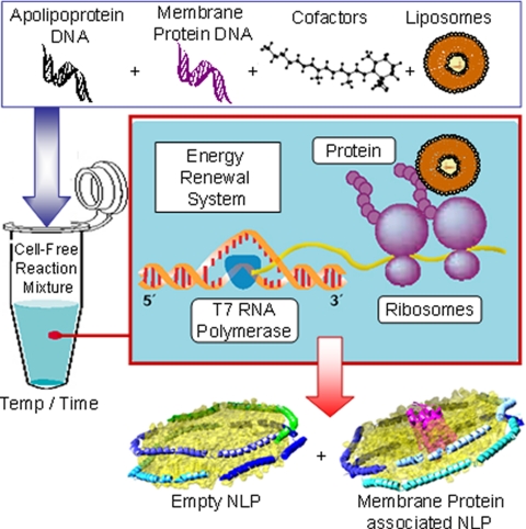
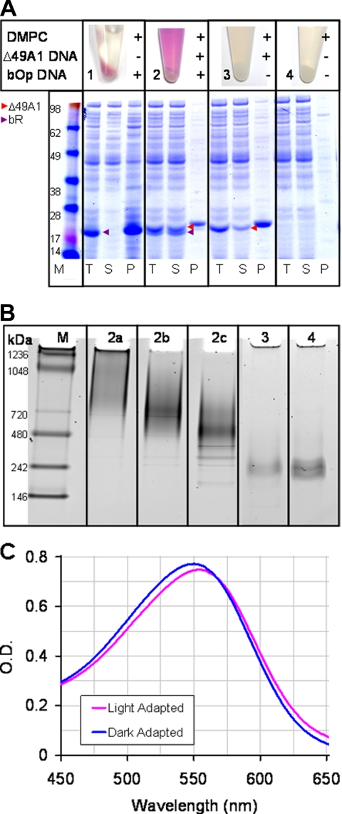
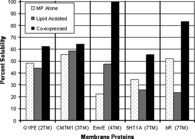
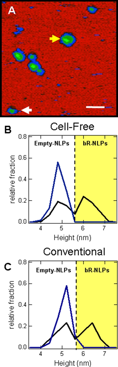
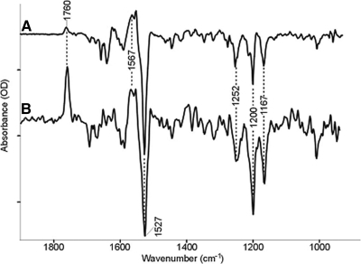
Here we demonstrate rapid production of solubilized and functional membrane protein by simultaneous cell-free expression of an apolipoprotein and a membrane protein in the presence of lipids, leading to the self-assembly of membrane protein-containing nanolipoprotein particles (NLPs). NLPs have shown great promise as a biotechnology platform for solubilizing and characterizing membrane proteins. However, current approaches are limited because they require extensive efforts to express, purify, and solubilize the membrane protein prior to insertion into NLPs. By the simple addition of a few constituents to cell-free extracts, we can produce membrane proteins in NLPs with considerably less effort. For this approach an integral membrane protein and an apolipoprotein scaffold are encoded by two DNA plasmids introduced into cell-free extracts along with lipids. For this study reported here we used plasmids encoding the bacteriorhodopsin (bR) membrane apoprotein and scaffold protein Delta1-49 apolipoprotein A-I fragment (Delta49A1). Cell free co-expression of the proteins encoded by these plasmids, in the presence of the cofactor all-trans-retinal and dimyristoylphosphatidylcholine, resulted in production of functional bR as demonstrated by a 5-nm shift in the absorption spectra upon light adaptation and characteristic time-resolved FT infrared difference spectra for the bR --> M transition. Importantly the functional bR was solubilized in discoidal bR.NLPs as determined by atomic force microscopy. A survey study of other membrane proteins co-expressed with Delta49A1 scaffold protein also showed significantly increased solubility of all of the membrane proteins, indicating that this approach may provide a general method for expressing membrane proteins enabling further studies.


Figures

F<sc>ig</sc>. 1.
Fig. 1.
Schematic of single step cell-free co-expression and stabilization of integral membrane proteins using an apolipoprotein scaffold. Constituents (DNA, lipid vesicles, cofactors, and cell-free lysates) are added together in a single reaction vial. The cell-free lysates take advantage of the T7-coupled transcription and translation system to produce a mixed population of self-assembled NLPs with and without associated integral membrane protein.
F<sc>ig</sc>. 2.
Fig. 2.
Single step production, purification, and characterization of MP·NLP complexes. A, Coomassie-stained SDS-PAGE gel image of total (T), soluble (S), and pellet (P) fractions from cell-free produced bR in the presence and absence of co-expressed apolipoprotein (Δ49A1). 1 μl of total, soluble, and resuspended pellet fractions were used for the gel. + indicates the addition of either DMPC, Δ49A1 DNA, or bOp DNA to the cell-free reaction; − denotes absence of additive. Red arrows indicate Δ49A1, and purple arrows indicate bR. Sample 1 (bOp and DMPC), bR is insoluble in the absence of co-expression of Δ49A1; Sample 2 (bOp and Δ49A1 co-expressed in the presence of DMPC), bR remains in the soluble fraction with co-expressed Δ49A1; Sample 3 (Δ49A1 and DMPC), production of empty NLPs; Sample 4, control cell-free reaction (no DNA) in the presence of DMPC only. All were expressed in the presence of 30–50 μm all-trans-retinal and 2 mg/ml DMPC. Purple color development observed in Samples 1 and 2 indicates incorporation of retinal into the bOp transcript representing proper folding of bR. B, native gel of size exclusion-purified NLPs prepared with Δ49A1 or other similar apolipoprotein as noted with and with out bR. Lane M, molecular weight marker; Lane 2a–2c, fractions from SEC-purified cell-free co-expressed bR·NLPs; Lane 2a, lipid-rich first fraction; Lane 2b, bR·NLP second fraction; Lane 2c, bR·NLP third fraction; Lane 3, cell-free produced empty NLPs; Lane 4, conventional assembly of empty NLPs with purchased Δ1–55 apolipoprotein A-I (Δ55A1). C, light/dark adaptation of affinity-purified bR·NLPs concentrated with a 100-kDa molecular mass cutoff filter and buffer-exchanged into TBS, pH 7.4. Blue, dark-adapted bR·NLPs with a λmax = 549 nm; magenta, light-adapted bR·NLP with a λmax = 554 nm. Dark-adapted spectra were collected after overnight adaptation. Light-adapted spectra were collected upon 15-min exposure to a white light-emitting diode source. Spectra were collected in a 50-μl masked quartz cuvette with a 1-cm path length. Absorbance maxima differ by a 5-nm shift between light- and dark-adapted bR.
F<sc>ig</sc>. 3.
Fig. 3.
Co-expression of membrane proteins with apolipoprotein Δ49A1 in the presence of lipid increases solubility of multiple membrane proteins. A comparison was made between the membrane protein expressed alone (MP Alone; gray), expression of the membrane protein in the presence of DMPC vesicles (Lipid Assisted; striped), and membrane protein co-expressed with apolipoprotein (Δ49ApoA1) in the presence of DMPC vesicles (Co-expressed; black). Membrane proteins with the number of transmembrane domain in parentheses are: glycophorin B (GYPE) (MNS blood group) (two TMs), chemokine-like factor (CKLF-like) MARVEL transmembrane domain (CMTM1) (three TMs), Escherichia coli small multidrug resistance (SMR) efflux transporter (EmrE) (four TMs), 5-hydroxytryptamine (serotonin) receptor (5HT1A) (seven TMs), and bR (seven TMs). In all cases the solubility of the membrane protein increased with co-expression of Δ49ApoA1. Data were generated from autoradiograms by the incorporation of [35S]methionine into the cell-free reaction (data not shown) quantified from SDS-PAGE using ImageJ software (National Institutes of Health).
F<sc>ig</sc>. 4.
Fig. 4.
AFM confirms the association between NLPs and bR. A, AFM image of NLPs produced through cell-free co-expression of Δ49A1 and bR in the presence of DMPC. The brighter green regions are NLPs with a higher height indicating the insertion and plausible location of bR in the lipid bilayer. Scale bars, 50 nm. The white arrow indicates expression of empty NLPs; the yellow arrow indicates the bR·NLP complex. B, height histogram of NLPs produced through conventional assembly of Δ55A1 with DMPC alone (blue) or in the presence of purple membrane bR and DMPC (black). The yellow shaded areas indicate populations with an increased height. C, height histogram of NLPs produced through cell-free expression of Δ49A1 with DMPC alone (blue) or co-expression of bR and Δ49A1 in the presence of DMPC (black). The yellow shaded areas indicate populations with an increased height. NLP heights were analyzed through cross-sectional analysis.
F<sc>ig</sc>. 5.
Fig. 5.
FTIR difference spectra of bR wild type (A) and bR·NLP (B). The largest peaks are 9.4 and 0.34 milli-optical density units, respectively. The positive bands represent vibrations in the M state, and the negative bands represent the ground state. Despite the smaller signal, the spectrum of BR·NLP clearly indicates functional protein that is stable over ∼104 laser flashes.

References

    1. Bayburt, T. H., Carlson, J. W., and Sligar, S. G. ( 1998) Reconstitution and imaging of a membrane protein in a nanometer-size phospholipid bilayer. J. Struct. Biol. 123, 37–44 - PubMed
    1. Bayburt, T. H., Grinkova, Y. V., and Sligar, S. G. ( 2006) Assembly of single bacteriorhodopsin trimers in bilayer nanodiscs. Arch. Biochem. Biophys. 450, 215–222 - PubMed
    1. Bayburt, T. H., and Sligar, S. G. ( 2003) Self-assembly of single integral membrane proteins into soluble nanoscale phospholipid bilayers. Protein Sci. 12, 2476–2481 - PMC - PubMed
    1. Leitz, A. J., Bayburt, T. H., Barnakov, A. N., Springer, B. A., and Sligar, S. G. ( 2006) Functional reconstitution of β2-adrenergic receptors utilizing self-assembling Nanodisc technology. BioTechniques 40, 601–602, 604, 606, passim. - PubMed
    1. Bayburt, T. H., Leitz, A. J., Xie, G., Oprian, D. D., and Sligar, S. G. ( 2007) Transducin activation by nanoscale lipid bilayers containing one and two rhodopsins. J. Biol. Chem. 282, 14875–14881 - PubMed

Publication types

MeSH terms

LinkOut - more resources