

Melanomagenesis: overcoming the barrier of melanocyte senescence
- PMID: 18604170
- PMCID: PMC2678050
- DOI: 10.4161/cc.7.13.6230
Melanomagenesis: overcoming the barrier of melanocyte senescence
Abstract
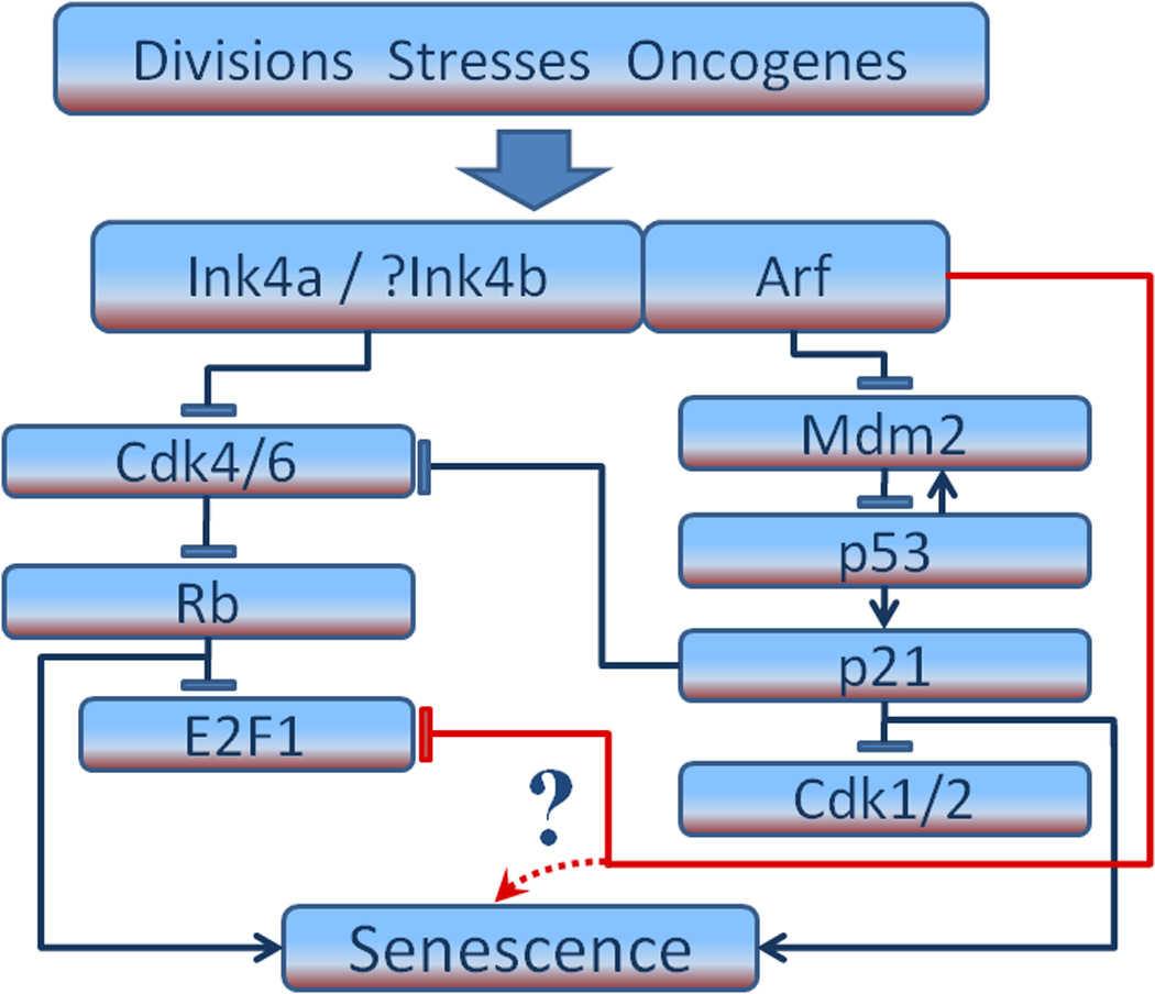
Although melanoma ultimately progresses to a highly aggressive and metastatic disease that is typically resistant to currently available therapy, it often begins as a benign nevus consisting of a clonal population of hyperplastic melanocytes that cannot progress because they are locked in a state of cellular senescence. Once senescence is overcome, the nevus can exhibit dysplastic features and readily progress to more lethal stages. Recent advances have convincingly demonstrated that senescence represents a true barrier to the progression of many types of cancer, including melanoma. Thus, understanding the mechanism(s) by which melanoma evades senescence has become a priority in the melanoma research community. Senescence in most cells is regulated through some combination of activities within the RB and p53 pathways. However, differences discovered among various tumor types, some subtle and others quite profound, have revealed that senescence frequently operates in a context-dependent manner. Here we review what is known about melanocyte senescence, and how such knowledge may provide a much-needed edge in our struggles to contain or perhaps vanquish this often-fatal malignancy.
Figures
References
-
- Hayflick L, Moorhead PS. The serial cultivation of human diploid cell strains. Exp Cell Res. 1961;25:585–621. - PubMed
-
- Ben Porath I, Weinberg RA. The signals and pathways activating cellular senescence. Int J Biochem Cell Biol. 2005;37:961–976. - PubMed
-
- Collado M, Blasco MA, Serrano M. Cellular senescence in cancer and aging. Cell. 2007;130:223–233. - PubMed
-
- Lloyd AC. Limits to lifespan. Nat Cell Biol. 2002;4:E25–E27. - PubMed
-
- Bartek J, Bartkova J, Lukas J. DNA damage signalling guards against activated oncogenes and tumour progression. Oncogene. 2007;26:7773–7779. - PubMed
Publication types
MeSH terms
Substances
Grants and funding
LinkOut - more resources
Full Text Sources
Other Literature Sources
Medical
Research Materials
Miscellaneous