. 2008 Aug;10(8):979-86.
doi: 10.1038/ncb1758. Epub 2008 Jul 6.

EphrinB1 controls cell-cell junctions through the Par polarity complex

Affiliations

EphrinB1 controls cell-cell junctions through the Par polarity complex

Hyun-Shik Lee et al. Nat Cell Biol. 2008 Aug.

Abstract

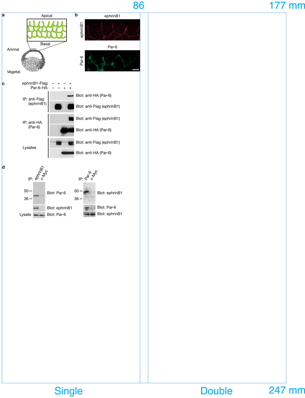
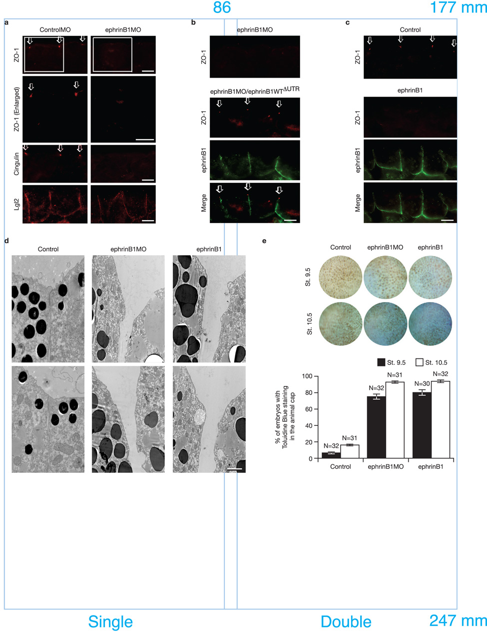
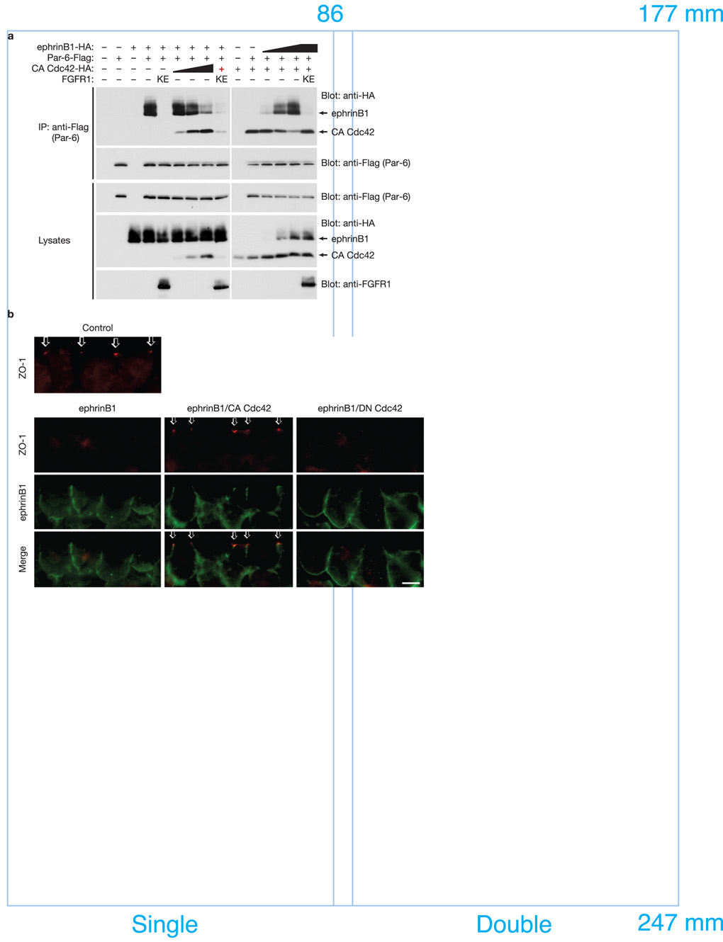
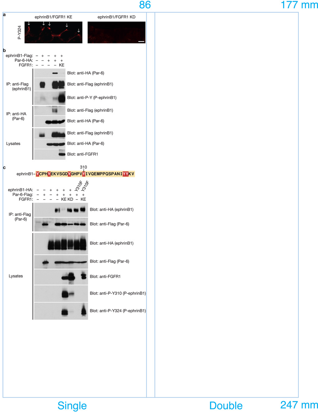
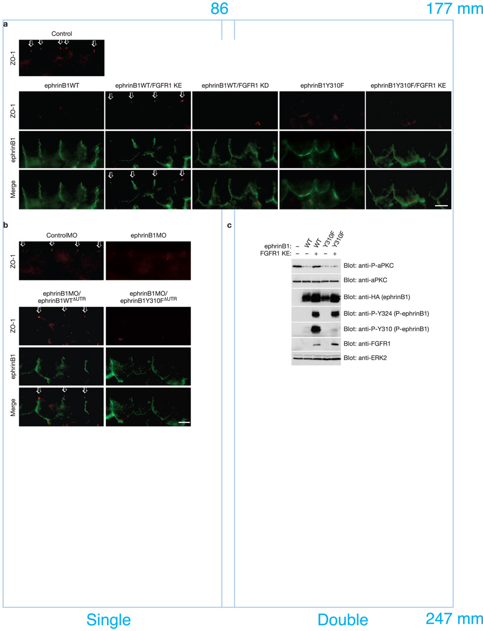
A body of evidence is emerging that shows a requirement for ephrin ligands in the proper migration of cells, and the formation of cell and tissue boundaries. These processes are dependent on the cell-cell adhesion system, which plays a crucial role in normal morphogenetic processes during development, as well as in invasion and metastasis. Although ephrinB ligands are bi-directional signalling molecules, the precise mechanism by which ephrinB1 signals through its intracellular domain to regulate cell-cell adhesion in epithelial cells remains unclear. Here, we present evidence that ephrinB1 associates with the Par polarity complex protein Par-6 (a scaffold protein required for establishing tight junctions) and can compete with the small GTPase Cdc42 for association with Par-6. This competition causes inactivation of the Par complex, resulting in the loss of tight junctions. Moreover, the interaction between ephrinB1 and Par-6 is disrupted by tyrosine phosphorylation of the intracellular domain of ephrinB1. Thus, we have identified a mechanism by which ephrinB1 signalling regulates cell-cell junctions in epithelial cells, and this may influence how we devise therapeutic interventions regarding these molecules in metastatic disease.


Figures

Figure 1
Figure 1. EphrinB1 phrinB1 binds Par-6 in vivo and in vitro
(a) Experimental scheme. Cartoon depicting region of embryo isolated and examined by immunofluorescence. (b) Immunofluorescence analysis shows that the localization of ephrinB1 overlaps with that of Par-6 along cell-cell boundaries and the basolateral domain in the gastrula stage (st.10.5) embryos. Each blastomere of 2-cell stage embryos was injected with 100 pg of ephrinB1 or 75 pg of HA-tagged Par-6 RNA where indicated. Embryos were sectioned and immunostained for ephrinB1 (C-18) or Par-6 (HA). The scale bar corresponds to 1 µm. (c) Oocytes were left uninjected (−) or injected (+) with Flag-tagged ephrinB1 (10 ng) and HA-tagged Par-6 (10 ng) RNAs where indicated. Oocyte lysates were IP’d with anti-Flag (top and second panels) and anti-HA (third and fourth panels) antibodies, then immunoblotted with anti-HA or anti-Flag antibodies to detect Par-6 and ephrinB1 proteins. (d) Immunoprecipitates using anti-ephrinB1 (rabbit), anti-Par-6 (rabbit) or anti-c-Myc antibodies (rabbit) in HT29 human colon carcinoma cell lysates were immunoblotted with anti-Par-6 (goat) and anti-ephrinB1 antibodies (goat). Lysates were analyzed directly by SDS-PAGE and immunoblotted with indicated antibodies to reveal endogenous expression levels of ephrinB1 and Par-6, respectively.
Figure 2
Figure 2. Inhibition and over-expression of ephrinB1 causes the disruption of TJ formation
(a) Each blastomere of 2-cell stage embryos was injected with 20 ng of ephrinB1MO or ControlMO where indicated. Embryos were sectioned and immunostained for ZO-1, Cingulin, and Lgl2. Boxed regions of first panel are enlarged in second panel. Arrows indicate the ZO-1 and Cingulin localization in TJs. The scale bar corresponds to 1 µm. (b) Each blastomere of 2-cell stage embryos was injected with 20 ng of ephrinB1MO and 150 pg of ephrinB1WTΔUTR-HA RNA where indicated. ΔUTR indicates use of a mutant RNA lacking a 5’ UTR sequence which is complementary to the ephrinB1MO, conferring resistance to the ephrinB1MO. Embryos were sectioned and immunostained for ZO-1 and ephrinB1 (HA). Arrows indicate the ZO-1 localization in TJs. The scale bar corresponds to 1 µm. (c) Each blastomere of 2-cell stage embryos was injected with 200 pg of ephrinB1-HA RNA. Embryos were sectioned and immunostained for ZO-1 and ephrinB1 (HA). Arrows indicate the ZO-1 localization in TJs. The scale bar corresponds to 1 µm. (d) Each blastomere of 2-cell stage embryos was injected with 20 ng of ephrinB1MO or 150 pg of ephrinB1-HA RNA and collected at Stage 10.5. Transmission electron microscopy shows disruption of TJs when ephrinB1 is inhibited or over-expressed in embryos. The scale bar corresponds to 0.5 µm. (e) Each blastomere of 2-cell stage embryos was injected with 20 ng of ephrinB1MO or 150 pg of ephrinB1-HA RNA. Embryos were collected at stage 9.5 and 10.5 and stained with 0.01% toluidine blue. At each stage, positive stained embryos were counted from each group and presented as a percentage (see histogram). Embryos were scored as positive when over 10% (St. 9.5) or over 50% (St. 10.5) of the ectodermal surface was stained. Note: More toluidine blue dye permeated the animal pole ectodermal layer of MO-injected (75.0±3.13% − 90.4±0.74% sem of embryos) and ephrinB1 over-expressing embryos (80.0±1.19% − 93.8±1.56% sem of embryos) than control (6.3±1.56% − 16.3±0.95% sem of embryos).
Figure 3
Figure 3. EphrinB1 and active Cdc42 compete for Par-6 binding
(a) Oocytes were left uninjected (−) or injected (+) with Par-6-Flag (10 ng) plus ephrinB1-HA (10 ng) and increasing amounts (1, 5, 10 ng) of CA Cdc42-HA RNAs or alternatively with Par-6-Flag (10 ng) plus CA Cdc42-HA (10 ng) and increasing amounts (1, 5, 10 ng) of ephrinB1-HA RNAs as indicated. Oocyte lysates were IP’d with anti-Flag antibody, then immunoblotted with anti-HA antibodies to detect ephrinB1 and active Cdc42 proteins. Oocyte lysates were analyzed directly by SDS-PAGE and immunoblotted with indicated antibodies. Note: the red + indicates the lowest amount of CA Cdc42-HA RNA (1 ng) was used. (b) Each blastomere of 2-cell stage embryos was injected with 200 pg of ephrinB1WT-Flag and 50 pg of CA Cdc42-HA or DN Cdc42-HA RNAs where indicated. Embryos were sectioned and immunostained for ZO-1 and ephrinB1 (Flag). Arrows indicate the ZO-1 localization in TJs. The scale bar corresponds to 1 µm.
Figure 4
Figure 4. FGF signaling blocks the interaction between ephrinB1 and Par-6 through the tyrosine phosphorylation of the intracellular domain of ephrinB1
(a) Each blastomere of 2-cell stage embryos was injected with 200 pg of ephrinB1-HA and 200 pg of FGFR1 KE or FGFR1 KD RNAs where indicated. Embryos were sectioned and immunostained for phosphorylated ephrinB1 (P-Y324). Arrows indicate the presence of phosphorylated ephrinB1 in apical cell junctions. The scale bar corresponds to 1 µm. (b) Oocytes were left uninjected (−) or injected (+) with ephrinB1-Flag (10 ng), Par-6-HA (10 ng) and FGFR1 KE (10 ng) RNAs as indicated. Oocyte lysates were IP’d with anti-Flag (top, second, and third panels) or anti-HA (fourth and fifth panels) antibodies, then immunoblotted with either anti-HA or anti-Flag antibodies to detect bound proteins. Oocyte lysates were analyzed directly by SDS-PAGE and immunoblotted with indicated antibodies. (c) Oocytes were left uninjected (−) or injected (+) with ephrinB1WT-HA (10 ng) or ephrinB1Y310F-HA (10 ng), Par-6-Flag (10 ng) and FGFR1 KE (10 ng) or FGFR1 KD (10 ng) RNAs as indicated. Oocyte lysates were IP’d with anti-Flag antibody, then immunoblotted with anti-HA antibody to detect ephrinB1 proteins. Oocyte lysates were analyzed directly by SDS-PAGE and immunoblotted with indicated antibodies.
Figure 5
Figure 5. Phosphorylation on tyrosine 310 regulates TJ formation by dissociating ephrinB1 from Par-6
(a) Each blastomere of 2-cell stage embryos was injected with 200 pg of ephrinB1WT-HA or ephrinB1Y310F-HA and 200 pg of FGFR1 KE or FGFR1 KD RNAs where indicated. Embryos were sectioned and immunostained for ZO-1 and ephrinB1 (HA). Arrows indicate the ZO-1 localization in TJs. The scale bar corresponds to 1 µm. (b) Each blastomere of 2-cell stage embryos was injected with 20 ng of ControlMO or ephrinB1MO and 150 pg of ephrinB1WTΔUTR-HA or ephrinB1Y310ΔUTR-HA RNAs where indicated. Embryos were sectioned and immunostained for ZO-1 and ephrinB1 (HA). Arrows indicate the ZO-1 localization in TJs. The scale bar corresponds to 1 µm. (c) Each blastomere of 2-cell stage embryos was injected with 200 pg of ephrinB1-HA or ephrinB1Y310-HA and 200 pg of FGFR1 KE RNAs where indicated. At stage 8.5, ectodermal explants were dissected, and harvested at stage 10.5. Lysates of these explants were analyzed directly by SDS-PAGE and immunoblotted with indicated antibodies.

Similar articles

Cited by

References

    1. Davy A, Aubin J, Soriano P. Ephrin-B1 forward and reverse signaling are required during mouse development. Genes Dev. 2004;18:572–583. - PMC - PubMed
    1. Cortina C, et al. EphB-ephrin-B interactions suppress colorectal cancer progression by compartmentalizing tumor cells. Nat Genet. 2007;39:1376–1383. - PubMed
    1. Heroult M, Schaffner F, Augustin HG. Eph receptor and ephrin ligand-mediated interactions during angiogenesis and tumor progression. Exp Cell Res. 2006;312:642–650. - PubMed
    1. Zhang J, Hughes S. Role of the ephrin and Eph receptor tyrosine kinase families in angiogenesis and development of the cardiovascular system. J Pathol. 2006;208:453–461. - PubMed
    1. Wimmer-Kleikamp SH, Lackmann M. Eph-modulated cell morphology, adhesion and motility in carcinogenesis. IUBMB Life. 2005;57:421–431. - PubMed

Publication types