. 2008 Aug;11(8):916-22.
doi: 10.1038/nn.2155. Epub 2008 Jul 6.

Light-sensitive neurons and channels mediate phototaxis in C. elegans

Affiliations

Light-sensitive neurons and channels mediate phototaxis in C. elegans

Alex Ward et al. Nat Neurosci. 2008 Aug.

Abstract

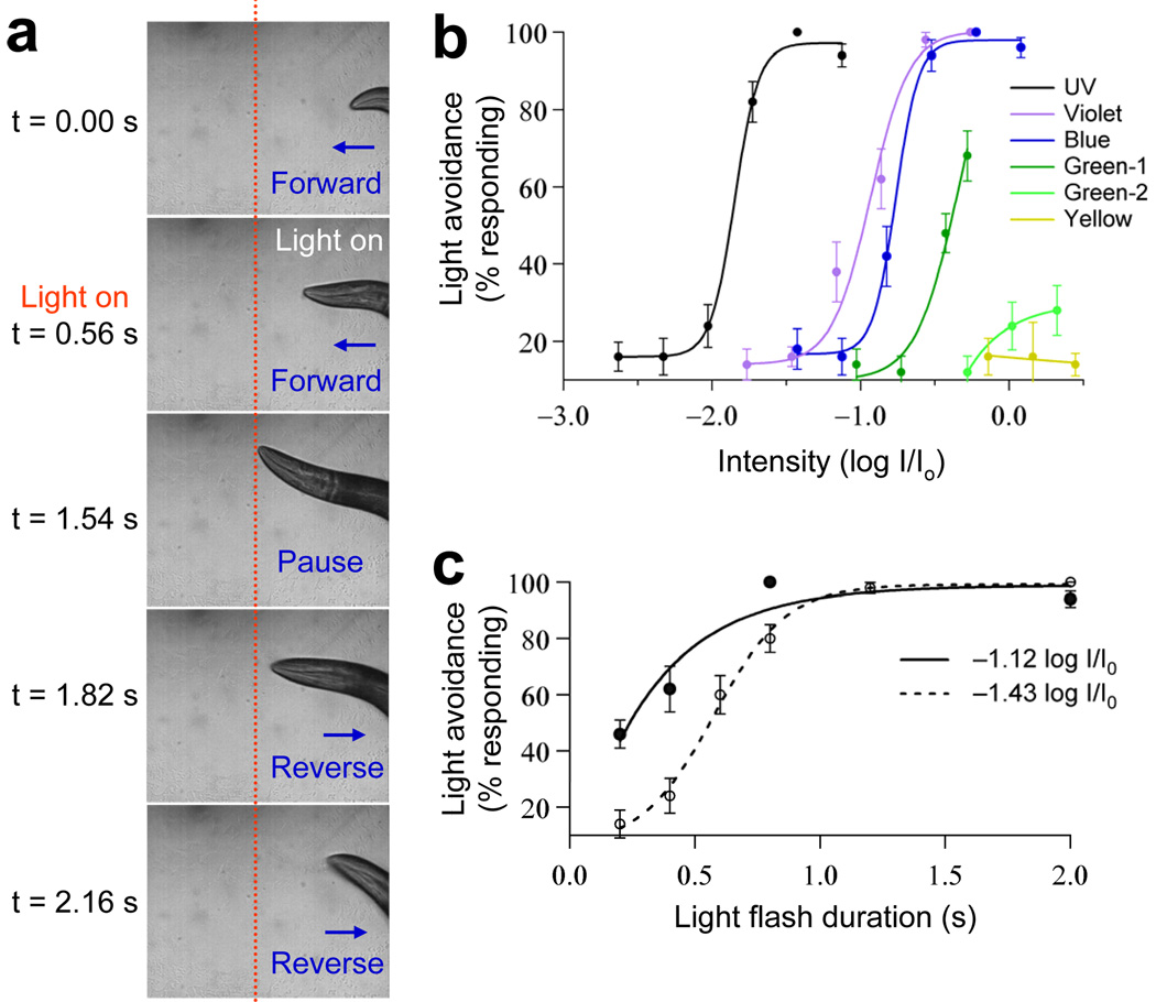
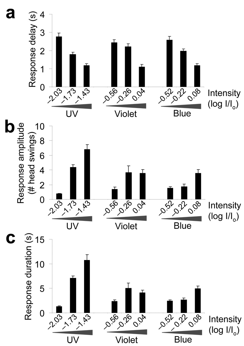
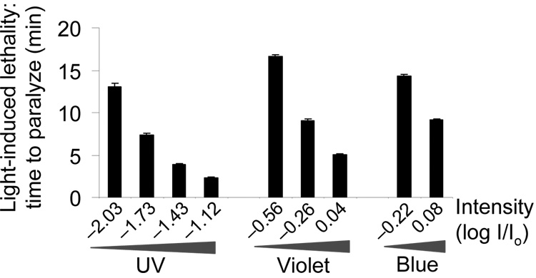
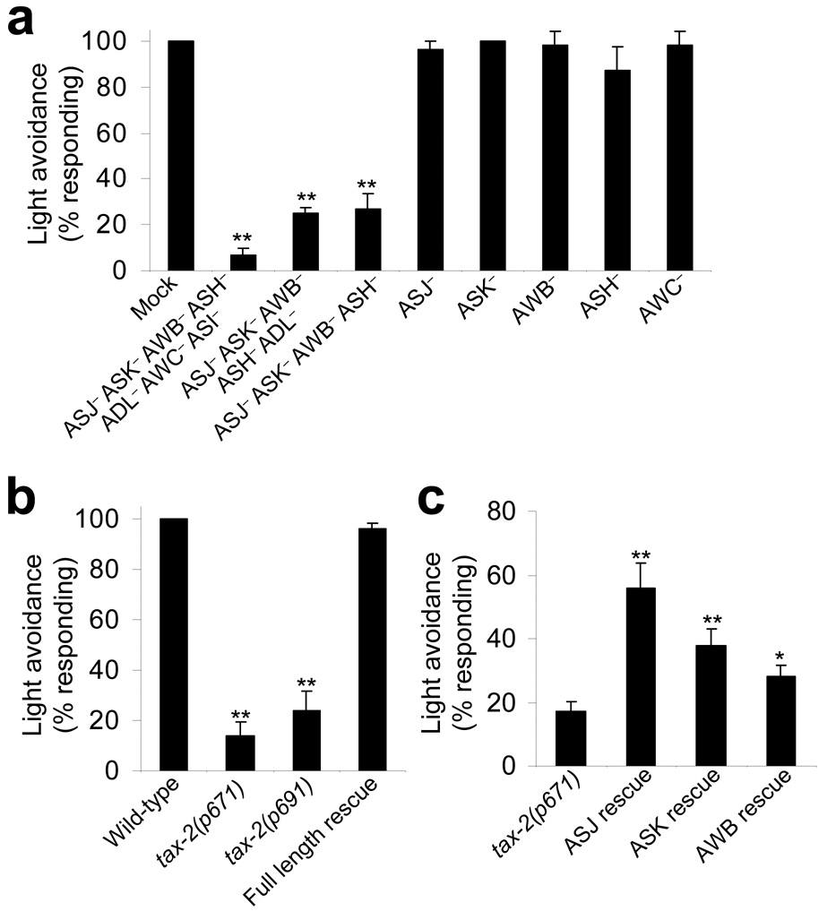
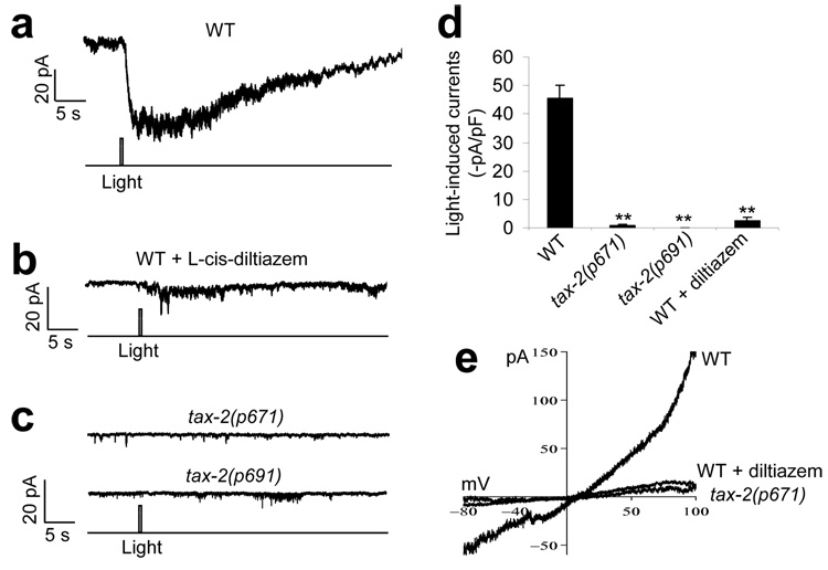
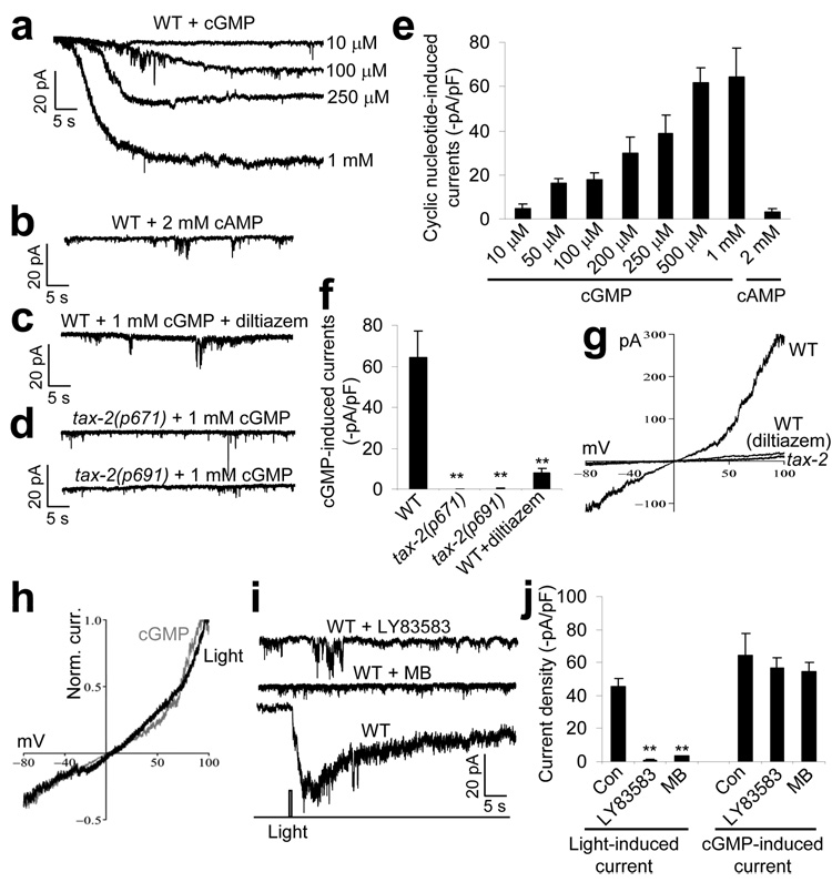
Phototaxis behavior is commonly observed in animals with light-sensing organs. C. elegans, however, is generally believed to lack phototaxis, as this animal lives in darkness (soil) and does not possess eyes. Here, we found that light stimuli elicited negative phototaxis in C. elegans and that this behavior is important for survival. We identified a group of ciliary sensory neurons as candidate photoreceptor cells for mediating phototaxis. Furthermore, we found that light excited photoreceptor cells by evoking a depolarizing conductance carried by cyclic guanosine monophosphate (cGMP)-sensitive cyclic nucleotide-gated (CNG) channels, revealing a conservation in phototransduction between worms and vertebrates. These results identify a new sensory modality in C. elegans and suggest that animals living in dark environments without light-sensing organs may not be presumed to be light insensitive. We propose that urbilaterians, the last common ancestor of bilaterians, might have already evolved a visual system that employs CNG channels and the second messenger cGMP for phototransduction.


Figures

Figure 1
Figure 1. Light stimulation evokes avoidance responses in C. elegans in a dose-dependent manner
(a) Snapshot images showing that a flash of light triggered an avoidance response in a worm moving forward. A flash of light (2 s, UV-A) was delivered by an objective to the head of a worm moving forward under a microscope. The animal quickly responded by stopping forward movement and initiating reversals. The dotted red line indicates the position of the worm in the field. (b) Worms responded to light in an intensity-dependent manner and were most sensitive to UV-A light. Light pulses (2 s) of varying intensity were tested for the head avoidance response and the percentage of worms that responded was scored (Io = 20 mW mm−2, n = 10). Error bars represent s.e.m. (c) Worms responded to light in a duration-dependent manner. Light pulses of varying duration were tested for the head avoidance response. Two different intensities of UV-A light were tested (−1.12log I/ Io and −1.43log I/Io, n = 10). We also examined violet and blue light (Supplementary Fig. 1). Error bars represent s.e.m.
Figure 2
Figure 2. Behavioral quantification of phototactic responses
(a) Quantification of the response delay. Worms responded to a flash of light by initiating reversals in as short as ~1 s, depending on the light intensity. The response delay was quantified as the time interval between the onset of light illumination and the time point at which the animal initiated backward movement. We tested three different intensities of UV-A, violet and blue light pulses (2 s, n = 10). Error bars represent s.e.m. (b) Quantification of the response amplitude. The assay was performed as described in a, and the number of head swings during backward movement was quantified (n = 10). Error bars represent s.e.m. (c) Quantification of the response duration. The assay was performed as described in a, and the duration of backward movement was quantified (n = 10). Error bars represent s.e.m.
Figure 3
Figure 3. Prolonged light exposure induces paralysis/lethality in worms
Worms were exposed to prolonged light illumination until death and the elapsed time was recorded. To keep the animal exposed to light continuously, we manually moved the stage to follow the animal to keep it in the field of illumination. Under this condition, worms were usually hyperactive at the beginning, but eventually ceased movement and pharyngeal pumping (n = 10). Error bars represent s.e.m.
Figure 4
Figure 4. Phototaxis in C. elegans requires ciliary sensory neurons and CNG channels
(a) Phototaxis in C. elegans required ciliary sensory neurons. Laser ablation of a group of ciliary sensory neurons led to a severe defect in light-induced avoidance responses. A 2-s light pulse (UV-A, −1.43log I/Io) was used. **P < 0.0002 compared with mock, n ≥ 4. Error bars represent s.e.m. (b) Phototaxis in C. elegans requires CNG channels. Mutations in the CNG channel homolog TAX-2 led to a severe defect in light-induced avoidance responses. Two different tax-2 mutant alleles (p671 and p691) were examined. Full-length rescue experiments were performed on tax-2(p691) mutant worms expressing a full length tax-2 genomic DNA described previously. **P < 0.000001 compared with wild type, n = 10. Error bars represent s.e.m. (c) Cell-specific rescue of tax-2 mutant phenotype indicated that CNG channels may act in ciliary sensory neurons to mediate phototaxis. The wild-type tax-2 cDNA was expressed as a transgene in ASJ, AWB or ASK of tax-2 mutant worms using cell-specific promoters (ASJ rescue, n = 10; ASK rescue, AWB rescue and tax-2 mutants, n ≥ 30). **P < 0.004 and *P < 0.04 compared with tax-2(p671). Error bars represent s.e.m.
Figure 5
Figure 5. Light stimulates the photoreceptor neuron ASJ by evoking an inward current carried by CNG channels
(a) Light evoked an inward current in the ASJ neuron of wild-type worms. The ASJ neuron from acutely dissected live worms was recorded by perforated voltage clamp (−70 mV). A flash of UV-A light (0.5 s, −1log I/Io) was used to stimulate the neuron. The same intensity and duration of UV-A light was used during the rest recordings unless otherwise indicated. Shown is a representative trace. (b) The light-induced current was sensitive to the CNG-channel inhibitor l-cis-diltiazem. Recording was performed as described in a. l-cis-diltiazem (100 µM) is membrane-permeable and was included in the bath solution. The inhibitory effect of this drug was reversible (Supplementary Fig. 5). Shown is a representative trace. (c) The light-induced current was absent in mutant worms lacking the CNG-channel homolog TAX-2. Recording was performed as in a. Two different tax-2 mutant alleles (p671 and p691) were examined. (d) Bar graph summarizing the data in ac. **P < 0.00001 compared with wild type, n ≥ 9. Error bars represent s.e.m. (e) IV relations of the light-induced conductance. Shown are voltage-ramp traces recorded from wild-type worms with and without l-cis-diltiazem and from tax-2(p671) mutant worms.
Figure 6
Figure 6. The light-sensitive CNG channels in the photoreceptor neuron ASJ are sensitive to cGMP
(a) cGMP induced an inward current in ASJ in a concentration-dependent manner. We dialyzed cGMP at varying concentrations into ASJ with the recording pipette. (b) cAMP failed to evoke an inward current in ASJ at concentrations of up to 2 mM. (c) The cGMP-induced current was sensitive to l-cis-diltiazem. The drug (100 µM) was included in the bath solution. (d) The cGMP-induced current was absent in tax-2 mutants. (e) Bar graph summarizing the cGMP- and cAMP-induced currents recorded from wild-type worms (n ≥ 5). (f) Bar graph summarizing the cGMP-induced currents recorded from tax-2 mutant worms. **P < 0.0001 compared with wild type, n ≥ 5. (g) IV relations of the cGMP-induced conductance. Shown are voltage-ramp traces recorded from wild-type worms with and without l-cis-diltiazem and tax-2(p671) mutant worms. (h) The light-induced and the cGMP-induced conductance shared a nearly identical IV relationship. The voltage-ramp traces from g and Figure 5e were normalized and superimposed. (i) The light-induced current was blocked by the guanylate cyclase inhibitors LY83583 and MB. LY83583 (100 µM) and MB (10 µM) were included in the bath solution. A control trace (drug free) is also shown. (j) Bar graph summarizing the effects of the guanlynate cyclase inhibitors on the light- and cGMP-induced currents. LY83853 and MB blocked the light-induced current, but had no significant effect on the cGMP-induced current. **P < 0.0003 compared with control, n ≥ 5. All error bars represent s.e.m.

Similar articles

Cited by

References

    1. Kandel ER. The neurobiology of behavior. In: Kandel ER, Schwartz JH, Jessell TM, editors. Principles of Neural Science. McGraw-Hill Medical; 2000. pp. 5–66.
    1. Bargmann CI. Comparative chemosensation from receptors to ecology. Nature. 2006;444:295–301. CrossRef. - PubMed
    1. Fu Y, Yau KW. Phototransduction in mouse rods and cones. Pflugers Arch. 2007;454:805–819. - PMC - PubMed
    1. Wang T, Montell C. Phototransduction and retinal degeneration in Drosophila. Pflugers Arch. 2007;454:821–847. - PubMed
    1. Berson DM. Phototransduction in ganglion-cell photoreceptors. Pflugers Arch. 2007;454:849–855. - PubMed

Publication types

MeSH terms